File:Sabin1909 fig04.jpg
From Embryology
Size of this preview: 511 × 599 pixels. Other resolution: 769 × 902 pixels.
Original file (769 × 902 pixels, file size: 97 KB, MIME type: image/jpeg)
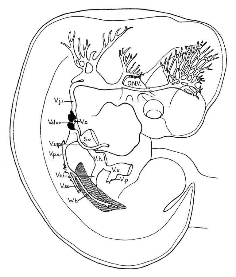
Fig 4. Human embryo measuring 10.5 mm
Mall collection, No. 109
Jugular Sacs
- The relation of the sac to the venous system as a whole is shown, which is a just external to the internal jugular vein reconstruction from serial sections.
- The sac lies external to the jugular vein and anterior to the primitive ulnar.
Approximately Carnegie stage 17 from the description and drawing.
Reference
Sabin FR. The lymphatic system in human embryos, with a consideration of the morphology of the system as a whole. (1909) Amer. J Anat. 9(1): 43–91.
Cite this page: Hill, M.A. (2026, October 31) Embryology Sabin1909 fig04.jpg. Retrieved from https://embryology.med.unsw.edu.au/embryology/index.php/File:Sabin1909_fig04.jpg
- © Dr Mark Hill 2026, UNSW Embryology ISBN: 978 0 7334 2609 4 - UNSW CRICOS Provider Code No. 00098G
File history
Yi efo/eka'e gwa ebo wo le nyangagi wuncin ye kamina wunga tinya nan
| Gwalagizhi | Nyangagi | Dimensions | User | Comment | |
|---|---|---|---|---|---|
| current | 14:08, 30 March 2011 | | 769 × 902 (97 KB) | S8600021 (talk | contribs) | ==Fig 4. Human embryo== ==Reference== Florence R. Sabin, The lymphatic system in human embryos, with a consideration of the morphology of the system as a whole. American Journal of Anatomy Volume 9, Issue 1, pages 43–91, 1909 [[Category:Historic Emb |
You cannot overwrite this file.
File usage
The following 3 pages use this file:


