File:Pohlman1911 plate1A.jpg

Original file (1,012 × 1,195 pixels, file size: 87 KB, MIME type: image/jpeg)
Plate 1A
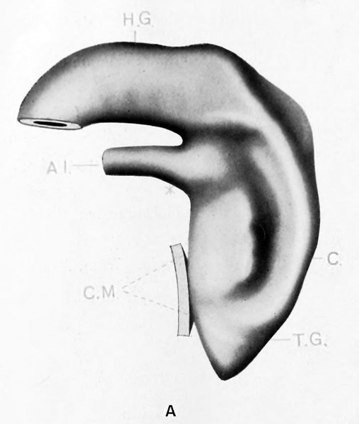
No. 3 (Mall 164; 3.5 mm. ; + ; 20; Good). Model A. x 100. Cloaca well developed and limited for caudal half of the ventral border by epithelial cloacal membrane. Allantois comes off at right angles and at lower border of dermal navel. Lateral furrow along later line of division. Carnegie stage 11
| Key to Lettering | ||
|---|---|---|
|
A., Allantois K., Kidney A.M., Position of anal membrane P.T., Precloacal mesodermic tissue C., Cloaca |
R.B., Renal bud CM., Cloacal membrane T.G., Tail cut C.S., Cloacal segment of Wolffian duct U., Ureter |
G.E., Genital eminence U.G., Urogenital sinus H.G., Hind gut U.M., Position of urogenital membrane * Probable position of point where allantois joins cloaca. |
All drawings represent 100 diameters enlargement except of Model F which is 50 diameters.
- Links: plate 1 | 1 A | 1 B | plate 2 | plate 2C | plate 2D | plate 3 | plate 3E | plate 3F | Pohlman 1911
Reference
Pohlman AG. The development of the cloaca in human embryos. (1911) Amer. J Anat. 12: 1-26.
Cite this page: Hill, M.A. (2026, October 4) Embryology Pohlman1911 plate1A.jpg. Retrieved from https://embryology.med.unsw.edu.au/embryology/index.php/File:Pohlman1911_plate1A.jpg
- © Dr Mark Hill 2026, UNSW Embryology ISBN: 978 0 7334 2609 4 - UNSW CRICOS Provider Code No. 00098G
File history
Yi efo/eka'e gwa ebo wo le nyangagi wuncin ye kamina wunga tinya nan
| Gwalagizhi | Nyangagi | Dimensions | User | Comment | |
|---|---|---|---|---|---|
| current | 15:13, 15 Haziran 2016 | | 1,012 × 1,195 (87 KB) | Z8600021 (talk | contribs) | ==Plate 1A== {{Pohlman1911 figures}} |
You cannot overwrite this file.
File usage
The following 2 pages use this file: