File:Keibel Mall 2 030.jpg
Original file (804 × 1,000 pixels, file size: 99 KB, MIME type: image/jpeg)
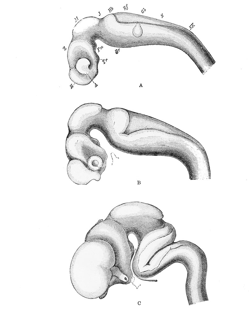
Fig. 30. Profile views of the brains of human embryos at third, fourth, and eighth weeks
Profile as seen during the third (A), fourth (B), and eighth (C) weeks, showing the conversion of the three primary cerebral vesicles into their chief subdivisions and the formation of the flexures of the neural tube.
A, optic vesicle; Br, pontine region; Gb, auditory vesicle; H, telencephalon; Hb, metencephalon; J, isthmus; M, mesencephalon; N, myelencephalon; NK, neck bend; Pm, mammillary recess; Rf, posterior medullary velum; Tr, infundibular recess; Z, diencephalon.
(After Wilhelm His (1831-1904))
Manual of Human Embryology II: Nervous System | Chromaffin Organs and Suprarenal Bodies | Sense-Organs | Digestive Tract and Respiration | Vascular System | Urinogenital Organs | Figures 2 | Manual of Human Embryology 1 | Figures 1 | Manual of Human Embryology 2 | Figures 2 | Franz Keibel | Franklin Mall | Embryology History
Cite this page: Hill, M.A. (2026, October 5) Embryology Keibel Mall 2 030.jpg. Retrieved from https://embryology.med.unsw.edu.au/embryology/index.php/File:Keibel_Mall_2_030.jpg
- © Dr Mark Hill 2026, UNSW Embryology ISBN: 978 0 7334 2609 4 - UNSW CRICOS Provider Code No. 00098G
File history
Yi efo/eka'e gwa ebo wo le nyangagi wuncin ye kamina wunga tinya nan
| Gwalagizhi | Nyangagi | Dimensions | User | Comment | |
|---|---|---|---|---|---|
| current | 11:45, 24 Ocak 2014 | | 804 × 1,000 (99 KB) | Z8600021 (talk | contribs) | {{Keibel_Mall 2 Images}} |
You cannot overwrite this file.
File usage
The following 4 pages use this file:


