File:Stage 9 SEM1.jpg: Difference between revisions
(Carnegie Stages 9 Features: embryonic disc, primitive node, primative streak, primative groove, somites, neural groove, brain plate region, connecting stalk, cut edge of amnion Facts: Week 3, 19 - 21 days, 1.5 - 2.5 mm, Somite Number 1 - 3 View 1: emb) |
No edit summary |
||
| Line 15: | Line 15: | ||
Original file name: 3-4SomiteSt9d20dorsal.jpg | Original file name: 3-4SomiteSt9d20dorsal.jpg | ||
'''Image Source:''' UNSW Embryology, no reproduction without permission. [http://embryology.med.unsw.edu.au/wwwhuman/Stages/stage9sem.htm UNSW Embryology Carnegie Stage 9 SEM] | '''Image Source:''' Prof Kathy Sulik scanning electron micrographs of the Carnegie stages of the early human embryo. UNSW Embryology, no reproduction without permission. [http://embryology.med.unsw.edu.au/wwwhuman/Stages/Stagesem.htm Carnegie Stages - Scanning Electron Micrography] | [http://embryology.med.unsw.edu.au/wwwhuman/Stages/stage9sem.htm UNSW Embryology Carnegie Stage 9 SEM] | ||
[[Category:Human Embryo]] [[Category:Carnegie Stage]] [[Category:Week 3]] [[Category:Mesoderm]] [[Category:Somite]] | [[Category:Human Embryo]] [[Category:Carnegie Stage]] [[Category:Week 3]] [[Category:Mesoderm]] [[Category:Somite]] [[Category:Scanning EM]] | ||
Revision as of 11:36, 14 August 2009
Carnegie Stages 9
Features: embryonic disc, primitive node, primative streak, primative groove, somites, neural groove, brain plate region, connecting stalk, cut edge of amnion
Facts: Week 3, 19 - 21 days, 1.5 - 2.5 mm, Somite Number 1 - 3
View 1: embryonic disc, showing the epiblast viewed from the amniotic (dorsal) side.
Events: Ectoderm - Neural plate brain region continues to expand, neural plate begins folding over the notochord. Mesoderm - segmentation of paraxial mesoderm begins (1 - 3 somite pairs), cardiac primordium forms
Identify: neural groove and neural folds, the mesoderm, which segments beside the neural groove to form somites but extends laterally to margin of embryonic disc lateral plate mesoderm, where it merges with the covering extraembryonic mesoderm.
The intra-embryonic coelom develops in the middle of the lateral plate mesoderm. Note amniotic ectoderm covered by extramebryonic mesoderm (empty spaces above and below the mesoderm are artefacts, as are the lateral folds in the ectoderm).
Original file name: 3-4SomiteSt9d20dorsal.jpg
Image Source: Prof Kathy Sulik scanning electron micrographs of the Carnegie stages of the early human embryo. UNSW Embryology, no reproduction without permission. Carnegie Stages - Scanning Electron Micrography | UNSW Embryology Carnegie Stage 9 SEM
File history
Yi efo/eka'e gwa ebo wo le nyangagi wuncin ye kamina wunga tinya nan
| Gwalagizhi | Nyangagi | Dimensions | User | Comment | |
|---|---|---|---|---|---|
| current | 10:51, 9 August 2009 | 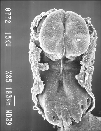 | 347 × 450 (42 KB) | S8600021 (talk | contribs) | Carnegie Stages 9 Features: embryonic disc, primitive node, primative streak, primative groove, somites, neural groove, brain plate region, connecting stalk, cut edge of amnion Facts: Week 3, 19 - 21 days, 1.5 - 2.5 mm, Somite Number 1 - 3 View 1: emb |
You cannot overwrite this file.