Episcopic Fluorescence Image Capture: Difference between revisions
(Created page with "{{Header}} ==Introduction== Episcopic Fluorescence Image Capture (EFIC). A microscopic imaging technique that serially sections embedded biological specimens and photogra...") |
mNo edit summary |
||
| Line 2: | Line 2: | ||
==Introduction== | ==Introduction== | ||
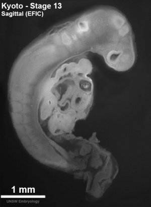
[[File:Stage 13 EFIC01.jpg|thumb|alt=|link=|EFIC sagittal image of embryo ([[Carnegie stage 13|stage 13]])]] | |||
[[Episcopic Fluorescence Image Capture]] (EFIC). A microscopic imaging technique that serially sections embedded biological specimens and photographs the tissue autofluorescence (epifluorescence) from the block surface. This generates an in register 2D image stack. Technique was described for the mouse in 2002.<ref name=PMID11743576><pubmed>11743576</pubmed></ref> | [[Episcopic Fluorescence Image Capture]] (EFIC). A microscopic imaging technique that serially sections embedded biological specimens and photographs the tissue autofluorescence (epifluorescence) from the block surface. This generates an in register 2D image stack. Technique was described for the mouse in 2002.<ref name=PMID11743576><pubmed>11743576</pubmed></ref> | ||
Revision as of 11:20, 17 August 2016
| Embryology - 27 Jun 2024 |
|---|
| Google Translate - select your language from the list shown below (this will open a new external page) |
|
العربية | català | 中文 | 中國傳統的 | français | Deutsche | עִברִית | हिंदी | bahasa Indonesia | italiano | 日本語 | 한국어 | မြန်မာ | Pilipino | Polskie | português | ਪੰਜਾਬੀ ਦੇ | Română | русский | Español | Swahili | Svensk | ไทย | Türkçe | اردو | ייִדיש | Tiếng Việt These external translations are automated and may not be accurate. (More? About Translations) |
Introduction
Episcopic Fluorescence Image Capture (EFIC). A microscopic imaging technique that serially sections embedded biological specimens and photographs the tissue autofluorescence (epifluorescence) from the block surface. This generates an in register 2D image stack. Technique was described for the mouse in 2002.[1]
References
- ↑ <pubmed>11743576</pubmed>
Reviews
<pubmed></pubmed> <pubmed></pubmed> <pubmed></pubmed> <pubmed></pubmed>
Articles
<pubmed>20503356</pubmed> <pubmed></pubmed> <pubmed></pubmed> <pubmed></pubmed>
Search PubMed
Search Pubmed: Episcopic Fluorescence Image Capture
Terms
External Links
External Links Notice - The dynamic nature of the internet may mean that some of these listed links may no longer function. If the link no longer works search the web with the link text or name. Links to any external commercial sites are provided for information purposes only and should never be considered an endorsement. UNSW Embryology is provided as an educational resource with no clinical information or commercial affiliation.
Glossary Links
- Glossary: A | B | C | D | E | F | G | H | I | J | K | L | M | N | O | P | Q | R | S | T | U | V | W | X | Y | Z | Numbers | Symbols | Term Link
Cite this page: Hill, M.A. (2024, June 27) Embryology Episcopic Fluorescence Image Capture. Retrieved from https://embryology.med.unsw.edu.au/embryology/index.php/Episcopic_Fluorescence_Image_Capture
- © Dr Mark Hill 2024, UNSW Embryology ISBN: 978 0 7334 2609 4 - UNSW CRICOS Provider Code No. 00098G