BGDB Sexual Differentiation - Fetal: Difference between revisions
mNo edit summary |
mNo edit summary |
||
| Line 147: | Line 147: | ||
| End of testis descent | | End of testis descent | ||
|} | |} | ||
==Fetal Kidney== | |||
'''Nephrogenesis''' is the formation of the functional nephron occurs in the fetal period with all of the nephrons formed by 30 to 34 weeks ({{GA}} 32 to 36 weeks). There are no new nephrons formed after this period. | |||
==Additional Information== | ==Additional Information== | ||
Revision as of 11:08, 31 May 2016
Introduction
In the previous section we observed late embryonic male genital development and now in fetal development we will observe early fetal female development. Then we will explore fetal development of the external genitalia and gonadal descent.
Week 10 Female

|

|
|

|

|
| Plane A (most lateral) | Plane B (lateral) |

|

|
| Plane C (medial) | Plane D (midline) |
Uterus and Vagina

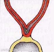
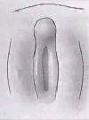
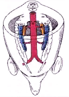
Mouse paramesonephric duct (Müllerian duct)[1] |
This mouse image shows the relationship between the mesonephric and paramesonephric ducts opening into the urogenital sinus.
|
| <mediaplayer width='490' height='500' image="http://php.med.unsw.edu.au/embryology/images/2/2d/Uterus_001_icon.jpg">File:Uterus_001.mp4</mediaplayer> | Female Uterus and Vagina (between week 9 and 20)
The uterus and broad ligament will eventulaly divide the pelvic cavity into two separate pouches.
|

|
This graph shows the growth during the fetal period of the uterus between week 19 and 38.[2]
|
External Genitalia
This next section will look at the development of the external genitalia using a series of animations and online resources.
Female External Genitalia
| <mediaplayer width='270' height='380' image="http://php.med.unsw.edu.au/embryology/images/3/37/Male_external_001_icon.jpg">File:Female_external_001.mp4</mediaplayer> | Animation showing the development of external female genitalia from the indifferent external structure (week 9 to 12 approximately).
|
Male External Genitalia

| <mediaplayer width='270' height='380' image="http://php.med.unsw.edu.au/embryology/images/3/37/Male_external_001_icon.jpg">File:Male_external_001.mp4</mediaplayer> | Animation showing the development of external male genitalia from the indifferent external structure (week 9 to 12 approximately).
The reduction of Testosterone to active metabolite, 5α-dihydrotestosterone (DHT) is carried out by the enzyme 5α-reductase expressed in the region or male external genitaila and prostate gland. Note that there are several 5α-reductase isoforms that differ in both tissue distribution and kinetics.
|
External Genitalia Comparison
Gonad Descent
| <mediaplayer width='296' height='430' image="http://php.med.unsw.edu.au/embryology/images/7/75/Gonad_blood_01_icon.jpg">File:Gonad blood 01.mp4</mediaplayer> | Animation shows the descent of the gonads and their blood supply.
|
Internal Gonad Descent
Testes Descent
| The linked animation shows the descent of the testes (between week 7 to 38, birth).
Descent of the testes into the scrotal sac begins generally during week 26 and may take several days.
Incomplete or failed descent can occur unilaterally or bilaterally, is more common in premature births, and can be completed postnatally. (see also cryptorchidism). |

|

|
| Start of testis descent | End of testis descent |
Fetal Kidney
Nephrogenesis is the formation of the functional nephron occurs in the fetal period with all of the nephrons formed by 30 to 34 weeks (GA 32 to 36 weeks). There are no new nephrons formed after this period.
Additional Information
| Additional Information - Content shown under this heading is not part of the material covered in this class. It is provided for those students who would like to know about some concepts or current research in topics related to the current class page. |
Testes Descent Timeline
Data from a study of male human fetal (between 10 and 35 weeks) gonad position.[3]
- 10 to 23 weeks - (9.45%) had migrated from the abdomen and were situated in the inguinal canal
- 24 to 26 weeks - (57.9%) had migrated from the abdomen
- 27 to 29 weeks - (16.7%) had not descended to the scrotum
A second study looked at the position of the testes[4]
- 33 weeks fetal testes had descended to the scrotum
- between 33 to 40 weeks (term) both testes have normally descended to the scrotum
Failure of descent (cryptorchidism) either unilateral or bilateral testicular descent, occurring in up to 30% premature and 3-4% term males.
Cryptorchidism in common eutherian mammals.[5]- Species comparison of descent timeline
Mouse Renal Stages

Histology showing the developmental stages of nephrogenesis.
<pubmed>21604189</pubmed>
Hyperfiltration Hypothesis
Developmental renal mass reduction could result in glomerular alterations that may have adverse effects in long-term renal health.
<pubmed>8743495</pubmed> <pubmed>22532329</pubmed>
Historic Genital Images
| Keith (1902) |
|---|
| Keith A. Human Embryology and Morphology. (1902) London: Edward Arnold.
Chapter 9 - The Uro-genital System
|


| Gary's Anatomy (1918) |
|---|
Gray H. Anatomy of the human body. (1918) Philadelphia: Lea & Febiger.
|


References
BGDB: Lecture - Gastrointestinal System | Practical - Gastrointestinal System | Lecture - Face and Ear | Practical - Face and Ear | Lecture - Endocrine | Lecture - Sexual Differentiation | Practical - Sexual Differentiation | Tutorial
Glossary Links
- Glossary: A | B | C | D | E | F | G | H | I | J | K | L | M | N | O | P | Q | R | S | T | U | V | W | X | Y | Z | Numbers | Symbols | Term Link
Cite this page: Hill, M.A. (2024, June 20) Embryology BGDB Sexual Differentiation - Fetal. Retrieved from https://embryology.med.unsw.edu.au/embryology/index.php/BGDB_Sexual_Differentiation_-_Fetal
- © Dr Mark Hill 2024, UNSW Embryology ISBN: 978 0 7334 2609 4 - UNSW CRICOS Provider Code No. 00098G