File:Respiratory histology 11.jpg: Difference between revisions
From Embryology
No edit summary |
mNo edit summary |
||
| Line 1: | Line 1: | ||
==Inferior Concha Respiratory Histology== | ==Inferior Concha Respiratory Histology== | ||
Inferior Concha, human | |||
{{Alcian blue}} | |||
{{van Gieson}} | |||
Layers from surface epithelium to underlying bone. | |||
===Respiratory epithelium=== | |||
* goblet cells | |||
* ciliated cells | |||
* basal cells | |||
===Lamina propria=== | |||
* connective tissue | |||
* cavernous sinusoids - large spaces (empty or filled with red blood cells) | |||
* glandular tissue - mucous glands (green) and muco-serous glands (brownish-green) | |||
===Bone=== | |||
* Lamellae and osteocytes in lacunae. | |||
* Haversian systems are rare or absent. | |||
{{Nasal_respiratory_links}} | |||
| Line 8: | Line 33: | ||
{{ | {{Blue Histology}} | ||
[[Category:Respiratory]] [[Category:Histology]] | [[Category:Respiratory]] [[Category:Histology]] | ||
Revision as of 13:18, 10 March 2013
Inferior Concha Respiratory Histology
Inferior Concha, human
(Stain - Alcian blue)
(Stain - van Gieson)
Layers from surface epithelium to underlying bone.
Respiratory epithelium
- goblet cells
- ciliated cells
- basal cells
Lamina propria
- connective tissue
- cavernous sinusoids - large spaces (empty or filled with red blood cells)
- glandular tissue - mucous glands (green) and muco-serous glands (brownish-green)
Bone
- Lamellae and osteocytes in lacunae.
- Haversian systems are rare or absent.
- Respiratory Histology: Bronchiole | Alveolar Duct | Alveoli | EM Alveoli septum | Alveoli Elastin | Trachea 1 | Trachea 2 | labeled lung | unlabeled lung | Respiratory Bronchiole | Lung Reticular Fibres | Nasal Inferior Concha | Nasal Respiratory Epithelium | Olfactory Region overview | Olfactory Region Epithelium | Histology Stains
Links: Histology | Histology Stains | Blue Histology images copyright Lutz Slomianka 1998-2009. The literary and artistic works on the original Blue Histology website may be reproduced, adapted, published and distributed for non-commercial purposes. See also the page Histology Stains.
Cite this page: Hill, M.A. (2024, June 18) Embryology Respiratory histology 11.jpg. Retrieved from https://embryology.med.unsw.edu.au/embryology/index.php/File:Respiratory_histology_11.jpg
- © Dr Mark Hill 2024, UNSW Embryology ISBN: 978 0 7334 2609 4 - UNSW CRICOS Provider Code No. 00098G
File history
Yi efo/eka'e gwa ebo wo le nyangagi wuncin ye kamina wunga tinya nan
| Gwalagizhi | Nyangagi | Dimensions | User | Comment | |
|---|---|---|---|---|---|
| current | 23:02, 28 February 2012 | 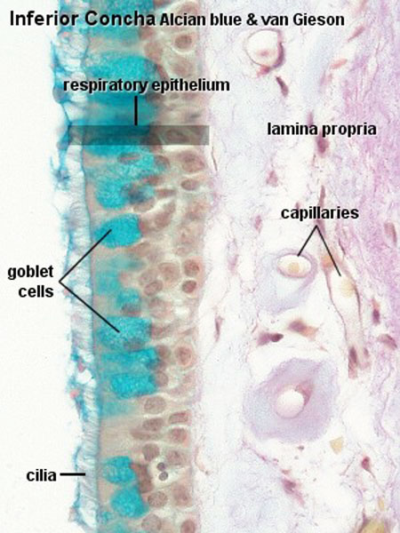 | 450 × 600 (65 KB) | Z8600021 (talk | contribs) |
You cannot overwrite this file.
File usage
The following 11 pages use this file:
- ANAT2241 Glandular Epithelia
- ANAT2241 Respiratory System
- ANAT2511 Respiratory System
- Draft 2016
- Histology
- Lecture - Respiratory Development
- Respiratory System - Histology
- Respiratory System - Postnatal
- Respiratory System - Upper Respiratory Tract
- SH Lecture - Respiratory System Development
- SH Practical - Respiratory