Category:Spinal Cord: Difference between revisions
From Embryology
No edit summary |
No edit summary |
||
| Line 1: | Line 1: | ||
This page lists UNSW Embryology content related to development of the spinal cord. | This page lists UNSW Embryology content related to development of the spinal cord. | ||
:'''Links:''' [[Neural_-_Spinal_Cord_Development|Spinal Cord]] | |||
[[Category:Neural]] | |||
Revision as of 17:49, 15 July 2012
This page lists UNSW Embryology content related to development of the spinal cord.
- Links: Spinal Cord
Pages in category 'Spinal Cord'
The following 36 pages are in this category, out of 36 total.
M
P
- Paper - Cell columns in the spinal cord of a human foetus of fourteen weeks (1941)
- Paper - Development of the innervation pattern in the upper limb of staged human embryos (1990)
- Paper - Factors Involved In The Formation Of The Filum Terminale
- Paper - The development and significance of the cell columns in the ventral horn of the cervical and upper thoracic spinal cord of the rabbit (1941)
- Paper - The early development of the meninges of the spinal cord in human embryos (1951)
- Paper - The structure of the spinal cord of the ostrich
R
- Template:Ref-Bardeen1903
- Template:Ref-DartShellshear1922
- Template:Ref-Hogg1945
- Template:Ref-Hoskins1914
- Template:Ref-Kunitomo1920
- Template:Ref-Miller1913
- Template:Ref-O’RahillyMuller1986
- Template:Ref-Romanes1941
- Template:Ref-Romanes1941a
- Template:Ref-Romanes1941b
- Template:Ref-ScharpenbergWindle1938
- Template:Ref-Sensenig1951
Media in category 'Spinal Cord'
The following 65 files are in this category, out of 65 total.
 Bailey385.jpg 787 × 683; 165 KB
 Bailey403.jpg 495 × 617; 118 KB
 Bailey407.jpg 793 × 695; 117 KB
 Caudal duplication syndrome.jpg 700 × 599; 47 KB
 Cervical vertebra.jpg 767 × 514; 71 KB
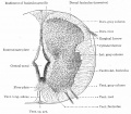
 Gray0664.jpg 696 × 500; 123 KB
 Gray0666.jpg 237 × 1,000; 46 KB
 Gray0666new.jpg 600 × 551; 47 KB
 Gray0670.jpg 800 × 504; 67 KB
 Gray0671.jpg 347 × 900; 54 KB
 Gray0675.jpg 615 × 600; 69 KB
 Gray0770.jpg 700 × 275; 68 KB
 Gray0804.jpg 550 × 700; 75 KB
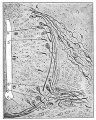
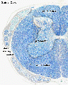
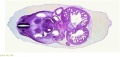
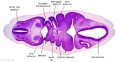
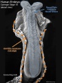
 Human Stage22 spinal cord01.jpg 1,044 × 889; 265 KB
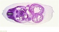
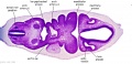
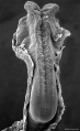
 Human Stage22 spinal cord02.jpg 1,044 × 889; 290 KB
 Human week 10 fetus 11.jpg 1,200 × 900; 304 KB
 Keith1921 fig064.jpg 908 × 565; 96 KB
 Keith1921 fig065.jpg 1,200 × 890; 219 KB
 Keith1921 fig066.jpg 1,000 × 1,067; 100 KB
 Keith1921 fig067.jpg 1,200 × 722; 150 KB
 Keith1921 fig068.jpg 903 × 784; 170 KB
 Keith1921 fig073a.jpg 884 × 822; 162 KB
 Keith1921 fig074.jpg 1,045 × 602; 134 KB
 Keith1921 fig075.jpg 856 × 636; 125 KB
 Keith1921 fig076.jpg 1,207 × 729; 265 KB
 Keith1921 fig077.jpg 629 × 640; 72 KB
 Keith1921 fig079.jpg 741 × 839; 109 KB
 Kollmann646.jpg 1,000 × 606; 122 KB
 Kollmann647.jpg 897 × 926; 188 KB
 McMurrich1930 fig81.jpg 1,280 × 796; 137 KB
 Mouse- spinal cord axons.jpg 600 × 693; 127 KB
 Neural tube SHH patterning cartoon.jpg 458 × 594; 92 KB
 Rugh 119.jpg 842 × 800; 127 KB
 Rugh 132.jpg 735 × 1,000; 195 KB
 Rugh 133.jpg 1,000 × 707; 141 KB
 Sensenig1951 plate01.jpg 1,979 × 2,591; 1.35 MB
 Sensenig1951 plate02.jpg 2,078 × 2,619; 1.52 MB
 Sensenig1951 plate03.jpg 1,963 × 2,664; 1.33 MB
 Sensenig1951 plate04.jpg 1,990 × 2,627; 1.29 MB
 Spinal cord delta notch model.png 599 × 457; 171 KB
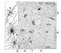
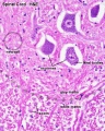
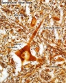
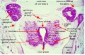
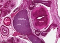
 Spinal cord histology 01.jpg 480 × 600; 116 KB
 Spinal cord histology 02.jpg 480 × 600; 121 KB
 Spinal cord histology 03.jpg 480 × 600; 103 KB
 Spinal cord histology 04.jpg 480 × 600; 119 KB
 Spinal cord histology 05.jpg 1,280 × 1,024; 463 KB
 Spinal cord histology 06.jpg 1,280 × 1,024; 318 KB
 Spinal cord histology 07.jpg 1,280 × 1,024; 365 KB
 Spinal cord histology 08.jpg 1,280 × 1,024; 418 KB
 Spinal cord histology 09.jpg 1,280 × 1,024; 227 KB
 Spinal cord histology 10.gif 480 × 600; 456 KB
 Spinal cord histology 11.jpg 481 × 600; 117 KB
 Spinal cord histology 12.jpg 480 × 600; 130 KB
 Spinal cord tracts.png 1,000 × 430; 204 KB
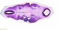 Stage 13 image 007.jpg 1,000 × 514; 93 KB
 Stage 13 image 022.jpg 1,000 × 473; 101 KB
 Stage 13 image 023.jpg 1,000 × 544; 110 KB
 Stage 13 image 057.jpg 1,000 × 511; 99 KB
 Stage 13 image 058.jpg 1,000 × 481; 94 KB
 Stage 13 image 059.jpg 1,000 × 513; 92 KB
 Stage 13 image 060.jpg 1,000 × 486; 96 KB
 Stage 13 image 061.jpg 1,000 × 600; 101 KB
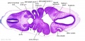
 Stage 22 image 176.jpg 1,000 × 659; 223 KB
 Stage10 sem6 annotated.jpg 720 × 960; 122 KB
 Stage10 sem6.jpg 614 × 1,000; 57 KB
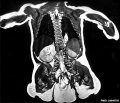
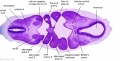
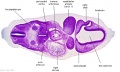
 Stage22 vertebra and spinal cord 1.jpg 1,000 × 725; 344 KB


