File:Respiratory histology 07.jpg: Difference between revisions
From Embryology
(==Respiratory Trachea== Respiratory histology 07.jpg lung, human H&E respiratory system, overview Original file name: lun04he.jpg {{Template:Blue Histology}} Category:Respiratory) |
No edit summary |
||
| Line 1: | Line 1: | ||
==Respiratory | ==Respiratory Lung Structure== | ||
Revision as of 16:27, 2 March 2011
Respiratory Lung Structure
Respiratory histology 07.jpg
lung, human H&E respiratory system, overview
Original file name: lun04he.jpg
Links: Histology | Histology Stains | Blue Histology images copyright Lutz Slomianka 1998-2009. The literary and artistic works on the original Blue Histology website may be reproduced, adapted, published and distributed for non-commercial purposes. See also the page Histology Stains.
Cite this page: Hill, M.A. (2024, June 27) Embryology Respiratory histology 07.jpg. Retrieved from https://embryology.med.unsw.edu.au/embryology/index.php/File:Respiratory_histology_07.jpg
- © Dr Mark Hill 2024, UNSW Embryology ISBN: 978 0 7334 2609 4 - UNSW CRICOS Provider Code No. 00098G
File history
Yi efo/eka'e gwa ebo wo le nyangagi wuncin ye kamina wunga tinya nan
| Gwalagizhi | Nyangagi | Dimensions | User | Comment | |
|---|---|---|---|---|---|
| current | 22:48, 28 February 2012 |  | 1,280 × 1,024 (255 KB) | Z8600021 (talk | contribs) | |
| 22:45, 28 February 2012 | 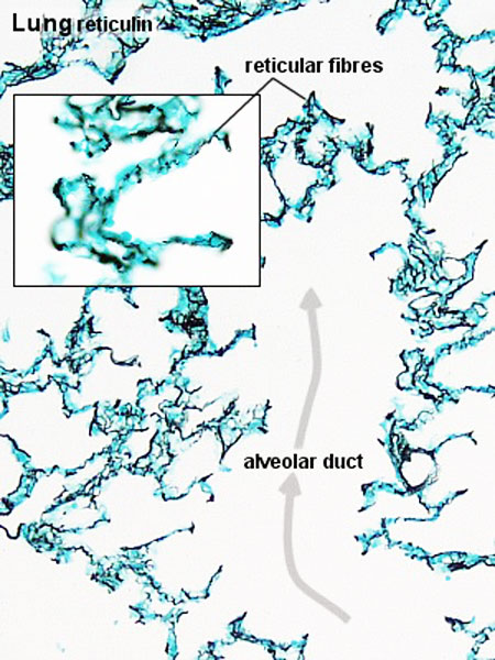 | 450 × 600 (82 KB) | Z8600021 (talk | contribs) | ==Fetal Lung Histology== * late canalicular period. * mucous connective tissue fills fairly wide spaces between the terminal sacs. {{Respiratory Histology}} {{Template:Blue Histology}} Category:Respiratory Category:Histology [[Category:Hum | |
| 16:22, 2 March 2011 | 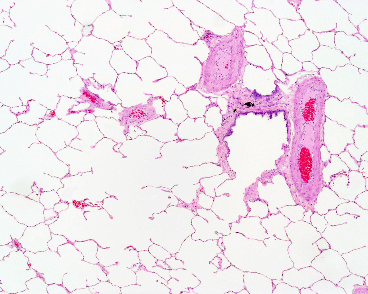 | 1,280 × 1,024 (255 KB) | S8600021 (talk | contribs) | ==Respiratory Trachea== Respiratory histology 07.jpg lung, human H&E respiratory system, overview Original file name: lun04he.jpg {{Template:Blue Histology}} Category:Respiratory |
You cannot overwrite this file.
File usage
The following 4 pages use this file: