File:Bailey388.jpg: Difference between revisions
From Embryology
(==Fig. 388. Ventral part of wall of lumbar cord of 70-hour duck embryo== Showing efferent root fibers first emerging from cord (combined from two sections) . Cajal. A, Spinal cord; B, perimedullary space; C, meningeal membrane; a, b, cones of radially di) |
No edit summary |
||
| Line 5: | Line 5: | ||
{{Template:Bailey 1921 Figures}} | {{Template:Bailey 1921 Figures}} | ||
[[Category:Neural]] | [[Category:Neural]] [[Category:Cajal]] | ||
Revision as of 01:07, 14 February 2011
Fig. 388. Ventral part of wall of lumbar cord of 70-hour duck embryo
Showing efferent root fibers first emerging from cord (combined from two sections) . Cajal. A, Spinal cord; B, perimedullary space; C, meningeal membrane; a, b, cones of radially directed axones; c, d, cones of transversely directed axones; Z>, bifurcated cone; E,F, cones crossing perimedullary space; G, aberrant cones.
- Text-Book of Embryology: Germ cells | Maturation | Fertilization | Amphioxus | Frog | Chick | Mammalian | External body form | Connective tissues and skeletal | Vascular | Muscular | Alimentary tube and organs | Respiratory | Coelom, Diaphragm and Mesenteries | Urogenital | Integumentary | Nervous System | Special Sense | Foetal Membranes | Teratogenesis | Gallery of All Figures
| Historic Disclaimer - information about historic embryology pages |
|---|
 |
Reference
Bailey FR. and Miller AM. Text-Book of Embryology (1921) New York: William Wood and Co.
Cite this page: Hill, M.A. (2024, June 10) Embryology Bailey388.jpg. Retrieved from https://embryology.med.unsw.edu.au/embryology/index.php/File:Bailey388.jpg
- © Dr Mark Hill 2024, UNSW Embryology ISBN: 978 0 7334 2609 4 - UNSW CRICOS Provider Code No. 00098G
File history
Click on a date/time to view the file as it appeared at that time.
| Date/Time | Thumbnail | Dimensions | User | Comment | |
|---|---|---|---|---|---|
| current | 00:38, 30 January 2011 | 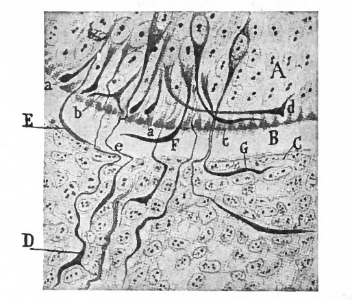 | 514 × 438 (68 KB) | S8600021 (talk | contribs) | ==Fig. 388. Ventral part of wall of lumbar cord of 70-hour duck embryo== Showing efferent root fibers first emerging from cord (combined from two sections) . Cajal. A, Spinal cord; B, perimedullary space; C, meningeal membrane; a, b, cones of radially di |
You cannot overwrite this file.
File usage
The following 4 pages use this file: