File:Foster135.jpg: Difference between revisions
From Embryology
No edit summary |
mNo edit summary |
||
| Line 1: | Line 1: | ||
==Fig. 135. Longitudinal section through intervertebral ligament and two vertebra== | |||
la | Longitudinal section through the intervertebral ligament and adjacent parts of two vertebra from the thoracic region of an advanced embryo of a {{sheep}}. (From Kolliker.) | ||
la. ligamentum longitudinale anterius ; Ip. ligamentum long, posterius ; li. ligamentum intervertebrale ; , k r . epiphysis of vertebra ; w. and w f . anterior and posterior vertebrae ; c. intervertebral dilatation of notochord ; c.' and c". vertebral dilatation of notochord. | |||
{{Template:Foster_1883_Figures_2}} | {{Template:Foster_1883_Figures_2}} | ||
[[Category:Sheep]] [[Category:Musculoskeletal]] | [[Category:Sheep]] [[Category:Musculoskeletal]][[Category:Axial Skeleton]] | ||
Latest revision as of 10:14, 20 Haziran 2018
Fig. 135. Longitudinal section through intervertebral ligament and two vertebra
Longitudinal section through the intervertebral ligament and adjacent parts of two vertebra from the thoracic region of an advanced embryo of a sheep. (From Kolliker.)
la. ligamentum longitudinale anterius ; Ip. ligamentum long, posterius ; li. ligamentum intervertebrale ; , k r . epiphysis of vertebra ; w. and w f . anterior and posterior vertebrae ; c. intervertebral dilatation of notochord ; c.' and c". vertebral dilatation of notochord.
Reference
Foster, M., Balfour, F. M., Sedgwick, A., & Heape, W. (1883). The Elements of Embryology. (2nd ed.). London: Macmillan and Co.
- Volume 1 - The History of the Chick: Egg structure and incubation beginning | Summary whole incubation | First day | Second day - first half | Second day - second half | Third day | Fourth day | Fifth day | Sixth day to incubation end | Figures 1
- Volume 2 - The History of the Mammalian Embryo: General Development | Embryonic Membranes and Yolk-Sac | Organs from Epiblast | Organs from Mesoblast | Alimentary Canal | Appendix | Figures 2
| Historic Disclaimer - information about historic embryology pages |
|---|
 |
File history
Yi efo/eka'e gwa ebo wo le nyangagi wuncin ye kamina wunga tinya nan
| Gwalagizhi | Nyangagi | Dimensions | User | Comment | |
|---|---|---|---|---|---|
| current | 19:17, 12 Ocak 2011 | 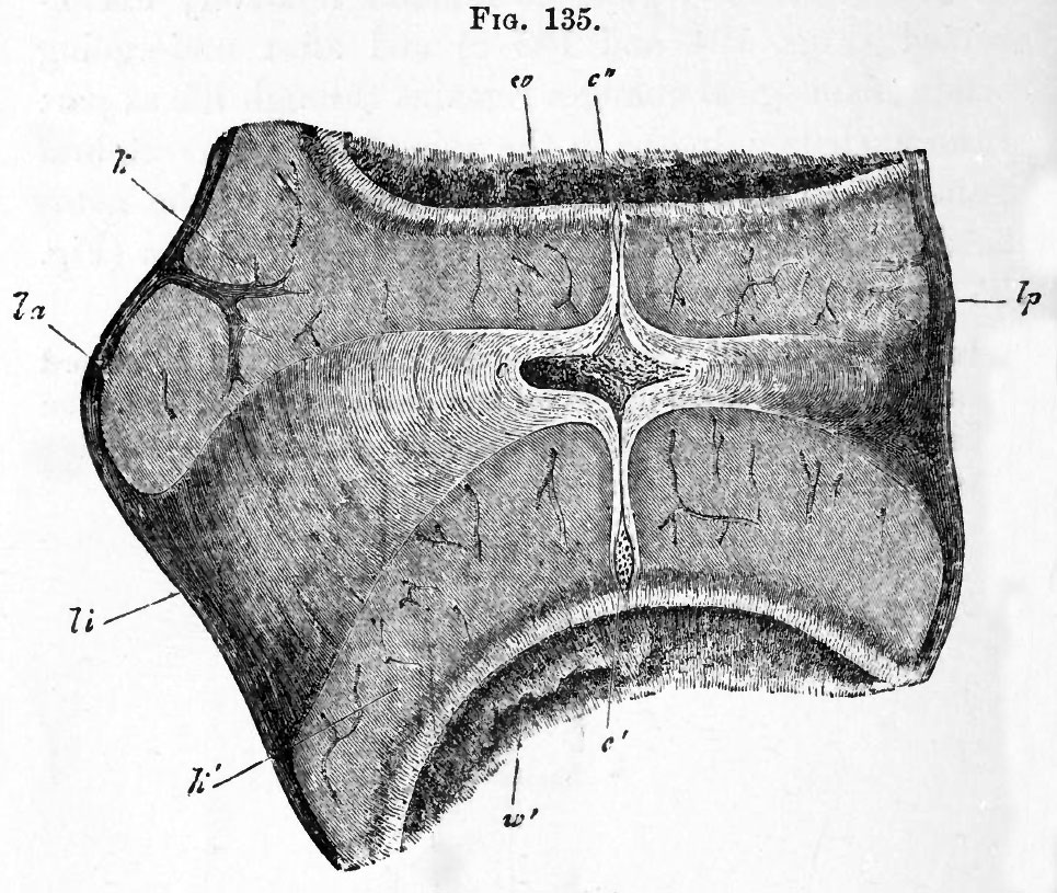 | 965 × 815 (230 KB) | S8600021 (talk | contribs) | {{Template:Foster_1883_Figures_2}} |
You cannot overwrite this file.
File usage
The following 2 pages use this file:


