File:Fetal brain MRI03.jpg: Difference between revisions
From Embryology
No edit summary |
mNo edit summary |
||
| Line 1: | Line 1: | ||
==Fetal Brain 28 Week MRI== | |||
Cortical surfaces for neonates at 28, 36 and 44 weeks PMA at scan with the labels overlaid. | |||
:'''Links:''' [[:File:Fetal brain MRI01.jpg|28-44 week MRI]] | [[:File:Fetal brain MRI02.jpg|28 week MRI]] | [[:File:Fetal brain MRI03.jpg|36 week MRI]] | [[:File:Fetal brain MRI04.jpg|44 week MRI]] | {{fetal neural}} | {{neural}} | {{MRI}} | |||
===Reference=== | |||
{{#pmid:26499811}} | |||
====Copyright==== | |||
https://creativecommons.org/licenses/by/4.0/ | |||
Fig. 8. resized and relabelled | |||
{{Footer}} | |||
[[Category:Neural]][[Category:Magnetic Resonance Imaging]] | |||
Revision as of 14:43, 7 July 2018
Fetal Brain 28 Week MRI
Cortical surfaces for neonates at 28, 36 and 44 weeks PMA at scan with the labels overlaid.
- Links: 28-44 week MRI | 28 week MRI | 36 week MRI | 44 week MRI | fetal neural | neural | MRI
Reference
Makropoulos A, Aljabar P, Wright R, Hüning B, Merchant N, Arichi T, Tusor N, Hajnal JV, Edwards AD, Counsell SJ & Rueckert D. (2016). Regional growth and atlasing of the developing human brain. Neuroimage , 125, 456-478. PMID: 26499811 DOI.
Copyright
https://creativecommons.org/licenses/by/4.0/
Fig. 8. resized and relabelled
Cite this page: Hill, M.A. (2024, June 27) Embryology Fetal brain MRI03.jpg. Retrieved from https://embryology.med.unsw.edu.au/embryology/index.php/File:Fetal_brain_MRI03.jpg
- © Dr Mark Hill 2024, UNSW Embryology ISBN: 978 0 7334 2609 4 - UNSW CRICOS Provider Code No. 00098G
File history
Yi efo/eka'e gwa ebo wo le nyangagi wuncin ye kamina wunga tinya nan
| Gwalagizhi | Nyangagi | Dimensions | User | Comment | |
|---|---|---|---|---|---|
| current | 14:41, 7 July 2018 | 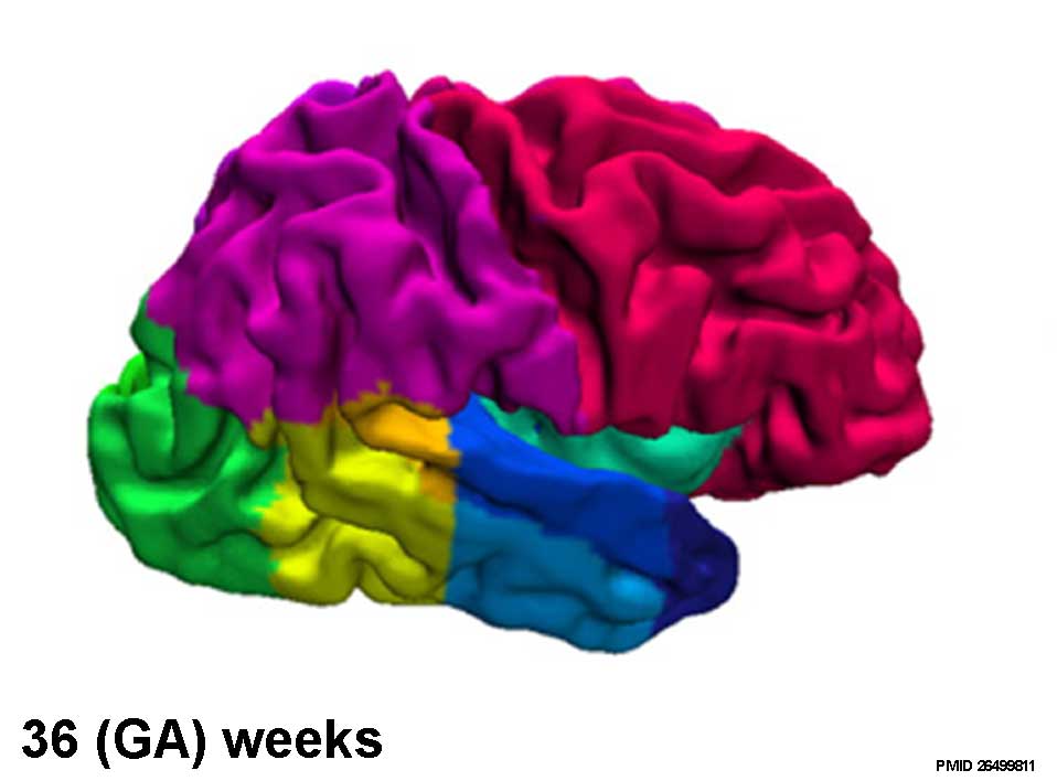 | 958 × 708 (34 KB) | Z8600021 (talk | contribs) |
You cannot overwrite this file.
File usage
The following page uses this file: