File:Ovary corpus luteum.jpg: Difference between revisions
(Z8600021 uploaded a new version of File:Ovary corpus luteum.jpg) |
(Z8600021 uploaded a new version of File:Ovary corpus luteum.jpg) |
(No difference)
| |
Revision as of 19:19, 5 May 2018
Ovary - Corpus Luteum
(Stain - Haematoxylin Eosin) Histology image shows the ovary in overview, the cortex and medulla of the ovary can be clearly seen.
Corpus luteum (yellow body) theca lutein cells and granulosa lutein cells. These cells work together in the production of ovarian hormones that support the initial pregnancy.
Corpus albicans (white body) lack of implantation and associated hCG will lead to this structure not producing hormones.
Atretic follicles are the degenerating follicles from various developmental stages that did not form the ovulating follicle and do not form the corpus luteum.
Connective Tissue
- fills most of the original follicular fluid-filled space.
Granulosa Lutein Cells
- the lighter stained cells.
- derived from the granulosa cells of the original follicle.
- contain aromatase enzyme.
- produce estrogen and progesterone from the androgens produced by the theca lutein cells.
Theca Lutein Cells
- the darker stained cells.
- derived from the theca interna of the original follicle.
- lack microvilli on the surface.
- lack the aromatase enzyme.
- produce androgens for the granulosa lutein cells to convert.
Cite this page: Hill, M.A. (2024, June 27) Embryology Ovary corpus luteum.jpg. Retrieved from https://embryology.med.unsw.edu.au/embryology/index.php/File:Ovary_corpus_luteum.jpg
- © Dr Mark Hill 2024, UNSW Embryology ISBN: 978 0 7334 2609 4 - UNSW CRICOS Provider Code No. 00098G
File history
Yi efo/eka'e gwa ebo wo le nyangagi wuncin ye kamina wunga tinya nan
| Gwalagizhi | Nyangagi | Dimensions | User | Comment | |
|---|---|---|---|---|---|
| current | 19:19, 5 May 2018 |  | 2,178 × 1,137 (376 KB) | Z8600021 (talk | contribs) | |
| 18:58, 5 May 2018 | 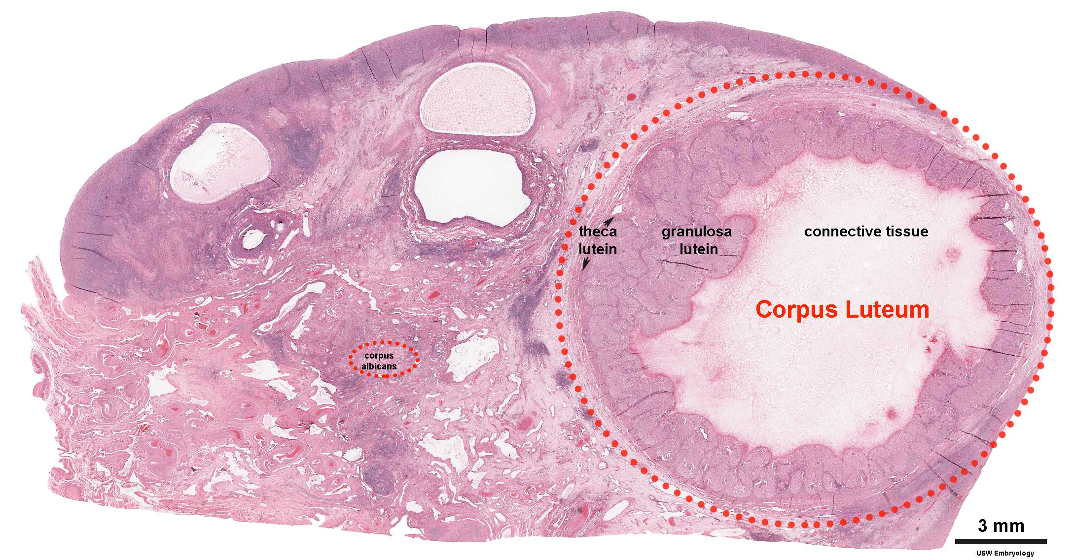 | 2,178 × 1,137 (375 KB) | Z8600021 (talk | contribs) | ||
| 18:50, 5 May 2018 | 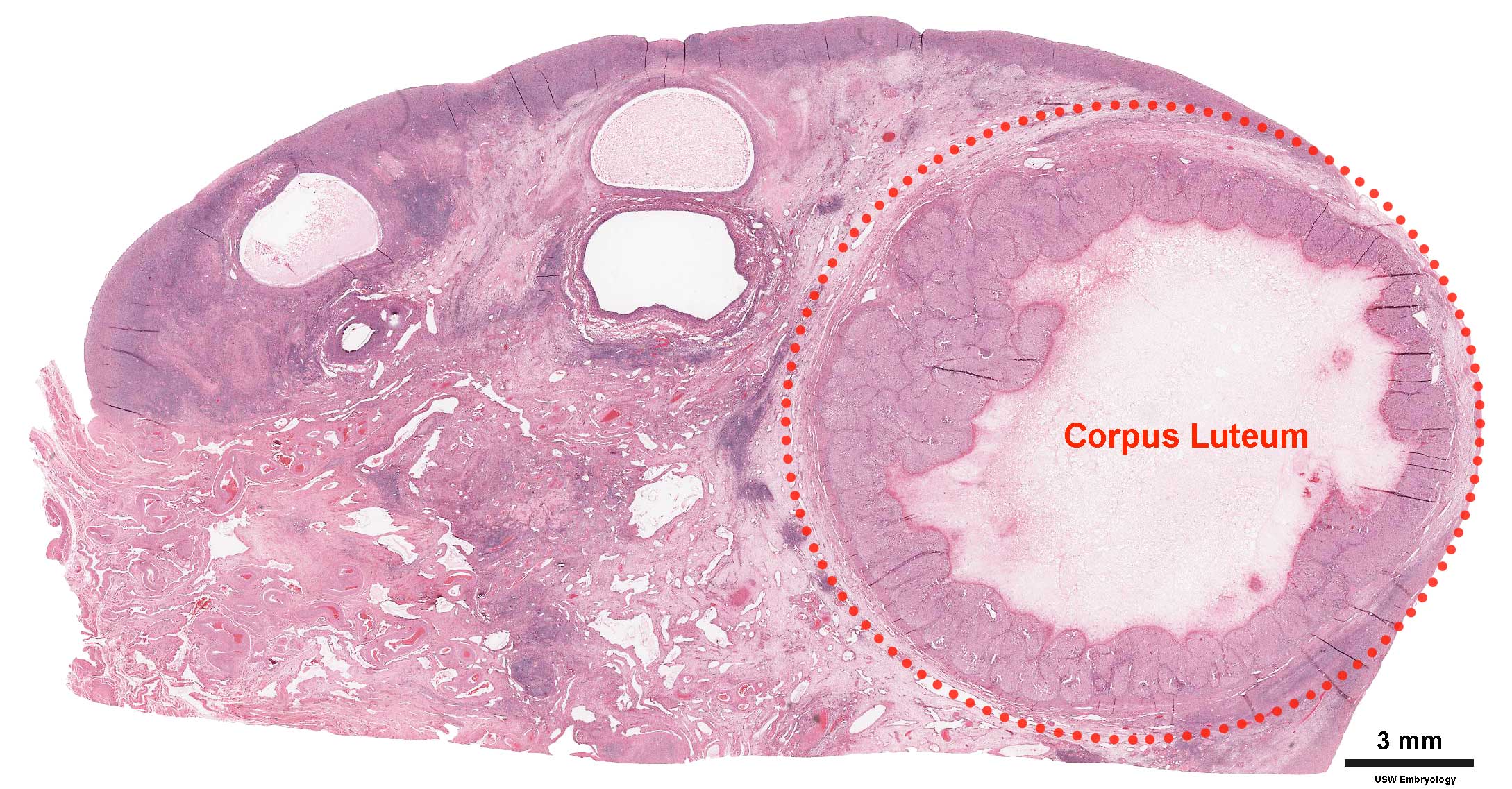 | 2,178 × 1,137 (369 KB) | Z8600021 (talk | contribs) | ||
| 10:10, 3 August 2009 |  | 800 × 455 (103 KB) | MarkHill (talk | contribs) | Ovary showing corpus luteum and atretic follicles. Histological image, H&E stained. Image Source: UNSW Embryology http://embryology.med.unsw.edu.au/Medicine/BGDlabfertilization6.htm |
You cannot overwrite this file.
File usage
The following 17 pages use this file:
- 2009 Lecture 3
- 2010 BGD Practical 3 - Implantation
- 2010 Foundations Lecture - Introduction to Human Development
- 2010 Lecture 3
- 2011 Lab 2 - Week 2
- ANAT2241 Female Reproductive System
- ANAT2341 Lab 2 - Week 2
- BGDA Practical - Female Reproductive Tract Histology
- BGDA Practical 3 - Implantation
- Corpus Luteum Development
- Foundations Lecture - Introduction to Human Development
- Foundations Practical - Week 1 and 2
- Lecture - Week 1 and 2 Development
- Menstrual Cycle
- Ovary Development
- Pre-Medicine Program - Embryology
- Talk:2011 Lab 2 - Week 2