Penis Development: Difference between revisions
mNo edit summary |
mNo edit summary |
||
| Line 16: | Line 16: | ||
|-bgcolor="F5FAFF" | |-bgcolor="F5FAFF" | ||
| | | | ||
* '''Complex epithelial remodeling underlie the fusion event in early fetal development of the human penile urethra'''<ref name=PMID27397682><pubmed>27397682</pubmed></ref> "The opening zipper progresses from proximal to distal along the shaft of the penis and clitoris into the glans in identical fashion in both sexes. The closing zipper mechanism is active only in males and is not a single process but rather a series of layered fusion events, uniquely different from the simple fusion of two epithelial surfaces as occurs in formation of the palate and neural tube." | |||
* '''Modifications of erectile tissue components in the penis during the fetal period'''<ref name=PMID25170760><pubmed>25170760</pubmed>| [http://journals.plos.org/plosone/article?id=10.1371/journal.pone.0106409 PLoS One.]</ref> "The penile erectile tissue has a complex microscopic anatomy with important functions in the mechanism of penile erection. The knowledge of such structures is necessary for understanding the normal physiology of the adult penis. Therefore, it is important to know the changes of these penile structures during fetal development. This study aims to analyze the development of the main components of the erectile tissue, such as collagen, smooth muscle fibers and elastic system fibers, in human fetuses. We found strong correlation between the elements analyzed with fetal age, both in corpus cavernosum and corpus spongiosum. The growth rate of these elements was more intense during the second trimester (13 to 24 WPC) of gestation, both in corpus cavernosum and in corpus spongiosum. There is greater proportional amount of collagen in the corpus spongiosum than in corpus cavernosum during all fetal period. In the corpus spongiosum, there is about four times more collagen than smooth muscle fibers and elastic system fibers, during all fetal period studied. " | * '''Modifications of erectile tissue components in the penis during the fetal period'''<ref name=PMID25170760><pubmed>25170760</pubmed>| [http://journals.plos.org/plosone/article?id=10.1371/journal.pone.0106409 PLoS One.]</ref> "The penile erectile tissue has a complex microscopic anatomy with important functions in the mechanism of penile erection. The knowledge of such structures is necessary for understanding the normal physiology of the adult penis. Therefore, it is important to know the changes of these penile structures during fetal development. This study aims to analyze the development of the main components of the erectile tissue, such as collagen, smooth muscle fibers and elastic system fibers, in human fetuses. We found strong correlation between the elements analyzed with fetal age, both in corpus cavernosum and corpus spongiosum. The growth rate of these elements was more intense during the second trimester (13 to 24 WPC) of gestation, both in corpus cavernosum and in corpus spongiosum. There is greater proportional amount of collagen in the corpus spongiosum than in corpus cavernosum during all fetal period. In the corpus spongiosum, there is about four times more collagen than smooth muscle fibers and elastic system fibers, during all fetal period studied. " | ||
|} | |} | ||
{| class="wikitable mw-collapsible mw-collapsed" | {| class="wikitable mw-collapsible mw-collapsed" | ||
! More recent papers | ! More recent papers | ||
Revision as of 16:04, 18 September 2016
Embryology - 20 Oct 2026    Expand to Translate Expand to Translate
|
|---|
| Google Translate - select your language from the list shown below (this will open a new external page) |
|
العربية | català | 中文 | 中國傳統的 | français | Deutsche | עִברִית | हिंदी | bahasa Indonesia | italiano | 日本語 | 한국어 | မြန်မာ | Pilipino | Polskie | português | ਪੰਜਾਬੀ ਦੇ | Română | русский | Español | Swahili | Svensk | ไทย | Türkçe | اردو | ייִדיש | Tiếng Việt These external translations are automated and may not be accurate. (More? About Translations) |
Introduction
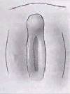
The external male genitalia consists of the penis and scrotum containing the testis.
Some Recent Findings
|
| More recent papers |
|---|
  This table allows an automated computer search of the external PubMed database using the listed "Search term" text link.
More? References | Discussion Page | Journal Searches | 2019 References | 2020 References Search term: Penis Embryology <pubmed limit=5>Penis Embryology</pubmed> |
Movies
|
|
|
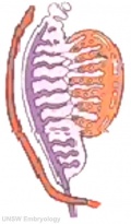
Mesonephic Duct Development
The paired mesonephic ducts (Wolffian ducts) go through a series of developmental changes to form the male internal genital tract.
Initiation
Invagination
Elongation
- Links:
Postnatal Development
Abnormalities
Endocrine Disruptors
Endocrine disruptors in female reproductive tract development and carcinogenesis.[3]
Additional Images
Historic
References
Reviews
<pubmed></pubmed> <pubmed></pubmed> <pubmed></pubmed>
Articles
<pubmed>5690451</pubmed> <pubmed></pubmed> <pubmed></pubmed>
Genital Links: genital | Lecture - Medicine | Lecture - Science | Lecture Movie | Medicine - Practical | primordial germ cell | meiosis | endocrine gonad | Genital Movies | genital abnormalities | Assisted Reproductive Technology | puberty | Category:Genital
| ||||
|
Glossary Links
- Glossary: A | B | C | D | E | F | G | H | I | J | K | L | M | N | O | P | Q | R | S | T | U | V | W | X | Y | Z | Numbers | Symbols | Term Link
Cite this page: Hill, M.A. (2026, October 20) Embryology Penis Development. Retrieved from https://embryology.med.unsw.edu.au/embryology/index.php/Penis_Development
- © Dr Mark Hill 2026, UNSW Embryology ISBN: 978 0 7334 2609 4 - UNSW CRICOS Provider Code No. 00098G

