Anatomy of the Human Body by Henry Gray: Difference between revisions
mNo edit summary |
|||
| (144 intermediate revisions by the same user not shown) | |||
| Line 1: | Line 1: | ||
{{Header}} | |||
{{Ref-Gray1918}} | |||
{{Historic Disclaimer}} | |||
==Introduction== | ==Introduction== | ||
[[File:Gray_1918.jpg|thumb]] | [[File:Gray_1918.jpg|thumb]] | ||
| Line 5: | Line 9: | ||
Classic anatomy textbook widely reproduced online, particularly the anatomical illustrations, due to the fact that the 1918 edition is out of copyright. W.H. Lewis edited the 20th edition published in September 1918, the current 40th edition was published in 2008. The majority of images were anatomical drawings with some cartoon simplifications. The text also includes earlier historic drawings, particularly in the embryology section that commences the text. | Classic anatomy textbook widely reproduced online, particularly the anatomical illustrations, due to the fact that the 1918 edition is out of copyright. W.H. Lewis edited the 20th edition published in September 1918, the current 40th edition was published in 2008. The majority of images were anatomical drawings with some cartoon simplifications. The text also includes earlier historic drawings, particularly in the embryology section that commences the text. | ||
Clicking the [[:Category:Gray's 1918 Anatomy|Category:Gray's 1918 Anatomy]] should display a list of the images available on this current website. Note that over time the image naming has varied and requires better standardisation. Images used here may be altered and edited from those appearing in the original textbook. | Clicking the [[:Category:Gray's 1918 Anatomy|Category:Gray's 1918 Anatomy]] should display a list of the images available on this current website. Note that over time the image naming has varied and requires better standardisation. Images used here may be altered and edited from those appearing in the original textbook. | ||
| Line 13: | Line 16: | ||
{| | {| | ||
| [[File: | | [[File:IBooks_icon.jpg|100px]] | ||
| '''iBooks''' | | '''iBooks''' | ||
| Line 19: | Line 22: | ||
|} | |} | ||
{{Grays Embryo iBook table}} | |||
ANATOMY OF THE HUMAN BODY | |||
==Textbook Introduction== | |||
THE term human anatomy comprises a consideration of the various structures which make up the human organism. In a restricted sense it deals merely with the parts which form the fully developed individual and which can be rendered evident to the naked eye by various methods of dissection. Regarded from such a standpoint it may be studied by two methods: (1) the various structures may be separately considered— systematic anatomy; or (2) the organs and tissues may be studied in relation to one another — topographical or regional anatomy. | |||
It is, however, of much advantage to add to the facts ascertained by nakedeye dissection those obtained by the use of the microscope. This introduces two fields of investigation, viz., the study of the minute structure of the various component parts of the body — histology — and the study of the human organism in its immature condition, i. e., the various stages of its intrauterine development from the fertilized ovum up to the period when it assumes an independent existence — embryology. Owing to the difficulty of obtaining material illustrating all the stages of this early development, gaps must be filled up by observations on the development of lower forms — comparative embryology, or by a consideration of adult forms in the line of human ancestry — comparative anatomy. The direct application of the facts of human anatomy to the various pathological conditions which may occur constitutes the subject of applied anatomy. Finally, the appreciation of structures on or immediately underlying the surface of the body is frequently made the subject of special study — smiace anatomy. | |||
===Systematic Anatomy=== | |||
The various systems of which the human body is composed are grouped under the following headings: | |||
# Osteology — the bony system or skeleton. | |||
# Syndesmology — the articulations or joints. | |||
# Myology — the muscles. With the description of the muscles it is convenient to include that of the fasciae which are so intimately connected with them. | |||
# Angiology — the vascular system, comprising the heart, bloodvessels, lymphatic vessels, and lymph glands. | |||
# Neurology — the nervous system. The organs of sense may be included in this system. | |||
# Splanchnology — the visceral system. Topographically the viscera form two groups, viz., the thoracic viscera and the abdomino-pelvic viscera. The heart, a thoracic viscus, is best considered with the vascular system. The rest of the viscera may be grouped according to their functions: (a) the respiratory apparatus; (6) the digestive apparatus; and (c) the urogenital apparatus. Strictly speaking, the third subgroup should include only such components of the urogenital apparatus as are included within the abdomino-pelvic cavity, but it is convenient to study under this heading certain parts which lie in relation to the surface of the body, e. g., the testes and the external organs of generation. | |||
For descriptive purposes. the body is supposed to be in the erect posture, with the arms hanging by the sides and the palms of the hands directed forward. The median plane is a vertical antero-posterior plane, passing through the center of the trunk. This plane will pass approximately through the sagittal suture of the skull, and hence any plane parallel to it is termed a sagittal plane. A vertical plane at right angles to the median plane passes, roughly speaking, through the central part of the coronal suture or through a line parallel to it; such a plane is known as a frontal plane or sometimes as a coronal plane. A plane at right angles to both the median and frontal planes is termed a transverse plane. | |||
The terms anterior or ventral, and posterior or dorsal, are employed to indicate the relation of parts to the front or back of the body or limbs, and the terms superior or cephalic, and inferior or caudal, to indicate the relative levels of different structures; structures nearer to or farther from the median plane are referred to as medial or lateral respectively. | |||
The terms superficial and deep are strictly confined to descriptions of the relative depth from the surface of the various structures; external and internal are reserved almost entirely for describing the walls of cavities or of hollow viscera. In the case of the limbs the words proximal and distal refer to the relative distance from the attached end of the limb. | |||
===Embryology=== | |||
THE term Embryology, in its widest sense, is applied to the various changes which take place during the growth of an animal from the egg to the adult condition: it is, however, usually restricted to the phenomena which occur before birth. Embryology may be studied from two aspects: | |||
# that of ontogeny, which deals only with the development of the individual; | |||
# that of phylogeny, which concerns itself with the evolutionary history of the animal kingdom. | |||
In vertebrate animals the development of a new being can only take place when a female germ cell or ovum has been fertilized by a male germ cell or spermatozoon. The ovum is a nucleated cell, and all the complicated changes by which the various tissues and organs of the body are formed from it, after it has been fertilized, are the result of two general processes, viz., segmentation and differentiation of cells. Thus, the fertilized ovum undergoes repeated segmentation into a number of cells which at first closely resemble one another, but are, sooner or later, differentiated into two groups: (1) somatic cells, the function of which is to build up the various tissues of the body; and (2) germinal cells, which become imbedded in the sexual glands — the ovaries in the female and the testes in the male — and are destined for the perpetuation of the species. | |||
Having regard to the main purpose of this work, it is impossible, in the space available in this section, to describe fully, or illustrate adequately, all the phenomena which occur in the different stages of the development of the human body. Only the principal facts are given, and the student is referred for further detaUs to one or other of the text-books^ on human embryology. | |||
Not all site images are included below. There may be several image versions (sizes, labeling, and formats gif, jpg, png). | Not all site images are included below. There may be several image versions (sizes, labeling, and formats gif, jpg, png). | ||
== | {{Historic Disclaimer}} | ||
==Development== | |||
<gallery> | <gallery> | ||
File:Gray0015.jpg|Neural Groove- series of sections dog embryo | File:Gray0015.jpg|15 Neural Groove- series of sections {{dog}} embryo | ||
File:Gray0020.jpg|Dorsal human embryo 2.11 mm | File:Gray0020.jpg|20 Dorsal human embryo 2.11 mm | ||
File:Gray0024. | File:Gray0024.jpg|24 Diagram showing earliest observed stage of human ovum | ||
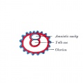
File:Gray0025. | File:Gray0025.jpg|25 Diagram illustrating early formation of allantois and differentiation of body-stalk | ||
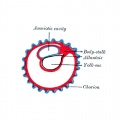
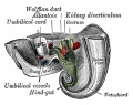
File:Gray0026. | File:Gray0026.jpg|26 Diagram showing later stage of allantoic development with commencing constriction of the yolk-sac | ||
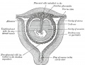
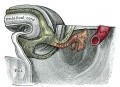
File:Gray0027. | File:Gray0027.jpg|27 Diagram showing the expansion of amnion and delimitation of the umbilicus | ||
File:Gray0031.jpg|Model of human embryo 1.3 mm | File:Gray0028.jpg|28 Diagram illustrating a later stage in the development of the umbilical cord | ||
File:Gray0032.jpg|Human Embryo Day 8 to 9 (week 3) | File:Gray0029.jpg|29 Diagram of a transverse section, showing the mode of formation of the amnion in the chick. | ||
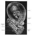
File:Gray0034.jpg|Uterus in the third and fourth month | File:Gray0030.jpg|30 Fetus of about eight weeks, enclosed in the amnion. | ||
File:Gray0036.jpg|Primary Chorionic Villi | File:Gray0031.jpg|31 Model of human embryo 1.3 mm | ||
File:Gray0037.jpg|Secondary Chorionic Villi | File:Gray0032.jpg|32 Human Embryo Day 8 to 9 (week 3) | ||
File:Gray0038.jpg|Fetus in Utero Between fifth and sixth months | File:Gray0033.jpg|33 Non-pregnant and pregnant uterus | ||
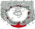
File:Gray0039.jpg|Placental circulation | File:Gray0034.jpg|34 Uterus in the third and fourth month | ||
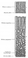
File:Gray0040.jpg| | File:Gray0035.jpg|35 Transverse section of a chorionic villus | ||
File:Gray0052.jpg| | File:Gray0036.jpg|36 Primary Chorionic Villi | ||
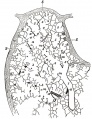
File:Gray0053.jpg| | File:Gray0037.jpg|37 Secondary Chorionic Villi | ||
File:Gray0058.jpg| | File:Gray0038.jpg|38 Fetus in Utero Between fifth and sixth months | ||
File:Gray0060.jpg| | File:Gray0039.jpg|39 Placental circulation | ||
File:Gray0063.jpg| | File:Gray0040.jpg|40 | ||
File:Gray0065.jpg| | File:Gray0041.jpg|41 Head end of human embryo about the end of the fourth week | ||
File:Gray0082.jpg| | File:Gray0042.jpg|42 Floor of Pharynx | ||
File:Gray0043.jpg|43 Head and neck of a human embryo eighteen weeks old with Meckel’s cartilage and hyoid bar exposed | |||
File:Gray0044.jpg|44 Under surface of the head of a human embryo about twenty-nine days old | |||
File:Gray0045.jpg|45 | |||
File:Gray0046.jpg|46 | |||
File:Gray0047.jpg|47 Head of a human embryo of about eight weeks | |||
File:Gray0048.jpg|48 Diagram showing the regions of the adult face and neck related to the fronto-nasal process and the branchial arches | |||
File:Gray0049.jpg|49 Primitive palate of a human embryo of thirty-seven to thirty-eight days | |||
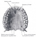
File:Gray0050.jpg|50 The roof of the mouth of a human embryo aged about two and a half months | |||
File:Gray0051.jpg|51 Frontal section of nasal cavities of a human embryo 28 mm long | |||
File:Gray0052.jpg|52 | |||
File:Gray0053.jpg|53 | |||
File:Gray0058.jpg|58 | |||
File:Gray0060.jpg|60 | |||
File:Gray0063.jpg|63 | |||
File:Gray0065.jpg|65 | |||
File:Gray0082.jpg|83 | |||
</gallery> | </gallery> | ||
<gallery> | <gallery> | ||
File:Gray0101.jpg|101 | |||
File:Gray0118.jpg|118 | |||
File:Gray0119.jpg|119 | |||
File:Gray0130.jpg|130 Human occipital bone, inner surface. | |||
File:Gray0176.jpg|176 Human adult mandible | |||
File:Gray0178.jpg|178 Human embryo CRL 24 mm outer aspect | |||
File:Gray0179.jpg|179 Human embryo CRL 24 mm inner aspect | |||
File:Gray0180.jpg|180 Human embryo CRL 95 mm outer aspect | |||
File:Gray0181.jpg|181 Human embryo CRL 95 mm inner aspect | |||
File:Gray0182.jpg|182 Mandible at birth | |||
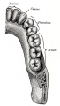
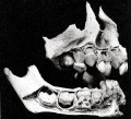
File:Gray0183.jpg|183 Mandible in childhood | |||
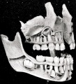
File:Gray0184.jpg|184 Mandible adult | |||
File:Gray0185.jpg|185 Mandible at old age | |||
File:Gray0193.jpg|193 Base of the skull | |||
</gallery> | </gallery> | ||
===301-400=== | ===301-400=== | ||
<gallery> | <gallery> | ||
File:Gray0301.jpg| | File:Gray0301.jpg|301 | ||
File:Gray0321.jpg| | File:Gray0321.jpg|321 Symphysis pubis exposed by a coronal section | ||
File:Gray0391.jpg|Adult human diaphragm (viewed from beneath) | File:Gray0379.jpg|379 Left orbicularis oculi (seen from behind) | ||
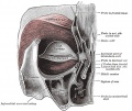
File:Gray0391.jpg|391 Adult human diaphragm (viewed from beneath) | |||
</gallery> | </gallery> | ||
===401-500 | ==Cardiovascular== | ||
401-500 | |||
<gallery> | <gallery> | ||
File:Gray0448.jpg|Artery and vein | File:Gray0448.jpg|448 Artery and vein | ||
File:Gray0458. | File:Gray0458.jpg|458 Diagram of the vascular channels in a human embryo of the second week | ||
File:Gray0462. | File:Gray0459.jpg|459 Human embryo of about fourteen days, with yolk-sac | ||
File:Gray0464. | File:Gray0460.jpg|460 Head of chick embryo of about thirty-eight hours' incubation | ||
File:Gray0467.jpg| | File:Gray0461.jpg|461 Diagram to illustrate the simple tubular condition of the heart | ||
File:Gray0468.jpg| | File:Gray0462.jpg|462 Heart of human embryo of about fourteen days | ||
File:Gray0470.jpg| | File:Gray0463.jpg|463 Heart of human embryo of about fifteen days | ||
File:Gray0472.jpg| | File:Gray0464.jpg|464 Dorsal surface of heart of human embryo of thirty-five days | ||
File:Gray0473. | File:Gray0465.jpg|465 Interior of dorsal half of heart from a human embryo of about thirty days | ||
File:Gray0474.jpg| | File:Gray0467.jpg|467 | ||
File:Gray0475.jpg| | File:Gray0468.jpg|468 | ||
File:Gray0476.jpg| | File:Gray0470.jpg|470 | ||
File:Gray0477.jpg| | File:Gray0472.jpg|472 | ||
File:Gray0478.jpg| | File:Gray0473.jpg|473 | ||
File:Gray0479.jpg| | File:Gray0474.jpg|474 | ||
File:Gray0480.jpg| | File:Gray0475.jpg|475 | ||
File:Gray0492.jpg| | File:Gray0476.jpg|476 | ||
File:Gray0498.jpg| | File:Gray0477.jpg|477 | ||
File:Gray0478.jpg|478 | |||
File:Gray0479.jpg|479 | |||
File:Gray0480.jpg|480 | |||
File:Gray0492.jpg|492 | |||
File:Gray0498.jpg|498 | |||
File:Gray0502.jpg|502 | |||
File:Gray0506.jpg|506 | |||
File:Gray0532.jpg|532 The celiac artery and its branches. | |||
File:Gray0533.jpg|533 The celiac artery and its branches. | |||
File:Gray0540.jpg|540 | |||
File:Gray0556.jpg|556 | |||
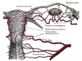
File:Gray0589.jpg|589 Vessels of the uterus and its appendages. | |||
</gallery> | </gallery> | ||
==Lymphatic== | |||
592 | |||
[[Immune System Development]] | |||
<gallery> | <gallery> | ||
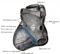
File: | File:Gray0592.jpg|592 Primary lymph sacs. | ||
File: | File:Gray0593.jpg|593 Lymph capillaries of the human conjunctiva. | ||
File: | File:Gray0594.jpg|594 Lymph capillaries from the human scrotum. | ||
File: | File:Gray0595.jpg|595 Lymph capillaries of the sole of the human foot. | ||
File: | File:Gray0596.jpg|596 Section through human tongue. | ||
File: | File:Gray0597.jpg|597 Lymph gland (Node). | ||
File: | File:Gray0598.jpg|598 Lymph gland tissue. | ||
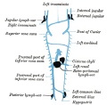
File: | File:Gray0599.jpg|599 Thoracic and right lymphatic ducts. | ||
File: | File:Gray0600.jpg|600 Modes of origin of thoracic duct. | ||
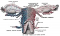
File: | File:Gray0601.jpg|601 Terminal collecting trunks of right side. | ||
File: | File:Gray0602.jpg|602 Lymph glands of the head. | ||
File:Gray0603.jpg|603 Lymphatics of pharynx. | |||
File:Gray0604.jpg|604 Lymphatics of the face. | |||
File:Gray0605.jpg|605 Lymphatics of the Tongue. | |||
File:Gray0606.jpg|606 Lymph glands of the upper extremity. | |||
File:Gray0607.jpg|607 Lymphatics of the mamma. | |||
File:Gray0608.jpg|608 Lymphatic vessels of the dorsal hand surface. | |||
File:Gray0609.jpg|609 Lymph glands of popliteal fossa. | |||
File:Gray0610.jpg|610 Superficial lymph glands and vessels of the lower extremity. | |||
File:Gray0611.jpg|611 Parietal lymph glands of the pelvis. | |||
File:Gray0612.jpg|612 Iliopelvic glands. | |||
File:Gray0613.jpg|613 Lymphatics of stomach. | |||
File:Gray0614.jpg|614 Lymphatics of stomach. | |||
File:Gray0615.jpg|615 Lymphatics of cecum and vermiform process. | |||
File:Gray0616.jpg|616 Lymphatics of cecum and vermiform process. | |||
File:Gray0617.jpg|617 Lymphatics of Colon. | |||
File:Gray0618.jpg|618 Lymphatic of the Bladder. | |||
File:Gray0619.jpg|619 Lymphatics of the Prostate. | |||
File:Gray0620.jpg|620 Lymphatics of the Uterus. | |||
File:Gray0621.jpg|621 Lymphatics of the thorax and abdomen. | |||
File:Gray0622.jpg|622 Tracheobronchial Lymph Glands | |||
</gallery> | </gallery> | ||
== | ==Neural== | ||
623 | |||
<gallery> | <gallery> | ||
File:Gray0702.jpg| | File:Gray0623.jpg|623 Neuroglia cells of brain. | ||
File:Gray0704.jpg| | File:Gray0649.jpg|649 Human Fetal Hindbrain (3 months) | ||
File:Gray0705.jpg| | File:Gray0651.jpg|651 Human Embryo Brain (week 4.5 exterior view) | ||
File:Gray0706.jpg| | File:Gray0652.jpg|652 Human Embryo Brain (week 5 exterior view) | ||
File:Gray0708.jpg | File:Gray0653.jpg|653 Human Embryo Brain (week 5 interior view) | ||
File:Gray0715.jpg| | File:Gray0654.jpg|654 Human Fetal Brain (3 months) | ||
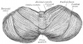
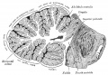
File:Gray0720.jpg|Median sagittal section of brain | File:Gray0655.jpg|655 Human Fetal Brain (4 months) | ||
File:Gray0732.jpg| | File:Gray0658.jpg|658 Human Fetal Brain (5 months) | ||
File:Gray0664.jpg|664 Transverse section of the medulla spinalis in the mid-thoracic region | |||
File:Gray0666.jpg|666 Transverse sections of the medulla spinalis at different levels | |||
File:Gray0666.jpg|666 (new layout) Transverse sections of the medulla spinalis at different levels | |||
File:Gray0670.jpg|670 Diagram showing afferent (sensory) and efferent fibers | |||
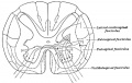
File:Gray0671.jpg|671 Spinal cord motor columns | |||
File:Gray0675.jpg|675 A spinal nerve with its anterior and posterior roots | |||
File:Gray0677.jpg|677 | |||
File:Gray0678.jpg|678 | |||
File:Gray0697.jpg|697 | |||
File:Gray0698.jpg|698 | |||
File:Gray0702.jpg|702 | |||
File:Gray0704.jpg|704 | |||
File:Gray0705.jpg|705 | |||
File:Gray0706.jpg|706 | |||
File:Gray0708.jpg|708 | |||
File:Gray0715.jpg|715 | |||
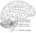
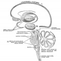
File:Gray0720.jpg|720 Median sagittal section of brain | |||
File:Gray0732.jpg|732 | |||
File:Gray0769.jpg|769 Section across the top of the skull | |||
File:Gray0770.jpg|770 Transverse section of medulla spinalis and its membranes | |||
File:Gray0778.jpg|778 Distribution of the maxillary and mandibular nerves, and the submaxillary ganglion | |||
File:Gray0781.jpg|781 Mandibular division of the Trigeminal Nerve | |||
File:Gray0784.jpg|784 Sensory areas of the head | |||
File:Gray0786.jpg|786 Oblique section through the right cavernous sinus | |||
File:Gray0788.jpg|788 Plan of the Facial and Intermediate Nerves and their Communication with Other Nerves | |||
File:Gray0790.jpg|790 The nerves of the scalp face and side of neck | |||
File:Gray0804.jpg|804 Cervical Plexus | |||
File:Gray0806.jpg|806 | |||
File:Gray0807.jpg|807 Branchial Plexus | |||
File:Gray0822.jpg|822 Lumbar Plexus | |||
</gallery> | </gallery> | ||
==Smell== | |||
852 | |||
<gallery> | <gallery> | ||
File: | File:Gray0852.jpg|852 Cartilages of the nose Side view | ||
File: | File:Gray0853.jpg|853 Cartilages of the nose, seen from below | ||
File:Gray0854.jpg|854 Bones and cartilages of septum of nose Right side | |||
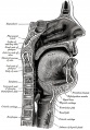
File:Gray0855.jpg|855 Lateral wall of nasal cavity | |||
File:Gray0856.jpg|856 | |||
File:Gray0857.jpg|857 | |||
File:Gray0858.jpg|858 | |||
File:Gray0859.jpg|859 Coronal section of nasal cavities | |||
File:Gray0860.jpg| | |||
File:Gray0861.jpg| | |||
File:Gray0862.jpg| | |||
</gallery> | </gallery> | ||
==Vision== | |||
== | |||
<gallery> | <gallery> | ||
File:Gray0863.jpg|863 | File:Gray0863.jpg|863 Chicken Optic Placode and Vesicle | ||
File:Gray0864.jpg|864 | File:Gray0864.jpg|864 Chicken Optic Placode and Vesicle | ||
File:Gray0865.jpg|865 | File:Gray0865.jpg|865 Human Optic Cup and Choroidal Fissure | ||
File:Gray0866.jpg|866 | File:Gray0866.jpg|866 Embryonic Rabbit Eye | ||
File:Gray0867.jpg|867 | File:Gray0867.jpg|867 Human Embryonic Eye | ||
File:Gray0868.jpg|868 | File:Gray0868.jpg|868 Section of developing eye of trout | ||
File:Gray0869.jpg|869 | File:Gray0869.jpg|869 Horizontal section of the eyeball | ||
File:Gray0870.jpg|870 | File:Gray0870.jpg|870 Cornea | ||
File:Gray0871.jpg|871 | File:Gray0871.jpg|871 Section of Human Cornea near the Margin | ||
File:Gray0872.jpg|872 | File:Gray0872.jpg|872 The Choroid and Iris | ||
File:Gray0873.jpg|873 | File:Gray0873.jpg|873 The Arteries of the Choroid and Iris | ||
File:Gray0874.jpg|874 | File:Gray0874.jpg|874 The Veins of the Choroid | ||
File:Gray0875.jpg|875 | File:Gray0875.jpg|875 Interior of anterior half of bulb of eye | ||
File:Gray0876.jpg|876 | File:Gray0876.jpg|876 Vessels of the choroid, ciliary processes, and iris of a child | ||
File:Gray0877.jpg|877 | File:Gray0877.jpg|877 Blood Vessels of the Eye | ||
File:Gray0878.jpg|878 | File:Gray0878.jpg|878 Iris front view | ||
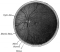
File:Gray0879.jpg|879 | File:Gray0879.jpg|879 Interior of posterior half of bulb of left eye | ||
File:Gray0880.jpg|880 | File:Gray0880.jpg|880 Optic Nerve entering Eyeball | ||
File:Gray0881.jpg|881 | File:Gray0881.jpg|881 Section of Retina | ||
File:Gray0882.jpg|882 | File:Gray0882.jpg|882 Plan of Retinal Neurons | ||
File:Gray0883.jpg|883 | File:Gray0883.jpg|883 Section through front of Eyeball | ||
File:Gray0884.jpg|884 | File:Gray0884.jpg|884 Lens Structure | ||
File:Gray0885.jpg|885 | File:Gray0885.jpg|885 Lens | ||
File:Gray0886.jpg|886 | File:Gray0886.jpg|886 Lens Profile | ||
File:Gray0887.jpg|887 | File:Gray0887.jpg|887 Lens Epithelium | ||
File:Gray0888.jpg|888 | File:Gray0888.jpg|888 Sagittal section of right orbital cavity | ||
File:Gray0889.jpg|889 | File:Gray0889.jpg|889 Muscles of the right orbit | ||
File:Gray0890.jpg|890 | File:Gray0890.jpg|890 Right Ocular Muscles | ||
File:Gray0891.jpg|891 | File:Gray0891.jpg|891 Right Eye Fascia Bulbi | ||
File:Gray0892.jpg|892 | File:Gray0892.jpg|892 Front of left eye with eyelids separated to show medial canthus | ||
File:Gray0893.jpg|893 Structure of the Eyelids | |||
File:Gray0894.jpg|894 The tarsi and their ligaments | |||
File:Gray0895.jpg|895 The Tarsal Glands | |||
File:Gray0896.jpg|896 The Lacrimal Gland | |||
File:Gray0897.jpg|897 Structures of the Lacrimal Gland | |||
</gallery> | </gallery> | ||
== | ==Hearing== | ||
<gallery> | <gallery> | ||
| Line 206: | Line 336: | ||
File:Gray0926.jpg|926 Right Human Membranous Labyrinth | File:Gray0926.jpg|926 Right Human Membranous Labyrinth | ||
File:Gray0927.jpg|927 Human Semicircular Canal and Duct | File:Gray0927.jpg|927 Human Semicircular Canal and Duct | ||
File:Gray0928.jpg|928 | File:Gray0928.jpg|928 Cross-section of Cochlea | ||
File:Gray0929.jpg|929 | File:Gray0929.jpg|929 Floor of Ductus Cochlearis | ||
File:Gray0930.jpg|930 | File:Gray0930.jpg|930 Limbus laminæ spiralis and membrana basilaris | ||
File:Gray0931.jpg|931 | File:Gray0931.jpg|931 Section through the spiral organ of Corti | ||
File:Gray0932.jpg|932 | File:Gray0932.jpg|932 The lamina reticularis and subjacent structures | ||
File:Gray0933.jpg|933 | File:Gray0933.jpg|933 Cochlear Division of the Acoustic Nerve | ||
</gallery> | </gallery> | ||
== | ==Somatosensory== | ||
<gallery> | <gallery> | ||
File:Gray0934.jpg|934 | File:Gray0934.jpg|934 End-bulb of Krause | ||
File:Gray0935.jpg|935 | File:Gray0935.jpg|935 Pacinian corpuscle | ||
File:Gray0936.jpg|936 | File:Gray0936.jpg|936 Papilla of the hand | ||
File:Gray0937.jpg|937 | File:Gray0937.jpg|937 Nerve ending of Ruffini | ||
File:Gray0938.jpg|938 | File:Gray0938.jpg|938 Organ of Golgi | ||
File:Gray0939.jpg|939 | File:Gray0939.jpg|939 Muscle Spindle | ||
</gallery> | </gallery> | ||
== | ==Integumentary== | ||
<gallery> | <gallery> | ||
File:Gray0943.jpg|943 | File:Gray0940.jpg|940 A diagrammatic sectional view of the skin | ||
File:Gray0947.jpg|947 | File:Gray0941.jpg|941 Section of Epidermis | ||
File:Gray0961.jpg|961 | File:Gray0942.jpg|942 The distribution of the bloodvessels in the skin of the sole of the foot | ||
File:Gray0943.jpg|943 Longitudinal section through nail and its nail groove | |||
File:Gray0944.jpg|944 | |||
File:Gray0945.jpg|945 | |||
File:Gray0946.jpg|946 | |||
</gallery> | |||
==Respiratory== | |||
<gallery> | |||
File:Gray0947.jpg|947 The head and neck human embryo thirty-two days seen from the ventral surface. | |||
File:Gray0948.jpg|948 Lung buds from a human embryo of about four weeks, showing commencing lobulations. | |||
File:Gray0949.jpg|949 Lungs of a human embryo more advanced in development than week 4. | |||
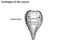
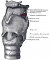
File:Gray0950.jpg|950 Cartilages of the larynx | |||
File:Gray0950 epiglottis cartilage.jpg|950 epiglottis cartilage | |||
File:Gray0950 thyroid cartilage.jpg|950 thyroid cartilage | |||
File:Gray0950 cricoid cartilage.jpg|950 cricoid cartilage | |||
File:Gray0950 arytenoid cartilage.jpg|950 arytenoid cartilage | |||
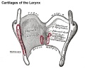
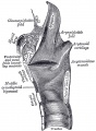
File:Gray0951.jpg|951 Ligaments of the larynx (anterior view) | |||
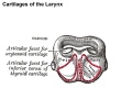
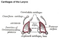
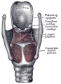
File:Gray0952.jpg|952 Ligaments of the larynx (posterior view) | |||
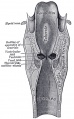
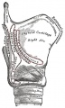
File:Gray0953.jpg|953 Larynx and upper part of the trachea | |||
File:Gray0954.jpg|954 | |||
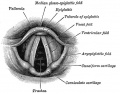
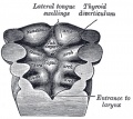
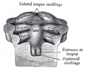
File:Gray0955.jpg|955 Larynx entrance | |||
File:Gray0956.jpg|956 | |||
File:Gray0957.jpg|957 | |||
File:Gray0958.jpg|958 | |||
File:Gray0959.jpg|959 | |||
File:Gray0960.jpg|960 | |||
File:Gray0961.jpg|961 Cartilages of larynx, trachea, and bronchi (front view) | |||
File:Gray0962.jpg|962 Bronchi and bronchioles | File:Gray0962.jpg|962 Bronchi and bronchioles | ||
File:Gray0963.jpg|963 | |||
File:Gray0964.jpg|964 | |||
File:Gray0965.jpg|965 | File:Gray0965.jpg|965 | ||
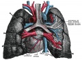
File:Gray0966.jpg|966 Lateral view of thorax, showing the relations of the pleuræ and lungs to the chest wall. Pleura in blue; lungs in purple. | File:Gray0966.jpg|966 Lateral view of thorax, showing the relations of the pleuræ and lungs to the chest wall. Pleura in blue; lungs in purple. | ||
| Line 237: | Line 396: | ||
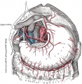
File:Gray0970.jpg|970 Front view of heart and lungs | File:Gray0970.jpg|970 Front view of heart and lungs | ||
File:Gray0971.jpg|971 Adult lungs | File:Gray0971.jpg|971 Adult lungs | ||
File:Gray0974.jpg|974 | File:Gray0974.jpg|974 Lung secondary lobule | ||
File:Gray0975.jpg| | File:Lung_secondary_lobule_01.jpg|974 relabeled version | ||
File:Gray0977.jpg| | File:Gray0975.jpg|975 Lung primary lobule | ||
File:Gray0982a.jpg| | File:Lung_primary_lobule_01.jpg|975 relabeled version | ||
File:Gray0982b.jpg| | File:Gray0976.jpg|976 Pig embryo lung | ||
File:Gray0983.jpg | </gallery> | ||
File:Gray0984.jpg | |||
File:Gray0985.jpg | ==Gastrointestinal== | ||
File:Gray0986.jpg| | <gallery> | ||
File:Gray0987.jpg | File:Gray0977.jpg|977 | ||
File:Gray0987a.jpg | File:Gray0978.jpg|978 | ||
File:Gray0987b.jpg | File:Gray0979.jpg|979 | ||
File:Gray0988.jpg | File:Gray0980.jpg|980 | ||
File:Gray0989.jpg | File:Gray0981.jpg|981 | ||
File:Gray0990.jpg| | File:Gray0982a.jpg|982a | ||
File:Gray0991.jpg| | File:Gray0982b.jpg|982b | ||
File:Gray0992.jpg| | File:Gray0983.jpg|983 | ||
File:Gray0993.jpg| | File:Gray0984.jpg|984 | ||
File:Gray0994.jpg| | File:Gray0985.jpg|985 | ||
File:Gray0996.jpg | File:Gray0986.jpg|986 | ||
File:Gray0987.jpg|987 | |||
File:Gray0987a.jpg|987a | |||
File:Gray0987b.jpg|987b | |||
File:Gray0988.jpg|988 | |||
File:Gray0989.jpg|989 | |||
File:Gray0990.jpg|990 | |||
File:Gray0991.jpg|991 | |||
File:Gray0992.jpg|992 | |||
File:Gray0993.jpg|993 | |||
File:Gray0994.jpg|994 | |||
File:Gray0996.jpg|996 | |||
File:Gray0997.jpg|997 | |||
File:Gray0998.jpg|998 | |||
File:Gray0999.jpg|999 | |||
File:Gray1000.jpg|1000 | |||
File:Gray1001.jpg|1001 | |||
File:Gray1002.jpg|1002 | |||
File:Gray1003.jpg|1003 | |||
File:Gray1004.jpg|1004 | |||
File:Gray1005.jpg|1005 | |||
File:Gray1006.jpg|1006 | |||
File:Gray1007.jpg|1007 | |||
File:Gray1008.jpg|1008 | |||
File:Gray1009.jpg|1009 | |||
File:Gray1010.jpg|1010 | |||
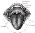
File:Gray1013.jpg|1013 The mouth cavity | |||
File:Gray1014.jpg|1014 The mouth cavity | |||
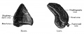
File:Gray1015.jpg|1015 Circumvallate papilla | |||
File:Gray1016.jpg|1016 Filiform papilla | |||
File:Gray1017.jpg|1017 Fungiform papilla | |||
File:Gray1018.jpg|1018 Mucous membrane of the tongue | |||
File:Gray1019.jpg|1019 Extrinsic muscles of the tongue | |||
File:Gray1020.jpg|1020 Intrinsic muscles of the tongue | |||
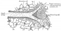
File:Gray1022.jpg|1022 Right parotid gland (posterior and deep aspects) | |||
File:Gray1023.jpg|1023 Right parotid gland (anterior and deep aspects) | |||
File:Gray1024.jpg|1024 Dissection showing salivary glands of right side | |||
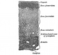
File:Gray1027.jpg|1027 Section through one of the crypts of the tonsil | |||
File:Gray1028.jpg|1028 Dissection of the Muscles of the Palate from behind | |||
File:Gray1029.jpg|1029 Front of nasa part of pharynx | |||
File:Gray1030.jpg|1030 Muscles of the Pharynx and Cheek | |||
File:Gray1033.jpg|1033 Section of the Human Esophagus | |||
File:Gray1039.jpg|1039 | |||
File:Gray1050.jpg|1050 Interior of the stomach | |||
File:Gray1095.jpg|1095 Gall bladder and bile ducts laid open | |||
File:Gray1096.jpg|1096 Gall bladder transverse section | |||
</gallery> | </gallery> | ||
== | ==Urogenital== | ||
<gallery> | <gallery> | ||
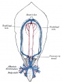
File: | File:Gray1108.jpg|1108 Broad ligament of adult showing Epoöphoron | ||
File: | File:Gray1109.jpg|1109 Urogenital Sinus of Female Human Embryo of 8.5 to 9 weeks old | ||
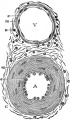
File: | File:Gray1111.jpg|1111 Transverse section of Human Embryo 8.5 to 9 Weeks Old | ||
File:Gray1112.jpg|1112 Longitudinal Section of Ovary of Cat Embryo of 9.4 cm long | |||
File:Gray1113.jpg|1113 Section of the Ovary of a Newly Born Child | |||
File:Gray1114.jpg|1114 Human Embryo (3.5 cm long) Testis Section of a Genital Cord | |||
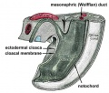
File:Gray1115.jpg|1115 Tail end of Human Embryo 25 to 29 Days Old | |||
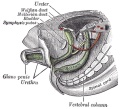
File:Gray1116.jpg|1116 Tail end of Human Embryo 32 to 33 Days Old | |||
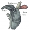
File:Gray1117.jpg|1117 Tail end of human embryo eight and a half to nine weeks old | |||
File:Gray1118.jpg|1118 Primitive Kidney and Bladder | |||
File:Gray1119.jpg|1119 Stages in the development of the external sexual organs in the male and female | |||
File:Gray1120.jpg|1120 Abdomen | |||
File:Gray1121.jpg|1121 Posterior abdominal wall | |||
File:Gray1122.jpg|1122 | |||
File:Gray1123.jpg|1123 | |||
File:Gray1124.jpg|1124 | |||
File:Gray1125.jpg|1125 | |||
File:Gray1126.png|1126 Retroperitoneal structures | |||
File:Gray1126.jpg|1126 Retroperitoneal structures | |||
File:Gray1127.jpg|1127 | |||
File:Gray1128.jpg|1128 | |||
File:Gray1129.jpg|1129 | |||
File:Gray1130.jpg|1130 | |||
File:Gray1131.jpg|1131 | |||
File:Gray1132.jpg|1132 | |||
File:Gray1133.jpg|1133 | |||
File:Gray1137.jpg|1137 | |||
File:Gray1138.jpg|1138 | |||
File:Gray1139.jpg|1139 | |||
File:Gray1140.jpg|1140 | |||
File:Gray1141.jpg|1141 | |||
File:Gray1152.jpg|1152 Fundus of the bladder with the vesiculæ seminales | |||
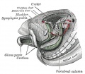
File:Gray1153.jpg|1153 Prostate and seminal vesicles | |||
File:Gray1160.jpg|1160 Prostate Gland | |||
File:Gray1161.jpg|1161 Uterus and right broad ligament | |||
File:Gray1163.jpg|1163 section of the ovary | |||
File:Gray1166.jpg|1166 | |||
File:Gray1170.jpg|1170 | |||
File:Gray1173.jpg|1173 | |||
</gallery> | </gallery> | ||
== | ==Endocrine== | ||
The Ductless Glands | |||
<gallery> | <gallery> | ||
File: | File:Gray1174.jpg|1174 The thyroid gland and its relations. | ||
File:Gray1175.jpg|1175 | |||
File: | File:Gray1181.jpg|1181 Pituitary - Median sagittal hypophysis adult monkey | ||
File: | File:Gray1182.jpg|1182 Vertical sections of the heads of early embryos of the rabbit | ||
File: | File:Gray1183.jpg|1183 | ||
File:Gray1184.jpg|1184 | |||
File: | File:Gray1185.jpg|1185 Section of a part of a suprarenal gland | ||
File: | File:Gray1192.jpg|1192 | ||
File: | |||
File:Gray1192.jpg| | |||
</gallery> | </gallery> | ||
== | |||
==Surface Anatomy== | |||
Surface Anatomy and Surface Markings | |||
<gallery> | <gallery> | ||
File:Gray1201.jpg| | File:Gray1201.jpg| | ||
File:Gray1202.jpg| | File:Gray1202.jpg| | ||
File:Gray1223.png|Abdomen Surface Markings for Liver, Stomach, and Great Intestine | File:Gray1223.png|Abdomen Surface Markings for Liver, Stomach, and Great Intestine | ||
File:Gray1235.jpg|Front of right upper extremity, showing surface markings for bones and nerves. | |||
File:Gray1236.jpg|Back of right upper extremity, showing surface markings for bones and nerves. | |||
File:Gray1237.jpg|Left Hand Palm, position of skin creases and bones, and surface markings for the volar arches | File:Gray1237.jpg|Left Hand Palm, position of skin creases and bones, and surface markings for the volar arches | ||
</gallery> | </gallery> | ||
| Line 299: | Line 532: | ||
{{ | {{Glossary}} | ||
{{ | |||
{{Footer}} | |||
[[Category:Historic Embryology]] [[Category:Gray's 1918 Anatomy]] | [[Category:Historic Embryology]] [[Category:Gray's 1918 Anatomy]][[Category:1910's]] | ||
Latest revision as of 13:29, 1 April 2019
| Embryology - 10 Jun 2024 |
|---|
| Google Translate - select your language from the list shown below (this will open a new external page) |
|
العربية | català | 中文 | 中國傳統的 | français | Deutsche | עִברִית | हिंदी | bahasa Indonesia | italiano | 日本語 | 한국어 | မြန်မာ | Pilipino | Polskie | português | ਪੰਜਾਬੀ ਦੇ | Română | русский | Español | Swahili | Svensk | ไทย | Türkçe | اردو | ייִדיש | Tiếng Việt These external translations are automated and may not be accurate. (More? About Translations) |
Gray H. Anatomy of the human body. (1918) Philadelphia: Lea & Febiger.
| Historic Disclaimer - information about historic embryology pages |
|---|
 |
Introduction


Classic anatomy textbook widely reproduced online, particularly the anatomical illustrations, due to the fact that the 1918 edition is out of copyright. W.H. Lewis edited the 20th edition published in September 1918, the current 40th edition was published in 2008. The majority of images were anatomical drawings with some cartoon simplifications. The text also includes earlier historic drawings, particularly in the embryology section that commences the text.
Clicking the Category:Gray's 1918 Anatomy should display a list of the images available on this current website. Note that over time the image naming has varied and requires better standardisation. Images used here may be altered and edited from those appearing in the original textbook.
| iBooks |
| iBook - Gray's Embryology | |
|---|---|

|
|
ANATOMY OF THE HUMAN BODY
Textbook Introduction
THE term human anatomy comprises a consideration of the various structures which make up the human organism. In a restricted sense it deals merely with the parts which form the fully developed individual and which can be rendered evident to the naked eye by various methods of dissection. Regarded from such a standpoint it may be studied by two methods: (1) the various structures may be separately considered— systematic anatomy; or (2) the organs and tissues may be studied in relation to one another — topographical or regional anatomy.
It is, however, of much advantage to add to the facts ascertained by nakedeye dissection those obtained by the use of the microscope. This introduces two fields of investigation, viz., the study of the minute structure of the various component parts of the body — histology — and the study of the human organism in its immature condition, i. e., the various stages of its intrauterine development from the fertilized ovum up to the period when it assumes an independent existence — embryology. Owing to the difficulty of obtaining material illustrating all the stages of this early development, gaps must be filled up by observations on the development of lower forms — comparative embryology, or by a consideration of adult forms in the line of human ancestry — comparative anatomy. The direct application of the facts of human anatomy to the various pathological conditions which may occur constitutes the subject of applied anatomy. Finally, the appreciation of structures on or immediately underlying the surface of the body is frequently made the subject of special study — smiace anatomy.
Systematic Anatomy
The various systems of which the human body is composed are grouped under the following headings:
- Osteology — the bony system or skeleton.
- Syndesmology — the articulations or joints.
- Myology — the muscles. With the description of the muscles it is convenient to include that of the fasciae which are so intimately connected with them.
- Angiology — the vascular system, comprising the heart, bloodvessels, lymphatic vessels, and lymph glands.
- Neurology — the nervous system. The organs of sense may be included in this system.
- Splanchnology — the visceral system. Topographically the viscera form two groups, viz., the thoracic viscera and the abdomino-pelvic viscera. The heart, a thoracic viscus, is best considered with the vascular system. The rest of the viscera may be grouped according to their functions: (a) the respiratory apparatus; (6) the digestive apparatus; and (c) the urogenital apparatus. Strictly speaking, the third subgroup should include only such components of the urogenital apparatus as are included within the abdomino-pelvic cavity, but it is convenient to study under this heading certain parts which lie in relation to the surface of the body, e. g., the testes and the external organs of generation.
For descriptive purposes. the body is supposed to be in the erect posture, with the arms hanging by the sides and the palms of the hands directed forward. The median plane is a vertical antero-posterior plane, passing through the center of the trunk. This plane will pass approximately through the sagittal suture of the skull, and hence any plane parallel to it is termed a sagittal plane. A vertical plane at right angles to the median plane passes, roughly speaking, through the central part of the coronal suture or through a line parallel to it; such a plane is known as a frontal plane or sometimes as a coronal plane. A plane at right angles to both the median and frontal planes is termed a transverse plane.
The terms anterior or ventral, and posterior or dorsal, are employed to indicate the relation of parts to the front or back of the body or limbs, and the terms superior or cephalic, and inferior or caudal, to indicate the relative levels of different structures; structures nearer to or farther from the median plane are referred to as medial or lateral respectively.
The terms superficial and deep are strictly confined to descriptions of the relative depth from the surface of the various structures; external and internal are reserved almost entirely for describing the walls of cavities or of hollow viscera. In the case of the limbs the words proximal and distal refer to the relative distance from the attached end of the limb.
Embryology
THE term Embryology, in its widest sense, is applied to the various changes which take place during the growth of an animal from the egg to the adult condition: it is, however, usually restricted to the phenomena which occur before birth. Embryology may be studied from two aspects:
- that of ontogeny, which deals only with the development of the individual;
- that of phylogeny, which concerns itself with the evolutionary history of the animal kingdom.
In vertebrate animals the development of a new being can only take place when a female germ cell or ovum has been fertilized by a male germ cell or spermatozoon. The ovum is a nucleated cell, and all the complicated changes by which the various tissues and organs of the body are formed from it, after it has been fertilized, are the result of two general processes, viz., segmentation and differentiation of cells. Thus, the fertilized ovum undergoes repeated segmentation into a number of cells which at first closely resemble one another, but are, sooner or later, differentiated into two groups: (1) somatic cells, the function of which is to build up the various tissues of the body; and (2) germinal cells, which become imbedded in the sexual glands — the ovaries in the female and the testes in the male — and are destined for the perpetuation of the species.
Having regard to the main purpose of this work, it is impossible, in the space available in this section, to describe fully, or illustrate adequately, all the phenomena which occur in the different stages of the development of the human body. Only the principal facts are given, and the student is referred for further detaUs to one or other of the text-books^ on human embryology.
Not all site images are included below. There may be several image versions (sizes, labeling, and formats gif, jpg, png).
| Historic Disclaimer - information about historic embryology pages |
|---|
|
Development
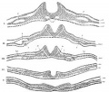
15 Neural Groove- series of sections dog embryo
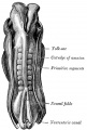
20 Dorsal human embryo 2.11 mm
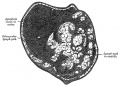
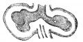
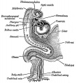
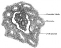
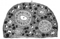
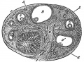
24 Diagram showing earliest observed stage of human ovum
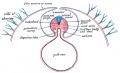
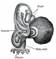
25 Diagram illustrating early formation of allantois and differentiation of body-stalk
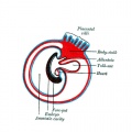
26 Diagram showing later stage of allantoic development with commencing constriction of the yolk-sac
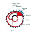
27 Diagram showing the expansion of amnion and delimitation of the umbilicus
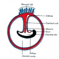
28 Diagram illustrating a later stage in the development of the umbilical cord
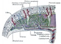
29 Diagram of a transverse section, showing the mode of formation of the amnion in the chick.
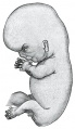
30 Fetus of about eight weeks, enclosed in the amnion.

31 Model of human embryo 1.3 mm
32 Human Embryo Day 8 to 9 (week 3)
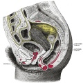
33 Non-pregnant and pregnant uterus
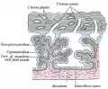
34 Uterus in the third and fourth month
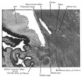
35 Transverse section of a chorionic villus
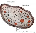
36 Primary Chorionic Villi
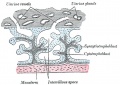
37 Secondary Chorionic Villi
38 Fetus in Utero Between fifth and sixth months
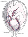
39 Placental circulation
40
41 Head end of human embryo about the end of the fourth week
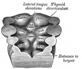
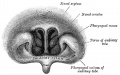
42 Floor of Pharynx
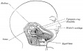
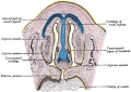
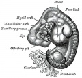
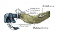
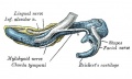
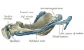
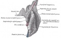
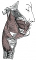
43 Head and neck of a human embryo eighteen weeks old with Meckel’s cartilage and hyoid bar exposed
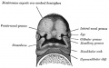
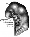
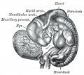
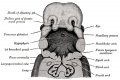
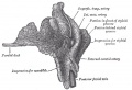
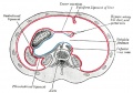
44 Under surface of the head of a human embryo about twenty-nine days old
45
46
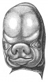
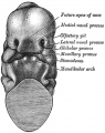
47 Head of a human embryo of about eight weeks
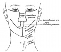
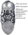
48 Diagram showing the regions of the adult face and neck related to the fronto-nasal process and the branchial arches
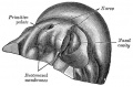
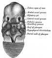
49 Primitive palate of a human embryo of thirty-seven to thirty-eight days
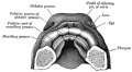
50 The roof of the mouth of a human embryo aged about two and a half months
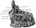
51 Frontal section of nasal cavities of a human embryo 28 mm long
52
53
58
60
63
65
83


101
118
119
130 Human occipital bone, inner surface.
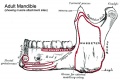
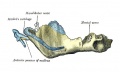
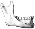
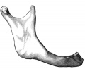
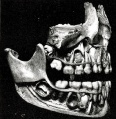
176 Human adult mandible
178 Human embryo CRL 24 mm outer aspect
179 Human embryo CRL 24 mm inner aspect
180 Human embryo CRL 95 mm outer aspect
181 Human embryo CRL 95 mm inner aspect
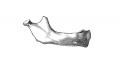
182 Mandible at birth
183 Mandible in childhood
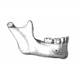
184 Mandible adult
185 Mandible at old age
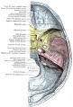
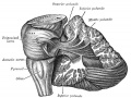
193 Base of the skull


301-400
301
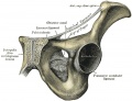
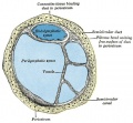
321 Symphysis pubis exposed by a coronal section
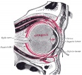
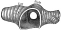
379 Left orbicularis oculi (seen from behind)
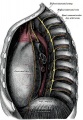
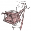
391 Adult human diaphragm (viewed from beneath)


Cardiovascular
401-500
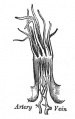
448 Artery and vein
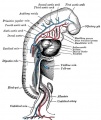
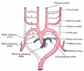
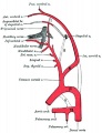
458 Diagram of the vascular channels in a human embryo of the second week
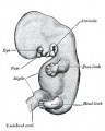
459 Human embryo of about fourteen days, with yolk-sac
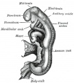
460 Head of chick embryo of about thirty-eight hours' incubation
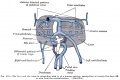
461 Diagram to illustrate the simple tubular condition of the heart
462 Heart of human embryo of about fourteen days
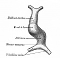
463 Heart of human embryo of about fifteen days
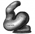
464 Dorsal surface of heart of human embryo of thirty-five days
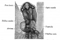
465 Interior of dorsal half of heart from a human embryo of about thirty days
467
468
470
472
473
474
475
476
477

478
479
480
492
498
502
506
532 The celiac artery and its branches.
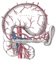
533 The celiac artery and its branches.
540
556
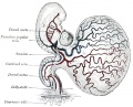
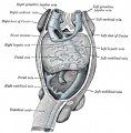
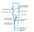
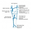
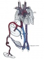
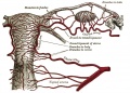
589 Vessels of the uterus and its appendages.


Lymphatic
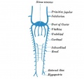
592 Primary lymph sacs.
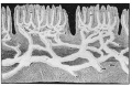
593 Lymph capillaries of the human conjunctiva.
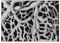
594 Lymph capillaries from the human scrotum.
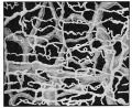
595 Lymph capillaries of the sole of the human foot.
596 Section through human tongue.
597 Lymph gland (Node).
598 Lymph gland tissue.
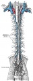
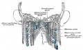
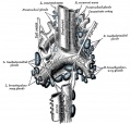
599 Thoracic and right lymphatic ducts.
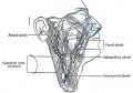
600 Modes of origin of thoracic duct.
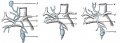
601 Terminal collecting trunks of right side.
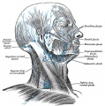
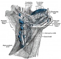
602 Lymph glands of the head.
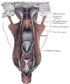
603 Lymphatics of pharynx.
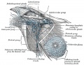
604 Lymphatics of the face.
605 Lymphatics of the Tongue.
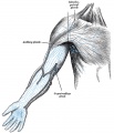
606 Lymph glands of the upper extremity.
607 Lymphatics of the mamma.
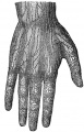
608 Lymphatic vessels of the dorsal hand surface.
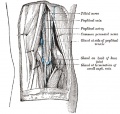
609 Lymph glands of popliteal fossa.
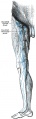
610 Superficial lymph glands and vessels of the lower extremity.
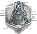
611 Parietal lymph glands of the pelvis.
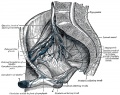
612 Iliopelvic glands.
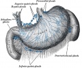
613 Lymphatics of stomach.
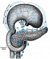
614 Lymphatics of stomach.
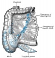
615 Lymphatics of cecum and vermiform process.
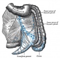
616 Lymphatics of cecum and vermiform process.
617 Lymphatics of Colon.
618 Lymphatic of the Bladder.
619 Lymphatics of the Prostate.
620 Lymphatics of the Uterus.
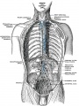
621 Lymphatics of the thorax and abdomen.
622 Tracheobronchial Lymph Glands


Neural
623
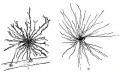
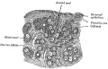
623 Neuroglia cells of brain.
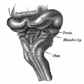
649 Human Fetal Hindbrain (3 months)
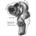
651 Human Embryo Brain (week 4.5 exterior view)
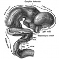
652 Human Embryo Brain (week 5 exterior view)
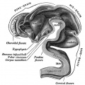
653 Human Embryo Brain (week 5 interior view)

654 Human Fetal Brain (3 months)
655 Human Fetal Brain (4 months)
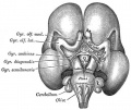
658 Human Fetal Brain (5 months)
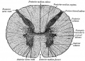
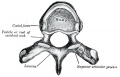
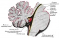
664 Transverse section of the medulla spinalis in the mid-thoracic region
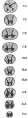
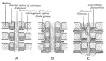
666 Transverse sections of the medulla spinalis at different levels
666 (new layout) Transverse sections of the medulla spinalis at different levels
670 Diagram showing afferent (sensory) and efferent fibers
671 Spinal cord motor columns
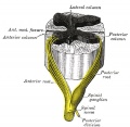
675 A spinal nerve with its anterior and posterior roots
677
678
697
698
702
704
705
706
708
715
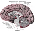
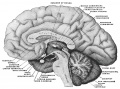
720 Median sagittal section of brain
732
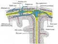
769 Section across the top of the skull
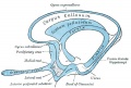
770 Transverse section of medulla spinalis and its membranes
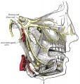
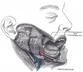
778 Distribution of the maxillary and mandibular nerves, and the submaxillary ganglion
781 Mandibular division of the Trigeminal Nerve
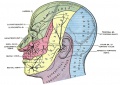
784 Sensory areas of the head
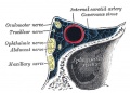
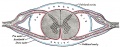
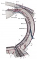
786 Oblique section through the right cavernous sinus
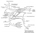
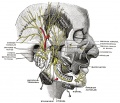
788 Plan of the Facial and Intermediate Nerves and their Communication with Other Nerves
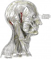
790 The nerves of the scalp face and side of neck
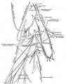
804 Cervical Plexus
806
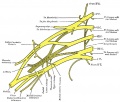
807 Branchial Plexus
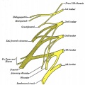
822 Lumbar Plexus


Smell
852
852 Cartilages of the nose Side view
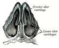
853 Cartilages of the nose, seen from below
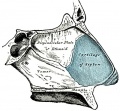
854 Bones and cartilages of septum of nose Right side
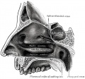
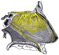
855 Lateral wall of nasal cavity
856
857
858
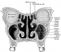
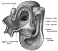
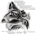
859 Coronal section of nasal cavities


Vision
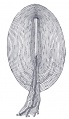
863 Chicken Optic Placode and Vesicle
864 Chicken Optic Placode and Vesicle
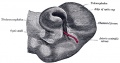
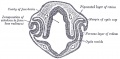
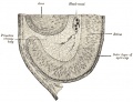
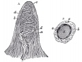
865 Human Optic Cup and Choroidal Fissure
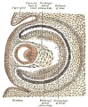
866 Embryonic Rabbit Eye
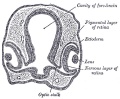
867 Human Embryonic Eye
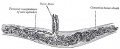
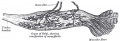
868 Section of developing eye of trout
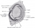
869 Horizontal section of the eyeball
870 Cornea
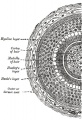
871 Section of Human Cornea near the Margin
872 The Choroid and Iris
873 The Arteries of the Choroid and Iris
874 The Veins of the Choroid
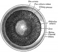
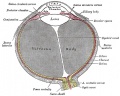
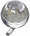
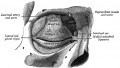
875 Interior of anterior half of bulb of eye

876 Vessels of the choroid, ciliary processes, and iris of a child
877 Blood Vessels of the Eye
878 Iris front view
879 Interior of posterior half of bulb of left eye
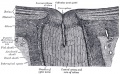
880 Optic Nerve entering Eyeball
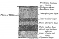
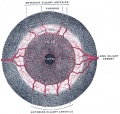
881 Section of Retina
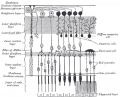
882 Plan of Retinal Neurons
883 Section through front of Eyeball
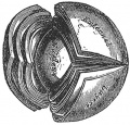
884 Lens Structure
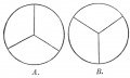
885 Lens
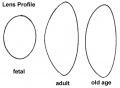
886 Lens Profile
887 Lens Epithelium
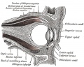
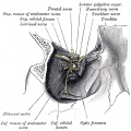
888 Sagittal section of right orbital cavity
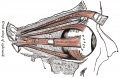
889 Muscles of the right orbit
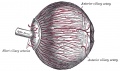
890 Right Ocular Muscles
891 Right Eye Fascia Bulbi
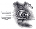
892 Front of left eye with eyelids separated to show medial canthus
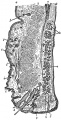
893 Structure of the Eyelids
894 The tarsi and their ligaments
895 The Tarsal Glands
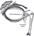
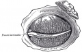
896 The Lacrimal Gland
897 Structures of the Lacrimal Gland


Hearing
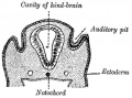
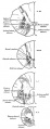
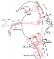
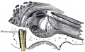
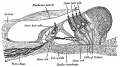
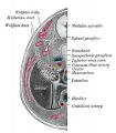
898 Section through human embryo head about twelve days old, in the region of the hind-brain
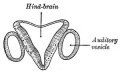
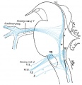
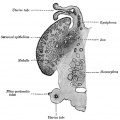
899 Section through hind-brain and auditory vesicles
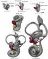
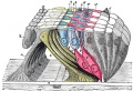
900 Lateral views of membranous labyrinth and acoustic complex
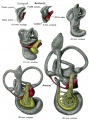
901 Median views of membranous labyrinth and acoustic complex in human embryos
902 Transverse section through head of fetal sheep in the region of the labyrinth
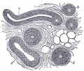
903 Transverse section of the cochlear duct of a fetal cat
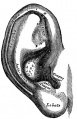
904 Auricula or Pinna
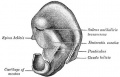
905 Cranial surface of cartilage of right auricula
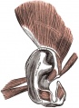
906 The muscles of the auricula
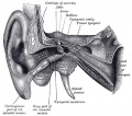
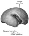
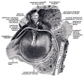
907 External and middle ear, opened from the front. Right side
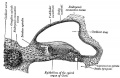
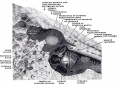
908 Horizontal section through left ear; upper half of section.
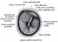
909 Right tympanic membrane
910 Tympanic membrane
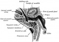
911 View of the inner wall of the tympanum
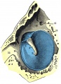
912 The right membrana tympani with the hammer and the chorda tympani, viewed from within, from behind, and from above.
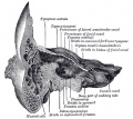
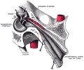
913 Coronal section of right temporal bone
914 Right Tympanic Cavity Walls
915 Auditory Tube
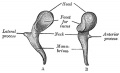
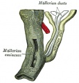
916 Middle Ear - Malleus
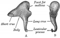
917 Middle Ear - Incus
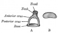
918 Middle Ear - Stapes
919 Chain of ossicles and their ligaments
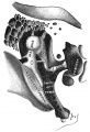
920 Right osseous labyrinth
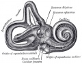
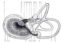
921 Right osseous labyrinth, Interior.
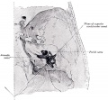
922 Position of the right bony labyrinth in the skull
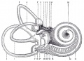
923 Inner Ear - Cochlea and Vestibule
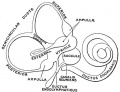
924 Inner Ear - The Membranous Labyrinth
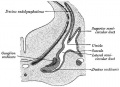
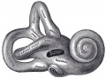
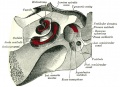
925 Right Human Membranous Labyrinth
926 Right Human Membranous Labyrinth
927 Human Semicircular Canal and Duct
928 Cross-section of Cochlea
929 Floor of Ductus Cochlearis
930 Limbus laminæ spiralis and membrana basilaris
931 Section through the spiral organ of Corti
932 The lamina reticularis and subjacent structures
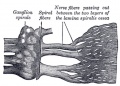
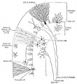
933 Cochlear Division of the Acoustic Nerve


Somatosensory
934 End-bulb of Krause
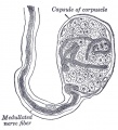
935 Pacinian corpuscle
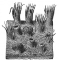
936 Papilla of the hand
937 Nerve ending of Ruffini
938 Organ of Golgi
939 Muscle Spindle


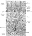
Integumentary
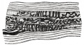
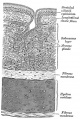
940 A diagrammatic sectional view of the skin
941 Section of Epidermis
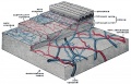
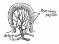
942 The distribution of the bloodvessels in the skin of the sole of the foot
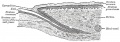
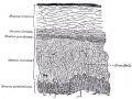
943 Longitudinal section through nail and its nail groove
944
945
946


Respiratory
947 The head and neck human embryo thirty-two days seen from the ventral surface.
948 Lung buds from a human embryo of about four weeks, showing commencing lobulations.
949 Lungs of a human embryo more advanced in development than week 4.
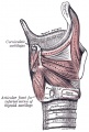
950 Cartilages of the larynx
950 epiglottis cartilage
950 thyroid cartilage
950 cricoid cartilage
950 arytenoid cartilage
951 Ligaments of the larynx (anterior view)
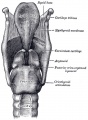
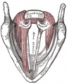
952 Ligaments of the larynx (posterior view)
953 Larynx and upper part of the trachea
954
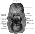
955 Larynx entrance
956
957
958
959
960
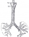
961 Cartilages of larynx, trachea, and bronchi (front view)
962 Bronchi and bronchioles
963
964
965
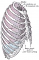
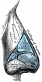
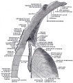
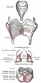
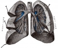
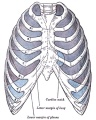
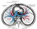
966 Lateral view of thorax, showing the relations of the pleuræ and lungs to the chest wall. Pleura in blue; lungs in purple.
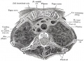
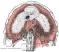
967 Transverse section through the upper margin of the second thoracic vertebra.
968
969
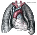
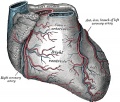
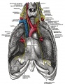
970 Front view of heart and lungs
971 Adult lungs
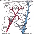
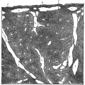
974 Lung secondary lobule
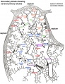
974 relabeled version
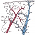
975 Lung primary lobule
975 relabeled version
976 Pig embryo lung


Gastrointestinal
977
978
979
980
981

982a

982b
983
984
985

986
987
987a
987b
988
989
990
991
992
993
994
996
997
998
999
1000

1001
1002
1003
1004
1005
1006
1007
1008
- Gray1009.jpg
1009
- Gray1010.jpg
1010
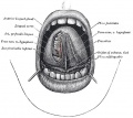
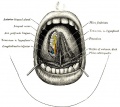
1013 The mouth cavity
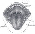
1014 The mouth cavity
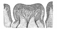
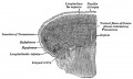
1015 Circumvallate papilla
1016 Filiform papilla
1017 Fungiform papilla
1018 Mucous membrane of the tongue
1019 Extrinsic muscles of the tongue
1020 Intrinsic muscles of the tongue
1022 Right parotid gland (posterior and deep aspects)
1023 Right parotid gland (anterior and deep aspects)
1024 Dissection showing salivary glands of right side

1027 Section through one of the crypts of the tonsil
1028 Dissection of the Muscles of the Palate from behind
1029 Front of nasa part of pharynx
1030 Muscles of the Pharynx and Cheek
1033 Section of the Human Esophagus
1039
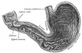
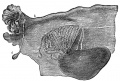
1050 Interior of the stomach
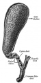
1095 Gall bladder and bile ducts laid open
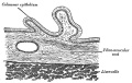
1096 Gall bladder transverse section


Urogenital
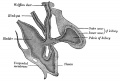
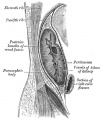
1108 Broad ligament of adult showing Epoöphoron
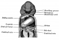
1109 Urogenital Sinus of Female Human Embryo of 8.5 to 9 weeks old
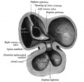
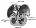
1111 Transverse section of Human Embryo 8.5 to 9 Weeks Old
1112 Longitudinal Section of Ovary of Cat Embryo of 9.4 cm long
1113 Section of the Ovary of a Newly Born Child
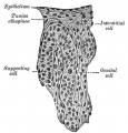
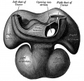
1114 Human Embryo (3.5 cm long) Testis Section of a Genital Cord
1115 Tail end of Human Embryo 25 to 29 Days Old
1116 Tail end of Human Embryo 32 to 33 Days Old
1117 Tail end of human embryo eight and a half to nine weeks old
1118 Primitive Kidney and Bladder
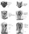
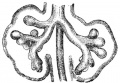
1119 Stages in the development of the external sexual organs in the male and female
1120 Abdomen
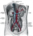
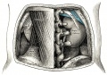
1121 Posterior abdominal wall

1122
1123
1124
1125
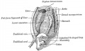
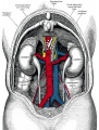
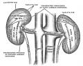
1126 Retroperitoneal structures

1126 Retroperitoneal structures
1127
1128
1129
1130
1131
1132
1133
1137
1138
1139
1140
1141
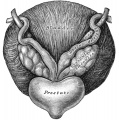
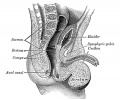
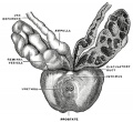
1152 Fundus of the bladder with the vesiculæ seminales
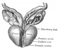
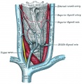
1153 Prostate and seminal vesicles
1160 Prostate Gland
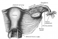
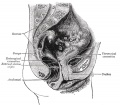
1161 Uterus and right broad ligament
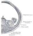
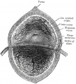
1163 section of the ovary
1166
1170
- Gray1173.jpg
1173


Endocrine
The Ductless Glands
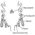
1174 The thyroid gland and its relations.
1175
1181 Pituitary - Median sagittal hypophysis adult monkey
1182 Vertical sections of the heads of early embryos of the rabbit
1183
1184
1185 Section of a part of a suprarenal gland
1192


Surface Anatomy
Surface Anatomy and Surface Markings
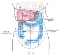
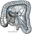
Abdomen Surface Markings for Liver, Stomach, and Great Intestine
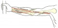
Front of right upper extremity, showing surface markings for bones and nerves.
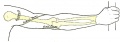
Back of right upper extremity, showing surface markings for bones and nerves.
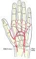
Left Hand Palm, position of skin creases and bones, and surface markings for the volar arches


Glossary Links
- Glossary: A | B | C | D | E | F | G | H | I | J | K | L | M | N | O | P | Q | R | S | T | U | V | W | X | Y | Z | Numbers | Symbols | Term Link
Cite this page: Hill, M.A. (2024, June 10) Embryology Anatomy of the Human Body by Henry Gray. Retrieved from https://embryology.med.unsw.edu.au/embryology/index.php/Anatomy_of_the_Human_Body_by_Henry_Gray
- © Dr Mark Hill 2024, UNSW Embryology ISBN: 978 0 7334 2609 4 - UNSW CRICOS Provider Code No. 00098G