ACPS Seminar 2014 - Implantation: Difference between revisions
m (→Summary) |
|||
| (42 intermediate revisions by the same user not shown) | |||
| Line 7: | Line 7: | ||
{| width=300px | {| width=300px | ||
| [[Image:Mark Hill.jpg|thumb|120px|left|Dr Mark Hill]] | | [[Image:Mark Hill.jpg|thumb|120px|left|Dr Mark Hill]] | ||
School of Medical Sciences | |||
E: m.hill@unsw.edu.au | |||
| [[File:Acps-logo.png|right|120px|link=http://www.acps.unsw.edu.au]] | | [[File:Acps-logo.png|right|120px|link=http://www.acps.unsw.edu.au]] | ||
|} | |} | ||
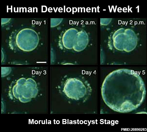
==Human Development - Week 1== | ==Human Development - Week 1== | ||
* Key developmental changes during Week 1 ({{GA}} week 3) | |||
* [[Bovine_Oocyte_Transport_Movie|Transport]] along uterine tube. | |||
{| border='0px' | {| border='0px' | ||
|- | |- | ||
| [[File:Week1_summary.jpg| | | [[File:Week1_summary.jpg|800px]] | ||
|- | |||
| valign="top" width=520px|<mediaplayer width='500' height='450' image="http://embryology.med.unsw.edu.au/embryology/images/0/07/Human_blastocyst_day_1-5.jpg">File:Human_blastocyst_day_3-6.mp4</mediaplayer> | | valign="top" width=520px|<mediaplayer width='500' height='450' image="http://embryology.med.unsw.edu.au/embryology/images/0/07/Human_blastocyst_day_1-5.jpg">File:Human_blastocyst_day_3-6.mp4</mediaplayer> | ||
<center>[[Media:Human_blastocyst_day_3-6.mp4|morula to blastocyst]] <ref name=PMID20890283><pubmed>20890283</pubmed>| [http://www.nature.com/nbt/journal/vaop/ncurrent/full/nbt.1686.html Nat Biotechnol.]</ref></center> | <center>[[Media:Human_blastocyst_day_3-6.mp4|morula to blastocyst]] <ref name=PMID20890283><pubmed>20890283</pubmed>| [http://www.nature.com/nbt/journal/vaop/ncurrent/full/nbt.1686.html Nat Biotechnol.]</ref></center> | ||
|} | |} | ||
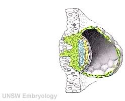
==Human Development - Week 2== | ==Human Development - Week 2== | ||
* Process of implantation during week 2 ({{GA}} week 4) of development in humans. | |||
{| | {| | ||
| width=300px|<mediaplayer width='250' height='260' image="http://php.med.unsw.edu.au/embryology/images/a/a9/Week2_001_icon.jpg">File:Week2_001.mp4</mediaplayer> | | width=300px|<mediaplayer width='250' height='260' image="http://php.med.unsw.edu.au/embryology/images/a/a9/Week2_001_icon.jpg">File:Week2_001.mp4</mediaplayer> | ||
| The beginning of the animation shows adplantation to the the uterus lining (endometrium epithelium). | |||
* '''green cells''' - trophoblast layer of the conceptus | * '''green cells''' - trophoblast layer of the conceptus | ||
| Line 34: | Line 35: | ||
* '''white cells''' - uterine endometrium epithelium | * '''white cells''' - uterine endometrium epithelium | ||
* '''red''' - maternal blood vessel | * '''red''' - maternal blood vessel | ||
|- | |||
| [[File:Stage5_bf08.jpg|300px]] | |||
| [[File:Stage5_bf04.jpg|200px]] | | [[File:Stage5_bf04.jpg|200px]] | ||
| | |- | ||
| Section view | |||
| Surface view | |||
|} | |} | ||
Corpus luteum supported by trophoblast secreted hCG. | |||
[[File:Human ovary - corpus luteum 01.jpg|500px]] | |||
==Early Implantation== | ==Early Implantation== | ||
| Line 43: | Line 51: | ||
{| | {| | ||
! Adplantation Signaling <ref><pubmed>23223073</pubmed>| [http://www.nature.com/nm/journal/v18/n12/full/nm.3012.html Nat Med]</ref> | ! Adplantation Signaling <ref name=PMID23223073><pubmed>23223073</pubmed>| [http://www.nature.com/nm/journal/v18/n12/full/nm.3012.html Nat Med]</ref> | ||
|- | |- | ||
| [[File:Implantation cartoon 01.jpg| | | [[File:Implantation cartoon 01.jpg|800px]] | ||
| | |- | ||
[[File:Implantation cartoon 02.jpg| | ! Early Implantation Signaling<ref name=PMID23223073><pubmed>23223073</pubmed>| [http://www.nature.com/nm/journal/v18/n12/full/nm.3012.html Nat Med]</ref> | ||
|- | |||
| [[File:Implantation cartoon 02.jpg|600px]] | |||
|} | |} | ||
| Line 55: | Line 64: | ||
{| border='0px' | {| border='0px' | ||
|- | |- | ||
| < | | <html5media height="550" width="660">File:Ectopic_01.mp4</html5media> | ||
| valign="top" | | | valign="top" |The movie shows an ectopic embryo (less than {{GA}} 10 weeks) implanted in the left uterine tube. | ||
The movie shows an ectopic embryo (less than 10 weeks | |||
* '''RT''' - Normal Right Tube, the movie starts by showing the normal right uterine tube. | * '''RT''' - Normal Right Tube, the movie starts by showing the normal right uterine tube. | ||
| Line 64: | Line 71: | ||
* '''L OV''' - Also visible is the left ovary | * '''L OV''' - Also visible is the left ovary | ||
* '''UT''' - uterus. | * '''UT''' - uterus. | ||
|} | |} | ||
{| | {| | ||
| Line 73: | Line 79: | ||
| valign=top|[[File:Stage20 bf12.jpg|400px]] | | valign=top|[[File:Stage20 bf12.jpg|400px]] | ||
'''Image:''' [[Contributors#Dr_Steven_O.27Connor|Dr Steven O'Connor]] (Houston, Texas) | |||
|} | |} | ||
{| | {| | ||
| Line 82: | Line 88: | ||
| [[File:Placental villi 4.jpg|300px]] | | [[File:Placental villi 4.jpg|300px]] | ||
|} | |} | ||
* Ectopic Model for implantation - some differences (e.g. Endocannabinoids G protein-coupled cannabinoid receptor CB1, ectopic pregnancy show down-regulation<ref><pubmed>19093002</pubmed></ref>) | |||
==Uterine Implantation== | ==Uterine Implantation== | ||
{| | {| | ||
| Line 92: | Line 101: | ||
==Tube Biobank== | ==Tube Biobank== | ||
[[File:Ectopic tubal cartoon.jpg|right|300px]] | |||
* Most published human studies are based upon only a very few tissue samples. | * Most published human studies are based upon only a very few tissue samples. | ||
* Our Biobank tissues will | * Our Biobank tissues will also be available to other implantation researchers. | ||
* Approval to collect tissue from multiple sites. | |||
[[File:HFTB_graph_4.jpg]] | [[File:HFTB_graph_4.jpg]] | ||
| Line 114: | Line 125: | ||
==Immune Protection== | ==Immune Protection== | ||
* Expression by fetal cells protects from maternal immune rejection. | * Expression by fetal cells protects from maternal immune rejection. | ||
'''HLA-G''' - Human leukocyte antigen G (HLA-G, HLA-6.0; HLA60, T-CELL A LOCUS; TCA) | '''HLA-G''' - Human leukocyte antigen G (HLA-G, HLA-6.0; HLA60, T-CELL A LOCUS; TCA) | ||
* HLA-G is a non-classical HLA class-Ib molecule expressed by the extravillous cytotrophoblasts (EVT) of the placenta. | * HLA-G is a non-classical HLA class-Ib molecule expressed by the extravillous cytotrophoblasts (EVT) of the placenta. | ||
* 338 AA protein, mRNA differential splicing gives rise to 7 isoforms: 4 membrane-bound forms (HLA-G1, G2, G3, G4) and 3 soluble forms (sHLA-G5, G6, G7) | * 338 AA protein, mRNA differential splicing gives rise to 7 isoforms: 4 membrane-bound forms (HLA-G1, G2, G3, G4) and 3 soluble forms (sHLA-G5, G6, G7). | ||
* miRNA regulated expression. | |||
{| | {| | ||
| Line 125: | Line 138: | ||
HLA-G expression (red) on trophoblast cell columns (CC) and extravillous trophoblasts.<ref><pubmed>22438923</pubmed>| [http://www.plosone.org/article/info%3Adoi%2F10.1371%2Fjournal.pone.0033395 PLoS One.]</ref> | HLA-G expression (red) on trophoblast cell columns (CC) and extravillous trophoblasts.<ref><pubmed>22438923</pubmed>| [http://www.plosone.org/article/info%3Adoi%2F10.1371%2Fjournal.pone.0033395 PLoS One.]</ref> | ||
|} | |} | ||
==Summary== | ==Summary== | ||
[[File:Early placental structure.jpg| | [[File:Early placental structure.jpg|right|500px]] | ||
* We have established a '''Uterine Tube Biobank''' at UNSW. | * We have established a '''Uterine Tube Biobank''' at UNSW. | ||
* First samples collected Sep 2013. | * First samples collected Sep 2013. | ||
| Line 140: | Line 152: | ||
** analysing expression of implantation associated proteins in these tissues. | ** analysing expression of implantation associated proteins in these tissues. | ||
* First study of a large number of first trimester implantation samples. | * First study of a large number of first trimester implantation samples. | ||
<br> | |||
<br> | |||
<br> | |||
<br> | |||
==Special Thanks== | ==Special Thanks== | ||
[[File:Galletti1770 birth.jpg|200px|right]] | |||
* '''Patients''' who have donated to this study. | * '''Patients''' who have donated to this study. | ||
* [https://med.unsw.edu.au/people/professor-william-ledger '''Prof William Ledger'''] my clinical collaborator. | * [https://med.unsw.edu.au/people/professor-william-ledger '''Prof William Ledger'''] my clinical collaborator. | ||
* '''Dr Anusha Hettiaratchi''' the Manager of Lowy Biorepository. | * '''Dr Anusha Hettiaratchi''' the Manager of Lowy Biorepository. | ||
* '''Dr Tatiana Zandanova''', '''Ngoc Chi Vo''' and '''Elizabeth Dalton''' three research students currently developing the project. | |||
* '''Australian Centre for Perinatal Science''' for supporting developmental research and researchers. | |||
<br> | |||
<br> | |||
<br> | |||
<br> | |||
<br> | |||
<br> | |||
<br> | |||
:'''Links:''' [[Implantation]] | [[Abnormal_Development_-_Ectopic_Implantation|Ectopic Implantation]] | [[Week 2]] | [[Trophoblast]] | |||
==External Links== | ==External Links== | ||
{{External Links}} | {{External Links}} | ||
| Line 158: | Line 184: | ||
<references/> | <references/> | ||
---- | |||
{{Footer}} | {{Footer}} | ||
Latest revision as of 08:42, 22 February 2016
Understanding Implantation and the new Uterine Tube Biobank
|
|
School of Medical Sciences E: m.hill@unsw.edu.au |

Human Development - Week 1

|
| <mediaplayer width='500' height='450' image="http://embryology.med.unsw.edu.au/embryology/images/0/07/Human_blastocyst_day_1-5.jpg">File:Human_blastocyst_day_3-6.mp4</mediaplayer>
|
Human Development - Week 2
- Process of implantation during week 2 (GA week 4) of development in humans.
| <mediaplayer width='250' height='260' image="http://php.med.unsw.edu.au/embryology/images/a/a9/Week2_001_icon.jpg">File:Week2_001.mp4</mediaplayer> | The beginning of the animation shows adplantation to the the uterus lining (endometrium epithelium).
|

|

|
| Section view | Surface view |
Corpus luteum supported by trophoblast secreted hCG.

Early Implantation
- Most data from animal models (mouse) of implantation.
- Very little human data for early (first trimester) events.
| Adplantation Signaling [2] |
|---|

|
| Early Implantation Signaling[2] |

|
Ectopic Implantation
| <html5media height="550" width="660">File:Ectopic_01.mp4</html5media> | The movie shows an ectopic embryo (less than GA 10 weeks) implanted in the left uterine tube.
|
| Ectopic GA Week 7 | Ectopic GA Week 10 |
|---|---|

|

Image: Dr Steven O'Connor (Houston, Texas) |
| First trimester placental villi | Term placental villi |
|---|---|

|

|
- Ectopic Model for implantation - some differences (e.g. Endocannabinoids G protein-coupled cannabinoid receptor CB1, ectopic pregnancy show down-regulation[3])
Uterine Implantation

|
|
Tube Biobank

- Most published human studies are based upon only a very few tissue samples.
- Our Biobank tissues will also be available to other implantation researchers.
- Approval to collect tissue from multiple sites.

Tube Analysis Techniques
- Analysing the proteins expressed at the site of implantation (fetal and maternal).
- Analysing the messenger RNA (mRNA) that encodes protein expression.
- Analysis of microRNA (miRNA) that regulates gene and protein expression.
- Later DNA genetic analysis (are there ectopic implantation associated genes?).
Protein Expression


mRNA Expression

Immune Protection
- Expression by fetal cells protects from maternal immune rejection.
HLA-G - Human leukocyte antigen G (HLA-G, HLA-6.0; HLA60, T-CELL A LOCUS; TCA)
- HLA-G is a non-classical HLA class-Ib molecule expressed by the extravillous cytotrophoblasts (EVT) of the placenta.
- 338 AA protein, mRNA differential splicing gives rise to 7 isoforms: 4 membrane-bound forms (HLA-G1, G2, G3, G4) and 3 soluble forms (sHLA-G5, G6, G7).
- miRNA regulated expression.
|
|

HLA-G expression (red) on trophoblast cell columns (CC) and extravillous trophoblasts.[4] |
Summary

- We have established a Uterine Tube Biobank at UNSW.
- First samples collected Sep 2013.
- We now have patient samples from:
- ectopic implantation tube
- control tube, from outside the implantation site
- control tube, from non-pregnant tube.
- Hope to also have control tube from hormonally induced pseudo-pregnacy.
- Three research students (Tatiana, Chi and Liz) working on the project.
- preparing protein, mRNA, miRNA and DNA.
- analysing expression of implantation associated proteins in these tissues.
- First study of a large number of first trimester implantation samples.
Special Thanks

- Patients who have donated to this study.
- Prof William Ledger my clinical collaborator.
- Dr Anusha Hettiaratchi the Manager of Lowy Biorepository.
- Dr Tatiana Zandanova, Ngoc Chi Vo and Elizabeth Dalton three research students currently developing the project.
- Australian Centre for Perinatal Science for supporting developmental research and researchers.
- Links: Implantation | Ectopic Implantation | Week 2 | Trophoblast
External Links
External Links Notice - The dynamic nature of the internet may mean that some of these listed links may no longer function. If the link no longer works search the web with the link text or name. Links to any external commercial sites are provided for information purposes only and should never be considered an endorsement. UNSW Embryology is provided as an educational resource with no clinical information or commercial affiliation.
- Prof William Ledger
- Australian Centre for Perinatal Science
- UNSW Biobank
- miRNA microRNA database | MicroRNA
References
Cite this page: Hill, M.A. (2024, June 14) Embryology ACPS Seminar 2014 - Implantation. Retrieved from https://embryology.med.unsw.edu.au/embryology/index.php/ACPS_Seminar_2014_-_Implantation
- © Dr Mark Hill 2024, UNSW Embryology ISBN: 978 0 7334 2609 4 - UNSW CRICOS Provider Code No. 00098G