Category:Vision: Difference between revisions
From Embryology
mNo edit summary |
mNo edit summary |
||
| (One intermediate revision by the same user not shown) | |||
| Line 1: | Line 1: | ||
This {{Embryology}} category shows pages and media related to the sensory system of vision (eye) development. | This {{Embryology}} category shows pages and media related to the sensory system of {{vision}} (eye) development. | ||
{{Vision Links}} | {{Vision Links}} | ||
[[Category:Senses]] | [[Category:Senses]] | ||
Latest revision as of 11:00, 9 May 2018
This Embryology category shows pages and media related to the sensory system of vision (eye) development.
Subcategories
This category has the following 4 subcategories, out of 4 total.
Pages in category 'Vision'
The following 143 pages are in this category, out of 143 total.
2
A
B
- Template:Bartelmez1922 figures
- BGDB Face and Ear - Early Embryo
- Book - A textbook of histology, including microscopic technic (1910) Special Histology 8
- Book - Biomicroscopy of the eye 2
- Book - Biomicroscopy of the eye 2-24
- Book - Biomicroscopy of the eye 2-25
- Book - Biomicroscopy of the eye 2-26
- Book - Manual of Human Embryology 16
- Book - Manual of Human Embryology 16-1
- Book - Manual of Human Embryology 16-2
- Book - Manual of Human Embryology 16-3
- Book - Manual of Human Embryology 16-4
- Book - Text-Book of Embryology 18
- Book - The Embryology Anatomy and Histology of the Eye 1
- Book - The Embryology Anatomy and Histology of the Eye 2
- Book - The Embryology Anatomy and Histology of the Eye 3
- Template:Brown 1906 Figures
- Template:Brown 1906 Footer
C
E
H
L
P
- Paper - A description of the histology of the eyes in two anencephalic fœtuses (1910)
- Paper - A phylogenetic consideration of the optic tectum
- Paper - A Specimen of Congenital Aphakia occurring in a Human Embryo of Five to Six Weeks
- Paper - Absence of the lens occurring in the human embryo (1922)
- Paper - Contribution to the structure and development of the vertebrate head
- Paper - Contribution to the structure and development of the vertebrate head 1
- Paper - Contribution to the structure and development of the vertebrate head 2
- Paper - Contribution to the structure and development of the vertebrate head 3
- Paper - On the development of the fissural and associated regions in the eye of the chick with some observations on the mammal (1921)
- Paper - On the development of the retina and optic nerve, and of the membranous labyrinth and auditory nerve
- Paper - On the growth of the human eyeball and optic nerve
- Paper - The change in position of the eyeballs during fetal life
- Paper - The developing third nerve nucleus in human embryos
- Paper - The development of the eyelids 1
- Paper - The development of the eyelids of the albino rat, until the completion of disjunction (1921)
- Paper - The development of the human iris
- Paper - The early development of the eye in staged human embryos
- Paper - The formation of neural crest from the primary optic vesicle in man
- Paper - The genesis and development of the nasolacrimal passages in man
- Paper - The history of the eye muscles (1917)
- Paper - The lachrymal gland (1916)
- Paper - The origin and development of the human extrinsic ocular muscles (1957)
- Paper - The Origin of the Otic and Optic Primordia in Man
- Paper - The process of differentiation of the retinal layers in vertebrates
- Paper - The process of retinal differentiation in man
- Paper - The relations of the hyaloid canal in the foetus and in the adult
- Template:Placode
- Template:Placodes
R
- Template:Ref-AddisonHow1921
- Template:Ref-Bartelmez1922
- Template:Ref-BartelmezBlount1954
- Template:Ref-Berliner1949
- Template:Ref-Bowen1922
- Template:Ref-Brown1906
- Template:Ref-Cameron1905a
- Template:Ref-Cameron1905b
- Template:Ref-Cameron1905c
- Template:Ref-DeDutta1939
- Template:Ref-Gilbert1957
- Template:Ref-HuberCrosby1933
- Template:Ref-KolbFernandezNelson2012
- Template:Ref-Mann1921b
- Template:Ref-Mann1922
- Template:Ref-Mann1925
- Template:Ref-Mann1927
- Template:Ref-Mann1927b
- Template:Ref-Mann1928
- Template:Ref-Mann1928b
- Template:Ref-Neal1917
- Template:Ref-O'Rahilly1965
- Template:Ref-Oakden1910
- Template:Ref-Pearson1980
- Template:Ref-ScammonArmstrong1925
- Template:Ref-Stockard1910b
- Template:Ref-Sundwall1916
- Template:Retina
S
V
Media in category 'Vision'
The following 116 files are in this category, out of 316 total.
(previous page) (next page) Lens-neural crest signaling 01.jpg 300 × 400; 24 KB
 Lens-neural crest signaling 02.jpg 521 × 522; 22 KB
 Lewis1906 fig425.jpg 1,000 × 1,191; 200 KB
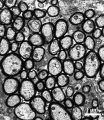
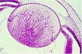
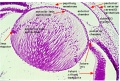
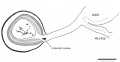
 Limbal palisades of Vogt PMID17211449.jpg 692 × 800; 86 KB
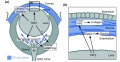
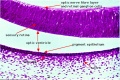
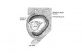
 Limbal stem cell niche cartoon PMID17211449.jpg 800 × 579; 52 KB
 Lizard embryo 10.jpg 1,200 × 900; 152 KB
 Lizard embryo 11.jpg 1,200 × 900; 122 KB
 Low 13.jpg 637 × 443; 52 KB
 McMurrich1930 fig82.jpg 1,280 × 955; 258 KB
 McMurrich1930 fig83.jpg 800 × 907; 108 KB
 Microphthalmia.jpg 600 × 504; 49 KB
 Morgan 1925 fig71.jpg 1,000 × 1,474; 64 KB
 Morgan 1925 fig72.jpg 1,000 × 362; 34 KB
 Mouse cornea development 01.jpg 1,200 × 880; 325 KB
 Mouse cornea E12.5.jpg 700 × 558; 123 KB
 Mouse cornea E13.5.jpg 700 × 550; 103 KB
 Mouse cornea E16.5.jpg 701 × 562; 113 KB
 Mouse cornea P0.jpg 703 × 561; 101 KB
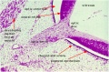
 Mouse eye and limbal region histology 01.jpg 1,200 × 467; 181 KB
 Mouse eye cell proliferation E13.5.jpg 875 × 1,244; 227 KB
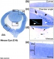 Mouse eye E18.jpg 884 × 977; 169 KB
 Mouse eye neural crest cornea 01.jpg 500 × 256; 34 KB
 Mouse eye neural crest cornea 02.jpg 800 × 655; 121 KB
 Mouse eye neural crest.jpg 1,086 × 1,509; 483 KB
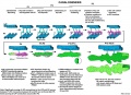
 Mouse eye TGF-beta model.jpg 1,000 × 519; 88 KB
 Mouse Schlemm's canal development 01.jpg 1,200 × 873; 161 KB
 Mouse-optic nerve axons.jpg 600 × 693; 126 KB
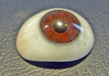 Ocular prosthesis.png 525 × 367; 359 KB
 Patten045.jpg 800 × 968; 156 KB
 Pax6 eye phenotypes.jpg 1,000 × 662; 152 KB
 Retinal patterning model.jpg 967 × 568; 120 KB
 Rugh 093.jpg 965 × 800; 176 KB
 Rugh 120.jpg 840 × 800; 118 KB
 Rugh 121.jpg 919 × 800; 161 KB
 Rugh 122.jpg 643 × 1,000; 187 KB
 Rugh 123.jpg 738 × 1,000; 219 KB
 Rugh 124.jpg 943 × 800; 144 KB
 Stage 13 image 053.jpg 1,000 × 423; 76 KB
 Stage 13 image 060.jpg 1,000 × 486; 96 KB
 Stage 13 image 061.jpg 1,000 × 600; 101 KB
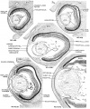
 Stage 22 image 008-eye.jpg 1,200 × 1,059; 555 KB
 Stage 22 image 103.jpg 1,000 × 670; 127 KB
 Stage 22 image 104.jpg 1,000 × 662; 172 KB
 Stage 22 image 105.jpg 1,000 × 665; 178 KB
 Stage 22 image 106.jpg 1,000 × 658; 213 KB
 Stage 22 image 152.jpg 1,000 × 670; 127 KB
 Stage 22 image 153.jpg 1,000 × 662; 191 KB
 Stage 22 image 154.jpg 1,000 × 665; 205 KB
 Stage 22 image 155.jpg 1,000 × 672; 239 KB
 Stage 22 image 206.jpg 1,200 × 754; 245 KB
 Stage 22 image 207.jpg 1,200 × 753; 208 KB
 Stage 22 image 208.jpg 1,200 × 903; 368 KB
 Stage 22 image 209.jpg 1,200 × 808; 305 KB
 Stage 22 image 211.jpg 1,200 × 760; 242 KB
 Stage 22 image 212.jpg 1,200 × 753; 269 KB
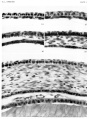
 Stage11 histology-optic pit.jpg 800 × 510; 157 KB
 Stage11 histology-optic vesicle-hindbrain.jpg 800 × 536; 158 KB
 Streeter1957 fig04-19.jpg 1,538 × 800; 95 KB
 Streeter1957 fig04-20.jpg 1,538 × 800; 78 KB
 Streeter1957 fig04-21.jpg 1,538 × 800; 95 KB
 Streeter1957 fig04-22.jpg 1,538 × 800; 105 KB
 Streeter1957 fig04-23.jpg 1,538 × 800; 115 KB
 Streeter1957 fig04.jpg 1,028 × 1,266; 205 KB
 Streeter1957 fig05.jpg 1,280 × 1,690; 546 KB
 Streeter1957 fig06-19.jpg 1,280 × 834; 120 KB
 Streeter1957 fig06-20.jpg 1,280 × 834; 133 KB
 Streeter1957 fig06-21.jpg 1,280 × 834; 185 KB
 Streeter1957 fig06-22.jpg 1,280 × 834; 181 KB
 Streeter1957 fig06-23.jpg 1,280 × 834; 161 KB
 Streeter1957 fig06.jpg 1,280 × 1,541; 622 KB
 Streeter1957 plate01.jpg 1,500 × 2,009; 486 KB
 Tectum signaling model 01.jpg 600 × 314; 41 KB
 Wen1928-Fig07.jpg 1,265 × 648; 166 KB
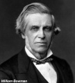 William Bowman.jpg 600 × 665; 49 KB
 Xenopus cornea development timeline.jpg 1,288 × 800; 77 KB
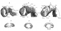
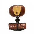
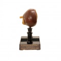
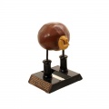
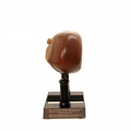
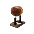
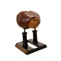
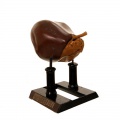
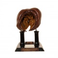
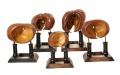
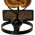
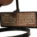
 Ziegler model 20.jpg 800 × 800; 38 KB
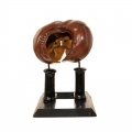
 Ziegler model 21.jpg 800 × 800; 44 KB
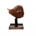
 Ziegler model 22.jpg 800 × 800; 41 KB
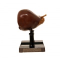
 Ziegler model 23.jpg 797 × 800; 49 KB
 Ziegler model 24.jpg 800 × 800; 43 KB
 Ziegler model 25.jpg 800 × 800; 46 KB
 Ziegler model 26.jpg 800 × 800; 48 KB
 Ziegler model 27.jpg 800 × 800; 52 KB
 Ziegler model 28.jpg 800 × 800; 49 KB
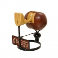
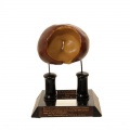
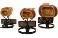 Ziegler model 29.jpg 881 × 582; 69 KB
 Ziegler model 30.jpg 800 × 800; 30 KB
 Ziegler model 31.jpg 800 × 800; 0 bytes
 Ziegler model 32.jpg 800 × 800; 41 KB
 Ziegler model 33.jpg 800 × 800; 28 KB
 Ziegler model 34.jpg 800 × 800; 34 KB
 Ziegler model 35.jpg 800 × 800; 29 KB
 Ziegler model 36.jpg 800 × 800; 34 KB
 Ziegler model 37.jpg 800 × 800; 29 KB
 Ziegler model 38.jpg 800 × 800; 30 KB
 Ziegler model 39.jpg 800 × 800; 38 KB
 Ziegler model 40.jpg 800 × 800; 40 KB
 Ziegler model 41.jpg 800 × 800; 30 KB
 Ziegler model 42.jpg 800 × 800; 33 KB
 Ziegler model 43.jpg 800 × 800; 38 KB
 Ziegler model 44.jpg 800 × 800; 43 KB
 Ziegler model 45.jpg 800 × 800; 33 KB
 Ziegler model 46.jpg 800 × 799; 35 KB
 Ziegler model 47.jpg 800 × 800; 40 KB
 Ziegler model 48.jpg 800 × 800; 46 KB
 Ziegler model 49.jpg 1,000 × 623; 73 KB
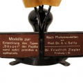 Ziegler model legend 21.jpg 500 × 500; 0 bytes
 Ziegler model legend 23.jpg 500 × 500; 0 bytes
 Ziegler model legend 26.jpg 500 × 499; 52 KB
 Ziegler model legend 33.jpg 800 × 800; 74 KB
 Ziegler model legend 35.jpg 800 × 800; 60 KB
 Ziegler model legend 37.jpg 800 × 800; 51 KB
 Ziegler model legend 38.jpg 800 × 800; 53 KB
 Ziegler model legend 41.jpg 800 × 800; 64 KB
 Ziegler model legend 45.jpg 800 × 799; 0 bytes
 Ziegler model legend 48.jpg 800 × 803; 74 KB


