File:Mouse interdigit apoptosis 01.jpg: Difference between revisions
No edit summary |
No edit summary |
||
| Line 7: | Line 7: | ||
==Reference== | ===Reference=== | ||
<pubmed>17194222</pubmed>| [http://www.pubmedcentral.nih.gov/articlerender.fcgi?artid=1713256 PMC1713256] | [http://www.plosgenetics.org/article/info%3Adoi%2F10.1371%2Fjournal.pgen.0020216 PLoS Genet.] | <pubmed>17194222</pubmed>| [http://www.pubmedcentral.nih.gov/articlerender.fcgi?artid=1713256 PMC1713256] | [http://www.plosgenetics.org/article/info%3Adoi%2F10.1371%2Fjournal.pgen.0020216 PLoS Genet.] | ||
Latest revision as of 16:31, 10 May 2012
Mouse Interdigit Apoptosis
Mouse embryo E15.5 hindlimb (wild-type) showing apoptotic cells in the interdigital mesenchyme. Insert is enlarged view of selected region (red box). Apoptosis was identified by acridine orange stain that appears yellow in figure.
Reference
<pubmed>17194222</pubmed>| PMC1713256 | PLoS Genet.
Copyright : © 2006 Bandyopadhyay et al. This is an open-access article distributed under the terms of the Creative Commons Attribution License, which permits unrestricted use, distribution, and reproduction in any medium, provided the original author and source are credited.
Original image name: Figure 2 (panel E and G extracted and resized from full image)
File history
Yi efo/eka'e gwa ebo wo le nyangagi wuncin ye kamina wunga tinya nan
| Gwalagizhi | Nyangagi | Dimensions | User | Comment | |
|---|---|---|---|---|---|
| current | 15:13, 14 November 2011 | 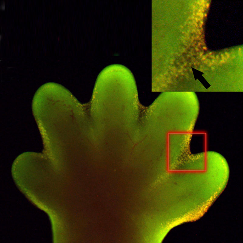 | 800 × 800 (81 KB) | S8600021 (talk | contribs) | ==Mouse Interdigit Apoptosis== Mouse embryo E15.5 hindlimb (wild-type) showing apoptotic cells in the interdigital mesenchyme. Insert is enlarged view of selected region (red box). Apoptosis was identified by acridine orange stain that appears yellow in |
You cannot overwrite this file.
File usage
The following 4 pages use this file: