File:Bailey370.jpg: Difference between revisions
No edit summary |
No edit summary |
||
| Line 1: | Line 1: | ||
==Fig. 370. A-F are sagittal sections showing structures lying in the median line and also paired structures (e.g., pallium) lying to one side of the median line== | |||
Edinger | |||
The cerebellum is black. It is doubtful whether the membranous roof in A indicated as pallium is strictly homologous with that structure in other forms, In B, Pallium indicates prepallial structures. | |||
Aq. SyL, Aquseductus Sylvii; Basis mesen., basis mesencephali; Bulb, olf., bulbus olfactorius; Corp. striat., corpus striatum; Epiph., epiphysis; G. h., ganglion habenulae; Hyp., hypophysis; Infund., infundibulum; Lam. t., lamina terminalis; Lob. elect., lobus electricus; L. vagi, lobus vagi; L. opt., mid-brain roof; Med. obi., medulla oblongata; Opt., optic nerve; Pl.chor., plexus chorioideus; Rec. inf., recessus infundibuli; Rec. mam., recessus mammillaris; Saccus vase., saccus vasculosus; Sp. c., spinal cord; ventr., ventricle; v. m. a., velum medullare anterius; v.m. p., velum medullare posterius. | |||
:G and H show the mesial surface of the cerebral hemispheres in a low (G) and high (H) Mammal. G. Elliot Smith, Edinger, slightly modified. | :G and H show the mesial surface of the cerebral hemispheres in a low (G) and high (H) Mammal. G. Elliot Smith, Edinger, slightly modified. | ||
Revision as of 16:49, 29 January 2011
Fig. 370. A-F are sagittal sections showing structures lying in the median line and also paired structures (e.g., pallium) lying to one side of the median line
Edinger
The cerebellum is black. It is doubtful whether the membranous roof in A indicated as pallium is strictly homologous with that structure in other forms, In B, Pallium indicates prepallial structures.
Aq. SyL, Aquseductus Sylvii; Basis mesen., basis mesencephali; Bulb, olf., bulbus olfactorius; Corp. striat., corpus striatum; Epiph., epiphysis; G. h., ganglion habenulae; Hyp., hypophysis; Infund., infundibulum; Lam. t., lamina terminalis; Lob. elect., lobus electricus; L. vagi, lobus vagi; L. opt., mid-brain roof; Med. obi., medulla oblongata; Opt., optic nerve; Pl.chor., plexus chorioideus; Rec. inf., recessus infundibuli; Rec. mam., recessus mammillaris; Saccus vase., saccus vasculosus; Sp. c., spinal cord; ventr., ventricle; v. m. a., velum medullare anterius; v.m. p., velum medullare posterius.
- G and H show the mesial surface of the cerebral hemispheres in a low (G) and high (H) Mammal. G. Elliot Smith, Edinger, slightly modified.
- Text-Book of Embryology: Germ cells | Maturation | Fertilization | Amphioxus | Frog | Chick | Mammalian | External body form | Connective tissues and skeletal | Vascular | Muscular | Alimentary tube and organs | Respiratory | Coelom, Diaphragm and Mesenteries | Urogenital | Integumentary | Nervous System | Special Sense | Foetal Membranes | Teratogenesis | Gallery of All Figures
| Historic Disclaimer - information about historic embryology pages |
|---|
 |
Reference
Bailey FR. and Miller AM. Text-Book of Embryology (1921) New York: William Wood and Co.
Cite this page: Hill, M.A. (2024, June 20) Embryology Bailey370.jpg. Retrieved from https://embryology.med.unsw.edu.au/embryology/index.php/File:Bailey370.jpg
- © Dr Mark Hill 2024, UNSW Embryology ISBN: 978 0 7334 2609 4 - UNSW CRICOS Provider Code No. 00098G
File history
Yi efo/eka'e gwa ebo wo le nyangagi wuncin ye kamina wunga tinya nan
| Gwalagizhi | Nyangagi | Dimensions | User | Comment | |
|---|---|---|---|---|---|
| current | 16:44, 29 January 2011 | 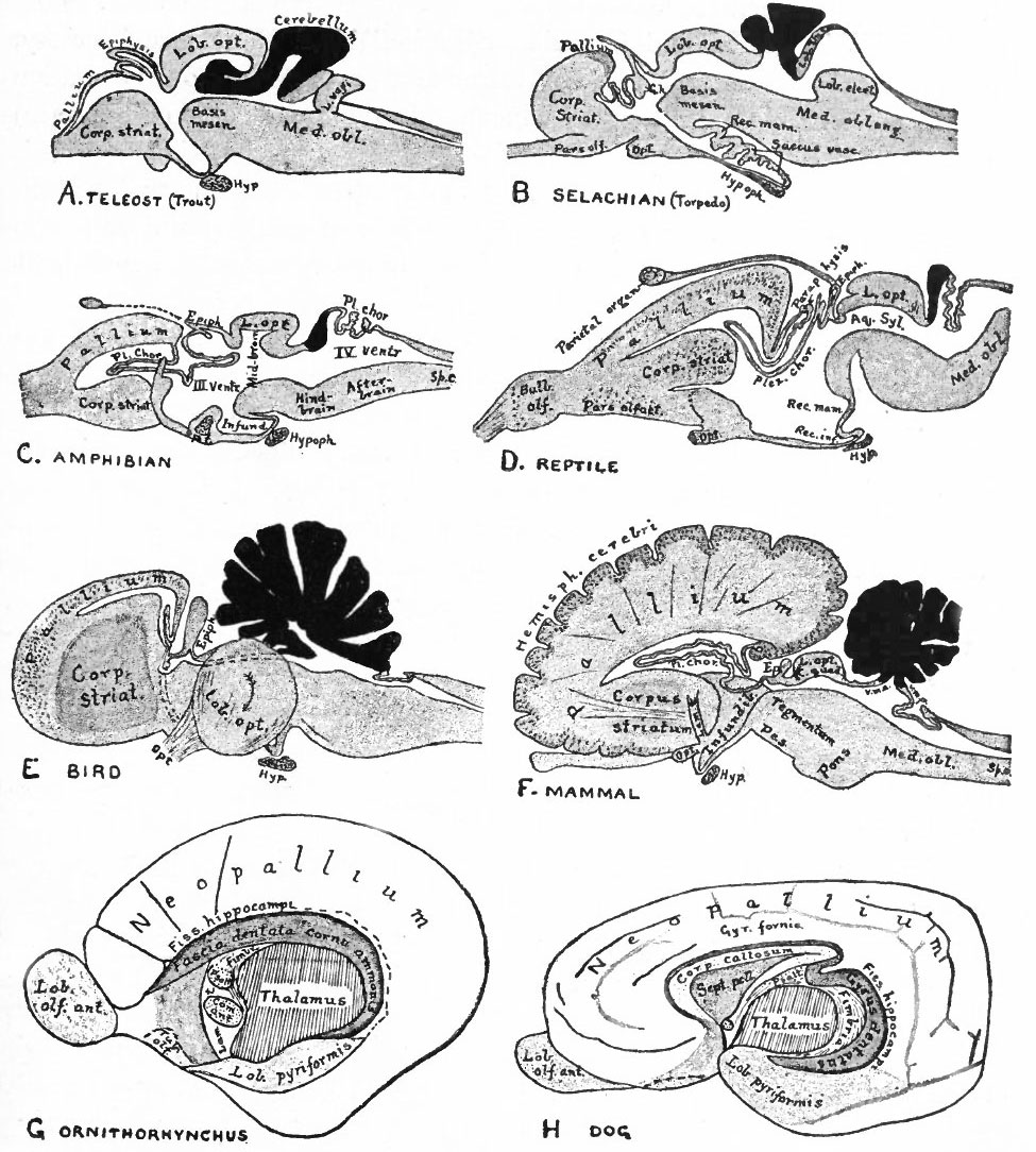 | 975 × 1,084 (242 KB) | S8600021 (talk | contribs) | {{Template:Bailey 1921 Figures}} Category:Neural |
You cannot overwrite this file.