Morula: Difference between revisions
No edit summary |
|||
| Line 4: | Line 4: | ||
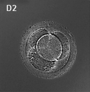
[[File:Human embryo day 2.jpg|thumb|300px|Human morula (day 2)<ref name="PMID19924284"><pubmed>19924284</pubmed>| [http://www.ncbi.nlm.nih.gov/pmc/articles/PMC2773928 PMC2773928] | [http://www.plosone.org/article/info%3Adoi%2F10.1371%2Fjournal.pone.0007844 PLoS One]</ref>]] | [[File:Human embryo day 2.jpg|thumb|300px|Human morula (day 2)<ref name="PMID19924284"><pubmed>19924284</pubmed>| [http://www.ncbi.nlm.nih.gov/pmc/articles/PMC2773928 PMC2773928] | [http://www.plosone.org/article/info%3Adoi%2F10.1371%2Fjournal.pone.0007844 PLoS One]</ref>]] | ||
(Latin, ''morula'' = mulberry) An early stage in post-fertilization development when cells have rapidly mitotically divided to produce a solid mass of cells (12-15 cells) with a "mulberry" appearance. This stage is followed by formation of a cavity in this cellular mass [[blastocyst]] stage. | (Latin, ''morula'' = mulberry) An early stage in post-fertilization development when cells have rapidly mitotically divided to produce a solid mass of cells (12-15 cells) with a "mulberry" appearance. This stage is followed by formation of a cavity in this cellular mass [[blastocyst]] stage. | ||
A key event prior to morula formation is "compaction", where the 8 cell embryo undergoes changes in cell morphology and cell-cell adhesion that initiates the formation of this solid ball of cells. | |||
In humans, morula stage of development occurs during the first days of the first week following [[fertilization]]. This developmental stage is followed by formation of a cavity, the blastocoel, which defines formation of the [[blastocyst]]. | In humans, morula stage of development occurs during the first days of the first week following [[fertilization]]. This developmental stage is followed by formation of a cavity, the blastocoel, which defines formation of the [[blastocyst]]. | ||
| Line 18: | Line 21: | ||
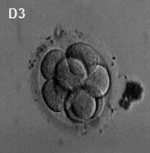
* '''Non-invasive imaging of human embryos before embryonic genome activation predicts development to the blastocyst stage'''<ref name="PMID20890283"><pubmed>20890283</pubmed>| [http://www.nature.com/nbt/journal/vaop/ncurrent/full/nbt.1686.html Nat Biotechnol.]</ref> "We report studies of preimplantation human embryo development that correlate time-lapse image analysis and gene expression profiling. By examining a large set of zygotes from in vitro fertilization (IVF), we find that success in progression to the blastocyst stage can be predicted with >93% sensitivity and specificity by measuring three dynamic, noninvasive imaging parameters by day 2 after fertilization, before embryonic genome activation (EGA)." | * '''Non-invasive imaging of human embryos before embryonic genome activation predicts development to the blastocyst stage'''<ref name="PMID20890283"><pubmed>20890283</pubmed>| [http://www.nature.com/nbt/journal/vaop/ncurrent/full/nbt.1686.html Nat Biotechnol.]</ref> "We report studies of preimplantation human embryo development that correlate time-lapse image analysis and gene expression profiling. By examining a large set of zygotes from in vitro fertilization (IVF), we find that success in progression to the blastocyst stage can be predicted with >93% sensitivity and specificity by measuring three dynamic, noninvasive imaging parameters by day 2 after fertilization, before embryonic genome activation (EGA)." | ||
|} | |} | ||
==Compaction== | |||
* E-cadherin mediated adhesion initiates at compaction at the 8-cell stage | |||
* regulated post-translationally via protein kinase C and other signalling molecules | |||
==Morulas in Other Species== | ==Morulas in Other Species== | ||
Revision as of 12:45, 14 October 2010
Introduction

(Latin, morula = mulberry) An early stage in post-fertilization development when cells have rapidly mitotically divided to produce a solid mass of cells (12-15 cells) with a "mulberry" appearance. This stage is followed by formation of a cavity in this cellular mass blastocyst stage.
A key event prior to morula formation is "compaction", where the 8 cell embryo undergoes changes in cell morphology and cell-cell adhesion that initiates the formation of this solid ball of cells.
In humans, morula stage of development occurs during the first days of the first week following fertilization. This developmental stage is followed by formation of a cavity, the blastocoel, which defines formation of the blastocyst.
- Links: Fertilization | Week 1 | Morula | Blastocyst
Some Recent Findings

|
Compaction
- E-cadherin mediated adhesion initiates at compaction at the 8-cell stage
- regulated post-translationally via protein kinase C and other signalling molecules
Morulas in Other Species
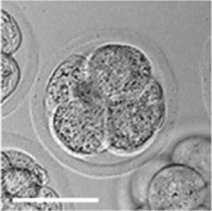
Mouse 4 cell morula stage development[3]
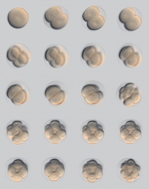
Sea Urchin early embryo cleavage pattern (SDB Gallery Images)
References
- ↑ 1.0 1.1 <pubmed>19924284</pubmed>| PMC2773928 | PLoS One
- ↑ <pubmed>20890283</pubmed>| Nat Biotechnol.
- ↑ <pubmed>20405021</pubmed>| PMC2854157 | PLoS
Articles
<pubmed>19289087</pubmed> <pubmed>20157423</pubmed>
Search PubMed
Search Pubmed: morula development | blastomere development |
Glossary Links
- Glossary: A | B | C | D | E | F | G | H | I | J | K | L | M | N | O | P | Q | R | S | T | U | V | W | X | Y | Z | Numbers | Symbols | Term Link
Cite this page: Hill, M.A. (2024, June 24) Embryology Morula. Retrieved from https://embryology.med.unsw.edu.au/embryology/index.php/Morula
- © Dr Mark Hill 2024, UNSW Embryology ISBN: 978 0 7334 2609 4 - UNSW CRICOS Provider Code No. 00098G

