BGDB Gastrointestinal - Activity 1: Difference between revisions
mNo edit summary |
mNo edit summary |
||
| (5 intermediate revisions by the same user not shown) | |||
| Line 12: | Line 12: | ||
<gallery> | <gallery> | ||
File:Week1 summary.jpg | File:Week1 summary.jpg|Week 1 and 2 | ||
File:Trilaminar embryo.jpg | File:Trilaminar embryo.jpg|Trilaminar Embryo | ||
File:Endoderm cartoon.jpg | File:Endoderm cartoon.jpg|Endoderm | ||
File:Mesoderm-cartoon4.jpg | File:Mesoderm-cartoon4.jpg|Mesoderm | ||
</gallery> | </gallery> | ||
==2. Folding== | ==2. Folding== | ||
| Line 79: | Line 79: | ||
==4. Early Abnormalities== | ==4. Early Abnormalities== | ||
===Meckel's Diverticulum== | ===Meckel's Diverticulum=== | ||
{| | {| | ||
| {{Meckel's diverticulum}} a midgut developmental abnormality, where the vitelline duct fails to degenerate completely. | | valign=top|{{Meckel's diverticulum}} a midgut developmental abnormality, where the vitelline duct fails to degenerate completely. | ||
This abnormality is a very common (incidence of 1–2% in the general population) and occurs from the improper closure and reabsorption of the yolk stalk (vitelline duct, omphalomesenteric duct). This transient connection lies between the early midgut region and the yolk sac. | This abnormality is a very common (incidence of 1–2% in the general population) and occurs from the improper closure and reabsorption of the yolk stalk (vitelline duct, omphalomesenteric duct). This transient connection lies between the early midgut region and the yolk sac. | ||
Named after Johann Friedrich Meckel (1781 – 1833) (the younger) a German anatomist who identified the abnormality origin in 1820. | Named after [[Embryology History - Johann Meckel|Johann Friedrich Meckel]] (1781 – 1833) (the younger) a German anatomist who identified the abnormality origin in 1820. | ||
<br><br> | |||
{| | {| | ||
|-bgcolor="FEF9E7" | |-bgcolor="FEF9E7" | ||
| Line 94: | Line 93: | ||
:''A congenital abnormality characterized by the outpouching or sac formation in the ileum. It is a remnant of the embryonic yolk sac in which the vitelline duct failed to close. During early gestation, the ompahlomesenteric or vitelline duct connects the fetal yolk sac to the primitive gut. By 7-8 weeks of gestation, this duct is normally completely obliterated. A Meckel diverticulum results when this structure fails to resorb completely.'' | :''A congenital abnormality characterized by the outpouching or sac formation in the ileum. It is a remnant of the embryonic yolk sac in which the vitelline duct failed to close. During early gestation, the ompahlomesenteric or vitelline duct connects the fetal yolk sac to the primitive gut. By 7-8 weeks of gestation, this duct is normally completely obliterated. A Meckel diverticulum results when this structure fails to resorb completely.'' | ||
|} | |} | ||
| [[File:Meckel's diverticulum 01.jpg| | | [[File:Meckel's diverticulum 01.jpg|250px]] | ||
A photograph of {{Meckel's diverticulum}} in the adult. The abnormality is shown as the blind-ended tube (diverticulum) lower right of image. | A photograph of {{Meckel's diverticulum}} in the adult. The abnormality is shown as the blind-ended tube (diverticulum) lower right of image. | ||
| Line 100: | Line 99: | ||
==Interactive Component== | ==Interactive Component== | ||
[https://moodle.telt.unsw.edu.au/mod/quiz/view.php?id=2260276 Attempt the Quiz] | |||
Latest revision as of 13:24, 6 Mayıs 2019
 |
Learning Activity 1
- Identify the different contributions of the trilaminar embryo to the formation of the gastrointestinal tract (GIT).
- Describe the development and folding of the yolk sac and endoderm.
- List the adult structures developed from the foregut, midgut and hindgut.
1. Trilaminar Embryo Contributions
The gastrointestinal tract has contributions from all 3 germ layers (ectoderm, mesoderm and endoderm). Identify the specific components that each germ layer contributes to the tract. We will discuss the associated organs later in Activity 3.
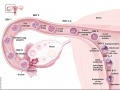
Week 1 and 2
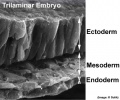
Trilaminar Embryo
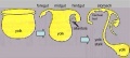
Endoderm
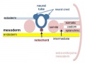
Mesoderm


2. Folding

In week 3 to 4 (GA week 5 and 6) folding of the entire embryonic disc occurs ventrally. The notochord forms a midline rod-like region running rostro-caudally.

Watch the two animations below and discuss the GIT folding that is occurring within the embryo.
| <html5media height="360" width="360">File:Endoderm 003.mp4</html5media> | <html5media height="540" width="390">File:Week3_folding.mp4</html5media> |
| Page | Play | Page | Play |
In relation to the notochord:
- Laterally (either side of the notochord) lies mesoderm.
- Rostrally (above the notochord end) lies the buccopharyngeal membrane, above this again is the mesoderm region forming the heart.
- Caudally (below the notochord end) lies the primitive streak (where gastrulation occurred), below this again is the cloacal membrane.
- Dorsally (above the notochord) lies the neural tube then ectoderm.
- Ventrally (beneath the notochord) lies the mesoderm then endoderm.
Folding of the embryonic disc "pinches off" part of the yolk sac forming the first primitive GIT.
| BM Bright Field | BM Scanning EM | BM Scanning EM | CM Scanning EM |
|---|---|---|---|

|

|

|

|
| Stage 11 | Stage 11 | Stage 11 | Stage 12 |
3. Gut Parts

Identify the 3 main subdivisions of the gastrointestinal tract.
- How were they defined in the early embryo?
- How are they later anatomically defined?
| Foregut | Midgut | Hindgut |
|---|---|---|
| trachea, respiratory tract, lungs
oesophagus (osophagus} gallbladder ( and bile ducts) pancreas (dorsal & ventral) upper duodenum |
lower duodenum
small intestines (jejunum, ileum) cecum Ascending colon proximal 2/3 of transverse colon |
distal 1/3 of transverse colon
descending colon sigmoid colon rectum Upper anal canal Urogenital sinus |
| celiac artery | superior mesenteric artery | inferior mesenteric artery |
4. Early Abnormalities
Meckel's Diverticulum
| Meckel's diverticulum a midgut developmental abnormality, where the vitelline duct fails to degenerate completely.
This abnormality is a very common (incidence of 1–2% in the general population) and occurs from the improper closure and reabsorption of the yolk stalk (vitelline duct, omphalomesenteric duct). This transient connection lies between the early midgut region and the yolk sac.
|

A photograph of Meckel's diverticulum in the adult. The abnormality is shown as the blind-ended tube (diverticulum) lower right of image. |
Interactive Component
|
Additional Information
| Additional Information - Content shown under this heading is not part of the material covered in this class. It is provided for those students who would like to know about some concepts or current research in topics related to the current class page. |
- gastrointestinal tract - also called the alimentary tract.
- Development of the oral cavity, palate and pharynx will be covered in BGDB Practical - Face and Ear Development.
| Gastrointestinal Tract Terms | ||
|---|---|---|
| ||
|

BGDB: Lecture - Gastrointestinal System | Practical - Gastrointestinal System | Lecture - Face and Ear | Practical - Face and Ear | Lecture - Endocrine | Lecture - Sexual Differentiation | Practical - Sexual Differentiation | Tutorial
Glossary Links
- Glossary: A | B | C | D | E | F | G | H | I | J | K | L | M | N | O | P | Q | R | S | T | U | V | W | X | Y | Z | Numbers | Symbols | Term Link
Cite this page: Hill, M.A. (2026, October 15) Embryology BGDB Gastrointestinal - Activity 1. Retrieved from https://embryology.med.unsw.edu.au/embryology/index.php/BGDB_Gastrointestinal_-_Activity_1
- © Dr Mark Hill 2026, UNSW Embryology ISBN: 978 0 7334 2609 4 - UNSW CRICOS Provider Code No. 00098G
