File:Robert Meyer.jpg: Difference between revisions
mNo edit summary |
mNo edit summary |
||
| Line 14: | Line 14: | ||
The Berlin Meyer collection of human embryos has been entirely lost. References to specific embryos in this collection are made in a number of historic publications. | The Berlin Meyer collection of human embryos has been entirely lost. References to specific embryos in this collection are made in a number of historic publications. | ||
===References=== | ===References=== | ||
Meyer, R. : Ueber die fetale Uterusschleimhaut, Zeit. f . Geburtsh. u. Gynak., vol. 38, 1898. Meyer, R. : Zur Entstehung des doppelten Uterus, Zeit. f . Geburtsh. u. Gynak., vol. 38, 1898b. | Meyer, R. : Ueber die fetale Uterusschleimhaut, Zeit. f . Geburtsh. u. Gynak., vol. 38, 1898. Meyer, R. : Zur Entstehung des doppelten Uterus, Zeit. f . Geburtsh. u. Gynak., vol. 38, 1898b. | ||
Latest revision as of 13:40, 5 May 2019
Robert Meyer
Robert Meyer (1864 – 1947) was a German embryologist and pathologist.
He studied medicine at the universities of Leipzig, Heidelberg and Strassburg, receiving his doctorate in 1889 (Strassburg). From 1890 to 1894 he was a medical practitioner in the community of Dedeleben, and afterwards worked as assistant to gynecologist Johann Veit in Berlin.
Head of the laboratory in the women's clinic at the Berlin Charité (1909 to 1911), and in 1912 succeeded Carl Arnold Ruge as chief of the pathological institute of the university women's clinic. In 1932 he became an honorary professor to the faculty of medicine at the university. Because of his Jewish ancestry, he was removed from his position at Berlin in 1935 and emigrated to the United States, settling in Minneapolis in 1939.
The "Weigert-Meyer law" is a rule concerning the anatomical relationship of the two ureters, named in conjunction with pathologist Carl Weigert.
Meyer Embryo Collection
The Berlin Meyer collection of human embryos has been entirely lost. References to specific embryos in this collection are made in a number of historic publications.
References
Meyer, R. : Ueber die fetale Uterusschleimhaut, Zeit. f . Geburtsh. u. Gynak., vol. 38, 1898. Meyer, R. : Zur Entstehung des doppelten Uterus, Zeit. f . Geburtsh. u. Gynak., vol. 38, 1898b.
Meyer, R. : Ueber epitheliale Gebilde im Myometrium des fetalen und kindlichen Uterus einsehl. des Gartner'schen Ganges, Berlin, 1899.
Meyer, R. : Ueber sogenannte Vornierenreste und das nephrogene Zwischenblastem bei mensehlichen Embryonen und ihre eventuelle pathologische Persistenz, Charite-Annalen, vol. 33, p. 649-656, with 2 text-figs., 1909.
Meyer, R. : Zur Kenntnis des Gartner'schen Ganges besonders in der Vagina und dem Hymen des Menschen, Arch. f. mikr. Anat., vol. 73, 1909.
Meyer, R. : Zur Entwicklungsgeschichte und Anatomie des utrieulus prostaticus beim Menschen, Arch. f. mikr. Anat. u. Entw., vol. 74, 1909b.
Meyer, R. : Ueber erosio portionis uteri, Deut. patholog. Gesellsch. Erlangen, 1910.
Meyer, R.: Die Erosion und Pseudoerosion des Erwachsenen, Arch. f. Gynak. vol. 91, part. 3, p. 1-35 with 1 plate and 3 text-figs., 1910.
Meyer, R. : Die Epithelentwicklung der cervix und portio vaginalis und die pseudoerosio congenita (Kongenital. histol. Ectropium), Arch. f. Gynak., vol. 90, part 3, p. 1-20 with 1 plate, 1910.
Meyer, R. : Beitrag zur Frage nach der Genese der im Urogenitalgebiet vorkommenden Mischgeschwiilste und Teratome, Reprint from the Charite Annalen, vol. 34, p. 1-23 with 10 text-figs., 1910.
Cite this page: Hill, M.A. (2024, June 25) Embryology Robert Meyer.jpg. Retrieved from https://embryology.med.unsw.edu.au/embryology/index.php/File:Robert_Meyer.jpg
- © Dr Mark Hill 2024, UNSW Embryology ISBN: 978 0 7334 2609 4 - UNSW CRICOS Provider Code No. 00098G
File history
Yi efo/eka'e gwa ebo wo le nyangagi wuncin ye kamina wunga tinya nan
| Gwalagizhi | Nyangagi | Dimensions | User | Comment | |
|---|---|---|---|---|---|
| current | 13:14, 5 May 2019 | 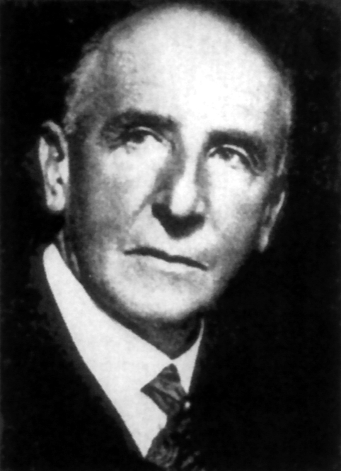 | 482 × 665 (127 KB) | Z8600021 (talk | contribs) | Robert Meyer (11 January 1864, in Hanover – 12 December 1947, in Minneapolis) was a German pathologist. He studied medicine at the universities of Leipzig, Heidelberg and Strassburg, receiving his doctorate at the latter institution in 1889. From 189... |
You cannot overwrite this file.
File usage
The following 6 pages use this file: