File:Human-retina-01.jpg: Difference between revisions
No edit summary |
mNo edit summary |
||
| (One intermediate revision by the same user not shown) | |||
| Line 1: | Line 1: | ||
==Adult Human Retina Histology== | ==Adult Human Retina Histology== | ||
Light micrograph of normal human retina stained with Richardson's methylene blue/azure II. Light enters the retina from the top of the image. | Light micrograph of normal human ((retina}} stained with Richardson's methylene blue/azure II. Light enters the retina from the top of the image. | ||
'''(a)''' Neural retina | '''(a)''' Neural retina | ||
| Line 9: | Line 9: | ||
'''(c)''' retinal pigment epithelium (RPE) | '''(c)''' retinal pigment epithelium (RPE) | ||
===Reference=== | |||
{{#pmid12186651}} | |||
From the Human Retina Teaching Set, Scheie Eye Institute, University of Pennsylvania, Philadelphia, USA. Courtesy of Ann Milam. | |||
====Copyright==== | |||
[http://www.biomedcentral.com/info/about/license BioMed Central Open Access] | |||
Original Image Name: Figure 1. http://genomebiology.com/2002/3/8/reviews/1022/figure/F1 | |||
{{Footer}} | |||
[[Category:Human]] [[Category:Vision]] [[Category:Neural]] [[Category:Senses]] [[Category:Histology]] | [[Category:Human]] [[Category:Vision]] [[Category:Neural]] [[Category:Senses]] [[Category:Histology]] | ||
Latest revision as of 12:07, 17 April 2018
Adult Human Retina Histology
Light micrograph of normal human ((retina}} stained with Richardson's methylene blue/azure II. Light enters the retina from the top of the image.
(a) Neural retina
(b) photoreceptor layer
(c) retinal pigment epithelium (RPE)
Reference
{{#pmid12186651}}
From the Human Retina Teaching Set, Scheie Eye Institute, University of Pennsylvania, Philadelphia, USA. Courtesy of Ann Milam.
Copyright
Original Image Name: Figure 1. http://genomebiology.com/2002/3/8/reviews/1022/figure/F1
Cite this page: Hill, M.A. (2026, October 20) Embryology Human-retina-01.jpg. Retrieved from https://embryology.med.unsw.edu.au/embryology/index.php/File:Human-retina-01.jpg
- © Dr Mark Hill 2026, UNSW Embryology ISBN: 978 0 7334 2609 4 - UNSW CRICOS Provider Code No. 00098G
File history
Yi efo/eka'e gwa ebo wo le nyangagi wuncin ye kamina wunga tinya nan
| Gwalagizhi | Nyangagi | Dimensions | User | Comment | |
|---|---|---|---|---|---|
| current | 15:56, 16 October 2010 | 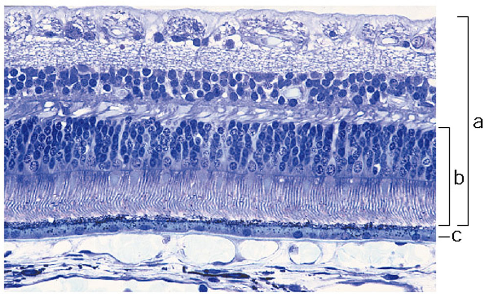 | 1,000 × 615 (197 KB) | S8600021 (talk | contribs) | ==Adult Human Retina Histology== Light micrograph of normal human retina stained with Richardson's methylene blue/azure II. Light enters the retina from the top of the image. (a) Neural retina (b) photoreceptor layer (c) retinal pigment epithelium (RP |
You cannot overwrite this file.
File usage
The following 2 pages use this file:


