Berlin Meeting 2017 - Digital Embryology Consortium: Difference between revisions
mNo edit summary |
m (→Summary) |
||
| (80 intermediate revisions by the same user not shown) | |||
| Line 1: | Line 1: | ||
{{Header}} | {{Header}} | ||
{| | |||
| colspan=2|Dr Mark Hill's two presentations from the [https://human-embryology.org/wiki/DEC_Berlin_Meeting_2017 '''December 2017 Berlin Meeting'''] | |||
|- | |||
| | |||
<br> | |||
# [[#Introduction|'''Introduction''']] | |||
# [[#Blechschmidt Collection|'''Blechschmidt Collection''']] | |||
<br> | |||
[https://human-embryology.org | [[File:DEC icons thumbnail.jpg|500px|link=https://human-embryology.org]] | ||
| valign=top|[[File:Mark Hill.jpg|150px]] | |||
Dr Mark Hill | |||
http://tiny.cc/DEC-Berlin_2017 | |||
[[File:QR-DEC-Berlin 2017.png|100px]] | |||
|} | |||
=Introduction= | |||
== | ==DEC Beginnings== | ||
===2013=== | ===2013=== | ||
| Line 19: | Line 30: | ||
! colspan=2|Research and Presentations | ! colspan=2|Research and Presentations | ||
|- | |- | ||
| [[File:Giere_Hill_-_Hill_Collection.jpg|300px|link=Hubrecht Collection)]] | |||
| [[File:Hill Viebahn Blechschmidt Collection.jpg|300px|link=https://human-embryology.org/wiki/Blechschmidt_Collection]] | | [[File:Hill Viebahn Blechschmidt Collection.jpg|300px|link=https://human-embryology.org/wiki/Blechschmidt_Collection]] | ||
|- | |- | ||
| [[Museum_of_Natural_History_Berlin - 2013_Seminar|Museum of Natural History Embryology Seminar]] | |||
| [[Anatomy and Embryology Goettingen - 2013_Seminar|Goettingen Anatomy and Embryology Seminar]] | | [[Anatomy and Embryology Goettingen - 2013_Seminar|Goettingen Anatomy and Embryology Seminar]] | ||
|} | |} | ||
| Line 72: | Line 83: | ||
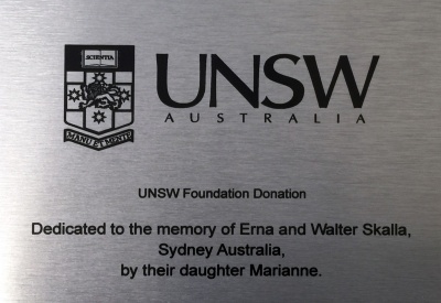
| [[File:UNSW Foundation donation plaque.jpg|400px|link=http://www.giving.unsw.edu.au/unsw-foundation-limited]] | | [[File:UNSW Foundation donation plaque.jpg|400px|link=http://www.giving.unsw.edu.au/unsw-foundation-limited]] | ||
|- | |- | ||
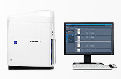
| [https://www.zeiss.com/microscopy/en_de/products/imaging-systems/axio-scan-z1.html Zeiss] [https://human-embryology.org/wiki/Slide_Scanning Axioscan.Z1] | | [https://www.zeiss.com/microscopy/en_de/products/imaging-systems/axio-scan-z1.html Zeiss] [https://human-embryology.org/wiki/Slide_Scanning Axioscan.Z1] | ||
(Described later in the meeting by Dr Thorsten Heupel) | |||
| [http://www.giving.unsw.edu.au/unsw-foundation-limited UNSW Foundation] donation plaque | | [http://www.giving.unsw.edu.au/unsw-foundation-limited UNSW Foundation] donation plaque | ||
|} | |} | ||
<br> | <br> | ||
<html5media width="480" height="360">https://www.youtube.com/embed/1RbeEqn9Rmo</html5media> | |||
[[Blechschmidt Collection]] scanning commences. | [[Blechschmidt Collection]] scanning commences. | ||
| Line 85: | Line 102: | ||
| [[File:Mark and Embryo.jpg|400px]] | | [[File:Mark and Embryo.jpg|400px]] | ||
|- | |- | ||
| [[Blechschmidt Collection]] slide scanning completed. | | [[Blechschmidt Collection]] slide scanning completed. (described later in the Berlin meeting by Hannes Sydow ) | ||
|} | |} | ||
[[Hinrichsen Collection]] slide scanning commences. (described later in the Berlin meeting by Dr Nenad Maricic.) | |||
<br> | <br> | ||
{| | {| | ||
| Line 94: | Line 113: | ||
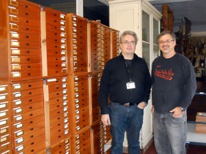
| width=260px|[[File:Hill Mirapeix Domenech-Mateu Collection.jpg|250px]] | | width=260px|[[File:Hill Mirapeix Domenech-Mateu Collection.jpg|250px]] | ||
Prof. Rosa Mirapeix | Mark Hill and Prof. Rosa Mirapeix | ||
| | | | ||
[https://human-embryology.org/wiki/Domenech-Mateu_Collection Domenech-Mateu Collection] – Universitat Autònoma de Barcelona. | [https://human-embryology.org/wiki/Domenech-Mateu_Collection Domenech-Mateu Collection] – Universitat Autònoma de Barcelona. | ||
(described later in the Berlin meeting by Prof. Rosa Mirapeix) | |||
<br><br> | <br><br> | ||
[https://human-embryology.org/index.php?title=HDBR_and_HuDSeN Collection HDBR and HuDSeN Collection] – Newcastle University. | [https://human-embryology.org/index.php?title=HDBR_and_HuDSeN Collection HDBR and HuDSeN Collection] – Newcastle University. | ||
(described later in the Berlin meeting by Dr Steven Lisgo) | |||
<br><br> | <br><br> | ||
[https://human-embryology.org/index.php?title=Amsterdam_Collection Amsterdam Collection] - Amsterdam University, Academic Medical Center | [https://human-embryology.org/index.php?title=Amsterdam_Collection Amsterdam Collection] - Amsterdam University, Academic Medical Center. | ||
<br | |||
(described later in the Berlin meeting by Prof. Wouter Lamers) | |||
<br> | |||
<br> | <br> | ||
[[Media:ANZSCDB-Poster-MH-Apr2016.pdf|ANZSCDB Meeting presentation]] | [[Media:ANZSCDB-Poster-MH-Apr2016.pdf|ANZSCDB Meeting presentation]] | ||
| Line 111: | Line 135: | ||
{| | {| | ||
|-bgcolor="FAF5FF" | |-bgcolor="FAF5FF" | ||
! colspan=2|Image Server | ! colspan=2|Image Server | ||
|- | |- | ||
| [[File:OmeroWeb.png|500px]] | | (Described by Dr Jean-Marie Burel later in the meeting) | ||
[[File:OmeroWeb.png|500px]] | |||
|- | |- | ||
| [[File:Ome-logo-400.png|200px|link=https://www.openmicroscopy.org|left]] Testing of online server [https://www.openmicroscopy.org/site The Open Microscopy Environment] [http://help.openmicroscopy.org OME Help] [http://help.openmicroscopy.org/web-client.html Using OMERO.web] | | [[File:Ome-logo-400.png|200px|link=https://www.openmicroscopy.org|left]] Testing of online server [https://www.openmicroscopy.org/site The Open Microscopy Environment] [http://help.openmicroscopy.org OME Help] [http://help.openmicroscopy.org/web-client.html Using OMERO.web] | ||
| Line 120: | Line 146: | ||
|} | |} | ||
<br> | <br> | ||
===Summary=== | ===Summary=== | ||
Between 2013 to 2016 following presentations, collection visits, and research the consortium was established. | Between 2013 to 2016 following presentations, collection visits, and research the consortium was established. | ||
| Line 146: | Line 173: | ||
|} | |} | ||
== | ==2017 DEC Current== | ||
=== | ===DEC Online=== | ||
'''Oct 2017''' - Software was updated to Omero Server (version 5.4). | |||
This topic will be covered in more detail in my presentation on the Blechschmidt collection. | |||
<br><br> | |||
'''The animation below shows the DEC server log-in process.''' | |||
<br><br> | |||
<html5media width="600" height="500">File:OMERO logging in.mp4</html5media> | |||
===Hinrichsen Collection=== | |||
{| | {| | ||
|-bgcolor="FAF5FF" | |-bgcolor="FAF5FF" | ||
! colspan=2|Slide Scanning | ! colspan=2|Slide Scanning | ||
|- | |- | ||
| [[Hinrichsen Collection]] | | [[Hinrichsen Collection]] - scanning completed. | ||
|- | |- | ||
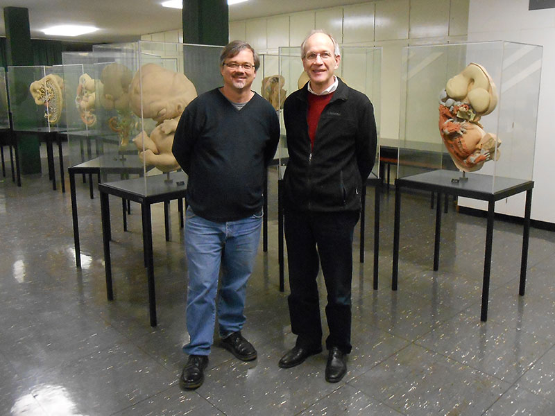
| [[File:Hinrichsen Collection Hill Brand-Saberi.jpg|300px]] | | [[File:Hinrichsen Collection Hill Brand-Saberi.jpg|300px]] | ||
Prof. Beate Brand-Saberi | Mark Hill and Prof. Beate Brand-Saberi | ||
| [[File: | |||
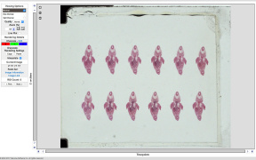
| [[File:Hinrichsen-Embryo514-Slide-100-Scene01.jpg|250px]] | |||
'''Embryo 514''', [[Carnegie stage 21]], CRL 23 mm<br>Sagittal plane, {{HE}}, [http://149.171.80.223:8080/webclient/?show=dataset-751 '''Slide 100'''] | |||
|- | |- | ||
|} | |} | ||
<br><br> | |||
(described later in the Berlin meeting by Dr. Nenad Maricic) | |||
* Jun 2017 - about 2500 slides scanned. | |||
* Nov 2017 - scans uploaded to online database. | |||
* Dec 2017 - scanning finished. | |||
<br> | |||
{{Hinrichsen Collection Jun2017scanning_collapsetable}} | |||
<br> | <br> | ||
===Embryological Collection MfN=== | |||
December 2017 - scanner delivered and installed. | |||
{| | {| | ||
| [[File:2017 Berlin install 01.JPG|400px]] | |||
| | | [[File:MfN DEC Scanner-room.jpg|400px]] | ||
|} | |} | ||
===Summary=== | ===Summary=== | ||
2017 completed | 2017 completed the second collection scanning and testing of the updated online database software. Delivered teh scanner to Berlin. | ||
{| class="wikitable mw-collapsible mw-collapsed" | {| class="wikitable mw-collapsible mw-collapsed" | ||
! Summary - DEC Current | ! Summary - DEC Current | ||
| Line 193: | Line 236: | ||
* Mar 2017 - software updated to import the JPGXR format files. | * Mar 2017 - software updated to import the JPGXR format files. | ||
* Server is currently being tested for remote uploading of files from international locations. | * Server is currently being tested for remote uploading of files from international locations. | ||
|- | |||
| [[File:DEC-database-stats-2017-12-11.jpg|800px]] | |||
|} | |||
<br> | |||
<br> | |||
<br> | |||
<br> | |||
(Presentation 2 below) | |||
=Blechschmidt Collection= | |||
[[File:Hill Viebahn Blechschmidt Collection.jpg|800px|link=https://human-embryology.org/wiki/Blechschmidt_Collection]] | |||
Mark Hill and Prof Christoph Viebahn | |||
==Prof. Erich Blechschmidt== | |||
{| | |||
| Prof. Erich Blechschmidt (1904 – 1992)<ref><pubmed>1476247</pubmed></ref> | |||
* German anatomist and director of Göttingen University’s Anatomical Institute from 1942 until 1973. | |||
* Alternate theories for embryogenesis based upon "morphogenetic fields" | |||
** believed that more than just genes might act to control development. | |||
* Wrote several embryology books and research articles (32 are listed in PubMed between 1955 to 1976) | |||
** all originally published in the German language. | |||
* The collection consists of about 120 human embryos approximately 200,000 serial sections. | |||
** In 1972 some embryos were incorporated into the [[Carnegie Collection]] (assigned Carnegie Nos. 10315-10434), but have since been returned to the University of Goettingen. | |||
* From the serial sections 64 models some more than 1 metre in size.<ref><pubmed>13227045</pubmed></ref> | |||
| [[File:Eric Blechschmidt.jpg|150px|alt= Erich Blechschmidt (1904 – 1992)]] | |||
Erich Blechschmidt<br>(1904 – 1992) | |||
|} | |} | ||
== | ==Embryo Models== | ||
[[File:Blechschmidt model collection room.jpg|600px]] | |||
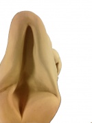
<gallery mode="packed-hover" caption="10 mm Embryo (stage17)"> | |||
File:Stage17_model_03.jpg|left | |||
File:Stage17_model_05.jpg|ventral | |||
File:Stage17_model_04.jpg|right | |||
File:Stage17_model_06.jpg|dorsal | |||
File:Stage17 model_01.jpg|cranial end | |||
File:Stage17 model_02.jpg|placental cord | |||
</gallery> | |||
Model (about 1 M) of Embryo 7.5 mm | |||
<html5media height="700" width="500">File:Embryo 7.5mm model 02.mp4</html5media> | |||
{| | |||
| valign="bottom"|{{Embryo 1.6mm movie 1}} | |||
| valign="bottom"|{{Embryo 3.1mm movie 1}} | |||
| valign="bottom"|{{Embryo 7.5mm movie 1}} | |||
| valign="bottom"|{{Embryo 10mm movie 1}} | |||
|} | |||
==DEC Online== | |||
{| | |||
| '''Oct 2017''' | |||
* Software has been updated to Omero Server (version 5.4). | |||
* OMERO version 5.4 now JPG XR compatible. | |||
* Server - Quad-Core Intel Xeon E5 Processor Speed - 3.7 GHz | |||
* Storage - 2 x Areca ARC-5028T2, Thunderbolt 2, 6-Bay RAID 5 - 72 TB | |||
* RAID 5 - consists of block-level striping with distributed parity. | |||
** Upon failure of a single drive, subsequent reads can be calculated from the distributed parity such that no data is lost. | |||
| width=210px|[[File:Ome-logo-400.png|200px|link=https://www.openmicroscopy.org/community/viewtopic.php?f=11&t=8284]] | |||
|} | |||
<br> | |||
{| class="wikitable mw-collapsible mw-collapsed" | {| class="wikitable mw-collapsible mw-collapsed" | ||
! Online Image Database - Access | ! Online Image Database - Access | ||
| Line 223: | Line 334: | ||
{{Embryo1951-09-01-Slide60 links}} | {{Embryo1951-09-01-Slide60 links}} | ||
====References==== | |||
<references/> | |||
{{Footer}} | {{Footer}} | ||
Latest revision as of 17:18, 11 December 2017
| Embryology - 14 Jun 2024 |
|---|
| Google Translate - select your language from the list shown below (this will open a new external page) |
|
العربية | català | 中文 | 中國傳統的 | français | Deutsche | עִברִית | हिंदी | bahasa Indonesia | italiano | 日本語 | 한국어 | မြန်မာ | Pilipino | Polskie | português | ਪੰਜਾਬੀ ਦੇ | Română | русский | Español | Swahili | Svensk | ไทย | Türkçe | اردو | ייִדיש | Tiếng Việt These external translations are automated and may not be accurate. (More? About Translations) |
| Dr Mark Hill's two presentations from the December 2017 Berlin Meeting | |
|
|

Dr Mark Hill
|

Introduction
DEC Beginnings
2013
| Research and Presentations | |
|---|---|
|

|
| Museum of Natural History Embryology Seminar | Goettingen Anatomy and Embryology Seminar |
2014
Establish the DEC.
| Kyoto Collection Research | |
|---|---|
| <html5media height="600" width="450">File:Stage20 MRI S05.mp4</html5media> | <html5media height="600" width="400">File:Stage20 MRI S02.mp4</html5media> |

Prof. Kohei Shiota, Dr Mark Hill, Prof. Shigehito Yamada and Prof. Mineo Yasuda |
|
2015
Planned with partners the digitization project.
| Presentations | |
|---|---|
| Kyoto (Nov 2015) | Washington (Dec 2015) |
|
|
| Purchased slide scanner | |
|
|
| Zeiss Axioscan.Z1
(Described later in the meeting by Dr Thorsten Heupel) |
UNSW Foundation donation plaque |
<html5media width="480" height="360">https://www.youtube.com/embed/1RbeEqn9Rmo</html5media>
Blechschmidt Collection scanning commences.
2016
| Slide Scanning | |
|---|---|

| |
| Blechschmidt Collection slide scanning completed. (described later in the Berlin meeting by Hannes Sydow ) | |
Hinrichsen Collection slide scanning commences. (described later in the Berlin meeting by Dr Nenad Maricic.)
| New collections join DEC | |
|---|---|

Mark Hill and Prof. Rosa Mirapeix |
Domenech-Mateu Collection – Universitat Autònoma de Barcelona. (described later in the Berlin meeting by Prof. Rosa Mirapeix)
(described later in the Berlin meeting by Dr Steven Lisgo)
(described later in the Berlin meeting by Prof. Wouter Lamers)
|
| Image Server | |
|---|---|
| (Described by Dr Jean-Marie Burel later in the meeting)
| |
| |

Summary
Between 2013 to 2016 following presentations, collection visits, and research the consortium was established.
| Summary - DEC Beginnings |
|---|
| Slide Scanner
A key to the project was to evaluate the slide scanning options.
|
| Online Database Software
A well supported and freely available software was required for this large image database. |
2017 DEC Current
DEC Online
Oct 2017 - Software was updated to Omero Server (version 5.4).
This topic will be covered in more detail in my presentation on the Blechschmidt collection.
The animation below shows the DEC server log-in process.
<html5media width="600" height="500">File:OMERO logging in.mp4</html5media>
Hinrichsen Collection
| Slide Scanning | |
|---|---|
| Hinrichsen Collection - scanning completed. | |

Mark Hill and Prof. Beate Brand-Saberi |

Embryo 514, Carnegie stage 21, CRL 23 mm |
(described later in the Berlin meeting by Dr. Nenad Maricic)
- Jun 2017 - about 2500 slides scanned.
- Nov 2017 - scans uploaded to online database.
- Dec 2017 - scanning finished.
| Hinrichsen Collection Scanning June 2017 | ||||||||||||
|---|---|---|---|---|---|---|---|---|---|---|---|---|
| Scan | Box | Serial | Slides | Stage | CRL (mm) | HDD | Cutting/Organ | Comp 85% |
JPEG | Staining | Slides | Test upload slides |
| 1 | EYO | 226 | 001-114 | 18 | 16 mm | 1 | Sagittal (good) | x | wdp | HE, Tri, Azan | 114 | |
| 2 | EYO | 226 | 5-100 | 18 | 16 mm | 1 | every 5th slide (good) | x | x | HE, Tri, Azan | 20 | 5 |
| 3 | EYO | 283 | 002-522 | 20 | 21 mm | 1 | Sagittal (good) | x | x | HE, Tri, Azan | 520 | |
| 4 | EYO | 294 | 001-174 | ? | 20 mm | 1 | Sagittal (good) | x | x | HE, Tri, Azan | 174 | |
| 5 | EYO | 335 | 001-270 | 20 | 22 mm | 1 | Frontal horizontal (very good) | x | x | HE, Tri, Azan | 270 | 2, 3, 4 |
| 6 | EYO | 364 | 001-131 | 14 | 6.5 mm | 1 | Sagittal (average) | x | x | HE, Tri, Azan | 131 | 1, 2, 3 |
| 7 | EYO | 388 | 009-280 | ? | 19mm | 1 | Sagittal (average) | x | x | HE, Tri, Azan | 271 | 9 |
| 8 | EYO | 514 | 001-330 | 21 | 23 mm | 1 | Sagittal | x | x | HE, Tri, Azan | 330 | 24, 67, 327 |
| 9 | EYO | 527 | 001-130 | ? | 20.0 mm | 1 | Transversal | x | HE, Tri, Azan | 130 | ||
| 10 | ME 32 | 536 | 001-335 | 20 | 22 mm | 1 | Frontal | x | HE, Tri, Azan | 335 | 1, 2 | |
| 11 | ME 53 | 564 | 006-228 | 18 | 16 mm | 1 | Frontal whole Embryo | x | HE | 52 | 132 | |
| 12 | ME 54 | 568 | 006-204 | 21 | 22.5 mm | 1 | Frontal whole embryo | x | HE | 46 | ||
| 13 | ME 65 | ? | 001-037 | 14 | 6.2 mm | 1 | Transverse (moderate)/Heart | x | Toluidinblue | 37 | 1, 6, 12, 20, 30 | |
| 14 | EYO | 457 | 40 | |||||||||
| Total | 2470 | |||||||||||
Embryological Collection MfN
December 2017 - scanner delivered and installed.

|

|
Summary
2017 completed the second collection scanning and testing of the updated online database software. Delivered teh scanner to Berlin.
| Summary - DEC Current |
|---|
Scanned Slide Files
This means there are always at least 3 separate copies of the original data in 3 different locations. |
Online Database
|

|
(Presentation 2 below)
Blechschmidt Collection
Mark Hill and Prof Christoph Viebahn
Prof. Erich Blechschmidt
Prof. Erich Blechschmidt (1904 – 1992)[1]
|

Erich Blechschmidt |
Embryo Models

- 10 mm Embryo (stage17)
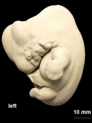
left
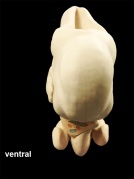
ventral
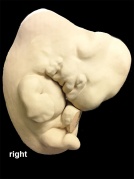
right

dorsal
cranial end
placental cord


Model (about 1 M) of Embryo 7.5 mm
<html5media height="700" width="500">File:Embryo 7.5mm model 02.mp4</html5media>
|
|
|
|
DEC Online
Oct 2017
|
| Online Image Database - Access |
|---|
| Please note that only registered users who have agreed to the DEC usage conditions can access the database images.
Therefore for non-registered viewers, the links below will show only a link to the OMERO log-in page. |

|
|
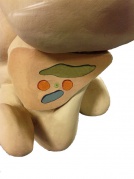
The images below are screenshots from the DEC online database.
- Embryo 1951-09-01 Slide 60 Scene 11
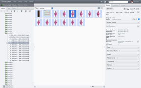
Slide menu
Slide sections
Slide label
Scene 11
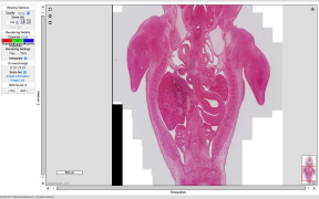
Zoom x0.6
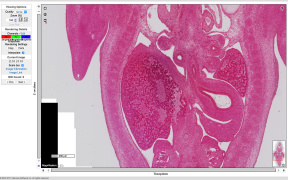
Zoom x1.25
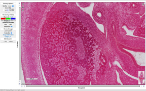
Zoom x2.5
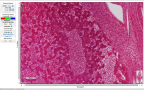
Zoom x5
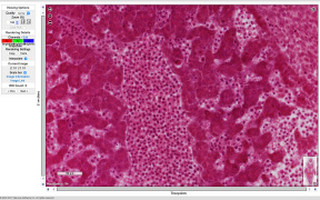
Zoom x10
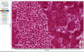
Zoom x20
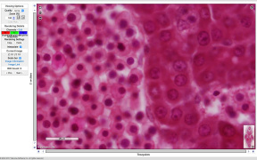
Zoom x40
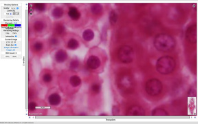
Zoom x80


Embryo 1951-01-09 CRL 11 mm - Week 6 stage 16 stage 17
- Slide 60 Links: Slide menu | Slide sections | Slide label | Scene 11 | Zoom x0.6 | Zoom x1.25 | Zoom x2.5 | Zoom x5 | Zoom x10 | Zoom x20 | Zoom x40 | Zoom x80
| Embryo 1951-09-01 Slide 60 Information |
|---|
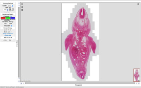
|
OMERO Blechschmidt Collection - Embryo 1951 09 01 Slide 60 Note these are only screenshots of the OMERO image database.
There are 12 serial sections on this single slide (76 slides in this embryo series). This single section pixel size is 24674H x 50841W - Image is as wide as more than 26 full HD TV screens (1920W×1080H) Pixels Size (XYZ) (µm): 0.22 x 0.22 |
References
Cite this page: Hill, M.A. (2024, June 14) Embryology Berlin Meeting 2017 - Digital Embryology Consortium. Retrieved from https://embryology.med.unsw.edu.au/embryology/index.php/Berlin_Meeting_2017_-_Digital_Embryology_Consortium
- © Dr Mark Hill 2024, UNSW Embryology ISBN: 978 0 7334 2609 4 - UNSW CRICOS Provider Code No. 00098G