Atlas of the Development of Man 1: Difference between revisions
mNo edit summary |
|||
| (19 intermediate revisions by 2 users not shown) | |||
| Line 1: | Line 1: | ||
{{Header}} | |||
{{Ref-Kollmann1907}} | |||
{{Historic Disclaimer}} | |||
[[File:Julius_kollmann.jpg|thumb|[[Embryology_History_-_Julius_Kollmann| Julius Konstantin Ernst Kollmann]] (1834-1918)]] | [[File:Julius_kollmann.jpg|thumb|[[Embryology_History_-_Julius_Kollmann| Julius Konstantin Ernst Kollmann]] (1834-1918)]] | ||
Handatlas der | =Handatlas der Entwicklungsgeschichte des Menschen - Volume 1= | ||
(Atlas of the Development of Man Volume 1) | |||
--[[User:S8600021|Mark Hill]] 09:10, 22 October 2011 (EST) This textbook was published in 1907 in the German language. The first volume is more of a textbook of early development, while the second volume is an atlas of images. None of the images from the first volume have yet been added, while the second volume images have all been added to the site as [[Atlas of the Development of Man 2]]. | --[[User:S8600021|Mark Hill]] 09:10, 22 October 2011 (EST) This textbook was published in 1907 in the German language. The first volume is more of a textbook of early development, while the second volume is an atlas of images. None of the images from the first volume have yet been added, while the second volume images have all been added to the site as [[Atlas of the Development of Man 2]]. | ||
:Links: [[Embryology_History_-_Julius_Kollmann| Julius Kollmann]] | {{KollmannAtlas1}} | ||
:'''Links:''' [[Atlas_of_the_Development_of_Man_1|Atlas (Volume 1)]] | [[Atlas_of_the_Development_of_Man_2|Atlas (Volume 2)]] | [[:Category:Kollmann|Category:Kollmann]] | [[Embryology_History_-_Julius_Kollmann| Julius Kollmann]] | |||
==Contents== | ==Contents== | ||
[[File:Kollmann-title page volume 1.jpg|thumb|400px]] | |||
[[Atlas of the Development of Man 1]] | |||
Introduction | |||
Predevelopment | ===Part 1. Predevelopment=== | ||
I. The egg | [[Atlas_of_the_Development_of_Man_1 - Part 1|Part 1. Predevelopment]] | ||
====I. The egg==== | |||
Structural unit yolk-rich egg (bird's egg) | Structural unit yolk-rich egg (bird's egg) | ||
II maturation the egg | ====II. maturation the egg==== | ||
III. Fertilization | ====III. Fertilization==== | ||
VI. Site of fertilisation | ====VI. Site of fertilisation==== | ||
a) Place of fertilization and pregnancy theories | a) Place of fertilization and pregnancy theories | ||
b) durability and resistance of the spermatozoa | b) durability and resistance of the spermatozoa | ||
c) ovulation | c) ovulation | ||
d) Detachment of the egg from the ovary, and the walk through the tubes | d) Detachment of the egg from the ovary, and the walk through the tubes | ||
===Part 2=== | ===Part 2. Ontogeny, Blastogenesis=== | ||
[[Atlas_of_the_Development_of_Man_1 - Part 2|Part 2. Ontogeny, Blastogenesis]] | |||
====I. Cleavage, Segmentation==== | |||
a) External appearances of cleavage | |||
b) Internal appearances of cleavage, mitosis | |||
# Occurrence and transformation of the dark chromatic nuclear division figure | |||
# Occurrence and transformation of the achromatic (colorless) core figure, called the mitotic spindle | |||
# Radiations in the yolk | |||
c) Amitosis or division of cells without the appearance of cell division | |||
c) Amitosis or division of cells without the appearance of cell division | |||
d) Growth and regeneration in conjunction with the process of cell division | d) Growth and regeneration in conjunction with the process of cell division | ||
II | ====II. Germinal vesicle (vesicula blastodermica) with the germinative (area embryonalis), viewed from outside==== | ||
====III. Germinal vesicle internal structure==== | |||
a) | a) Theory of endoderm origin and the gastrulation | ||
b) | b) Germ layer, blastosphere and blastula. Homology of the primary germ layers | ||
The Fundamental organs | |||
====IV. Primitive streak and neurenteric canal==== | |||
====V. Notochord==== | |||
# Front and rear end of the notochord | |||
# Sub-chordal line | |||
# Origin of the notochord | |||
====VI. Middle germ layer, mesoderm==== | |||
a) | a) root zone and parietal zone | ||
b) the origin of the middle germ layer | |||
c) Histogenetic significance of germ layers | |||
d) Homology of the middle germ | |||
e) The term germ layers | |||
=== | ====VII. Somites, Protovertebrae and their derivatives: myotome, sclerotome; head cavities==== | ||
The | a) The somites, protovertebrae (somite) | ||
* The first unit of the vertebra and the emergence of its derivatives, the myotome and the sclerotome | |||
* Special features of the trunk myotomes | |||
b) head cavities and somites of the head, somites | |||
ii mesenchyme | |||
====VIII. The boundaries of the bead==== | |||
===Part 3. The fetal membranes=== | |||
[[Atlas_of_the_Development_of_Man_1 - Part 3|Part 3. The fetal membranes]] | |||
====A. Fetal attachments==== | |||
I. The amnion and the serosa | |||
Amniotic fluid, cerebrospinal fluid | |||
II. Chorion | |||
III. The yolk sac vesicle omphalomesenterica | |||
IV. Allantois | |||
a) | a) The vesicular allantois | ||
b) | b) The allantois without bladder, abdominal stalk | ||
====B. The fetal envelopes==== | |||
I. The deciduous membranes, Membrana deciduae | |||
a) decidua vera | |||
b) Decidua reflexa | |||
II | II. Placenta | ||
===Part 4. Developmental stages of body shape=== | |||
[[Atlas_of_the_Development_of_Man_1 - Part 4|Part 4. Developmental stages of body shape]] | |||
====I. The human embryo until the end of the neural canal==== | |||
====II. human embryos until the onset of neck flexion==== | |||
I. Month (age 15-21 days) | |||
====III. Human embryos, I month (age 21-30 days)==== | |||
====IV. human embryos, II months (age 5 weeks)==== | |||
====V. Human embryos, II months (age 6 weeks)==== | |||
====VI. Human embryos, II months (age 7 - 8 weeks)==== | |||
# The specific physiognomy of embryos | |||
# Palingenesis and Cänogenesis | |||
# Prototype of vertebrates | |||
# Criteria for distinguishing normal and pathologic human embryos | |||
# Length and age provisions of the embryos and foetuses | |||
5. | ===Part 5. Development of systems and organs=== | ||
[[Atlas_of_the_Development_of_Man_1 - Part 5|Part 5. Development of systems and organs]] | |||
====I. Development of the skeletal system==== | |||
General | |||
Development of | a) Development of the head skeleton, Craniogenesis | ||
# Intramembranous bones of the head skeleton; fontanelles and switching-bone | |||
# Development of the bony canals for vessels and nerves the base of the skull | |||
# Metamerism of the vertebrate head | |||
b) Development of the trunk skeleton | |||
c) Development of the extremities and masses of the limb cytoskeleton | |||
# | # Upper limb | ||
# Development of the | # Lower limb | ||
# | # Development of the joints | ||
# Differences between arm and leg | |||
# Origin of the five-pointed extremities of vertebrates and of humans | |||
====II. Development of the muscular system==== | |||
General | |||
a) muscles of the trunk | |||
# Development of the dorsal trunk muscles | |||
# Development of the ventral trunk muscles | |||
# Muscles of the head, eyes and tympanic cavity | |||
# Development of the neck muscles | |||
# Muscles of the thorax | |||
# Diaphragm | |||
# Muscles of the abdominal wall | |||
b) muscles of the extremities | |||
* General | |||
# Upper limb | |||
# Lower extremity | |||
====III. The intestinal system==== | |||
a) General and appearance of the head, middle and end intestine | |||
b) Further outline of the intestinal system in the head, front, middle, pelvic and caudal | |||
* Foregut with branchial arch and branchial arch slits | |||
* Development the face with the help of the frontal process and the first branchial arch | |||
d) The Sharks | |||
Development of the oral cavity | |||
* | f) The organs of the oral cavity | ||
* | * Tongue | ||
* Tonsils | |||
* Glands of the foregut, salivary glands | |||
* Principle of development of glands | |||
g) Development of the intestinal tube | |||
Foregut | |||
* The stomach | |||
* The duodenum | |||
* The midgut | |||
The Endoderm | |||
h) development of the teeth | |||
i) glands of the intestinal system | |||
Thyroid and thymus | |||
The thyroid gland, thyroid gland | |||
Thymus | |||
In addition to the thyroid gland | |||
Carotid gland | |||
The Liver | |||
The pancreas | |||
k) the airways and lungs | |||
Respiratory system | |||
l) peritoneum | |||
Omentum and mesentery commune | |||
m) coelom | |||
====Development of the urogenital system==== | |||
I. The renal systems | |||
* General | |||
* The pronephros, pronephros | |||
* The mesonephros. Mesonephros, usually called Wolffian body | |||
* The metonephros, usually called permanent kidney | |||
* The adrenal glands | |||
II. Development of the genital organs | |||
* The gonads | |||
* Testis, epididymis and Wolffian | |||
* Development of the spermatozoa | |||
* Development of the ovary from the indifferent gonad | |||
Development of | III. Development of gender-transitions | ||
* Rudimentary organs, which are descended from the primitive kidney | |||
* | * The urogenital, urinary bladder and the outer genitalia | ||
* The | * Development of the anal opening | ||
* | * The anal region of the embryo in its earliest form | ||
* The | * Descent of the ovaries and testes | ||
* | |||
====IV. Development of the vascular system==== | |||
* General | |||
a) The heart | |||
* The | * The embryonic heart | ||
* | * Sinus venosus and its transformation | ||
* The | * The earliest system of the heart | ||
b) Development of the arterial system | |||
* | * The aortic arch | ||
* The arteries of the mesonephros and the kidney | |||
c) development of the venous system | |||
* | * Veins of the extremities | ||
* | * Veins of the upper extremity | ||
* | * Veins of the lower extremity | ||
d) The circulation of the fetus until birth | |||
* The | * The cycle after birth | ||
e) blood, blood vessels and lymphatics | |||
* | * The lymphatic system | ||
====V. Development - the nervous system==== | |||
a) The Central Nervous System | |||
* | * General | ||
* Inner expansion of the primitive neural canal | |||
* Formation of the anterior motor roots | |||
* Origin of dorsal root ganglia and the posterior sensory roots | |||
b) The expansion of the three brain vesicles | |||
c) The expansion of the posterior cerebral vesicle | |||
* The cerebellum, cerebellum | |||
d) The middle cerebral vesicle | |||
e) The anterior cerebral vesicle | |||
f) The hemispheres of the cerebrum | |||
* | * Expansion of the inner hemisphere vesicles | ||
* Corpus striatum | |||
g) Rhinencephalon, olfactory bulb | |||
* The ependyma of the anterior cerebral vesicle, choroid plexus and the lateral ventricle | |||
* Exterior remodeling of the hemisphere sulci and gyri | |||
* Primary amphibians, sulci primarii | |||
ii Peripheral Nervous System | |||
* | * General | ||
* Cranial nerve | |||
* Olfactory nerve | |||
* Hypoglossal | |||
* Formation of the head, cephalogenesis | |||
1) Nerve Trunk | |||
iii) Sympathetic nervous system | |||
* Sympathetic nerve in the head | |||
* Neck portion of the sympathetic | |||
* The chest portion of the sympathetic trunk | |||
====Integnment and sense organs==== | |||
I. integument | |||
I. integument | |||
* Development of hair | * Development of hair | ||
* Sebaceous glands | * Sebaceous glands | ||
* | * Sweat glands | ||
* Mammary glands | * Mammary glands | ||
* The nails | * The nails | ||
II. | II. Development of the eye | ||
* Development of the lens | * Development of the lens | ||
* The vitreous, vitreous body | * The vitreous, vitreous body | ||
* Further development of optic cup, and fetal ocular fissure | * Further development of optic cup, and fetal ocular fissure | ||
* The retina | |||
* Choroid and development of the iris, sclera and cornea | |||
* The development of the accessory organs | |||
* The lachrymal | |||
* General remarks on the eye | |||
III. Development of the Ear | |||
III. Development of the Ear | |||
* The otic vesicle | |||
* Internal development of the labyrinth | |||
* The internal design of the sacs and ampoules | |||
* The wrappings of the labyrinth | |||
* Development of the middle ear | |||
* Development of the auricle and external auditory ganges | |||
* Conversion of the first gill pouch | |||
IV development of the olfactory organ | |||
V. development of the taste organ | |||
====From the birth and development after birth==== | |||
About Inheritance | |||
a) Laws of Inheritance | |||
Factors of Inheritance | |||
b) inheritance of acquired characteristics | |||
c) theories of inheritance | |||
Subject Index | |||
{{GoogleTranslate}} | |||
==Sample Images== | ==Sample Images== | ||
| Line 431: | Line 416: | ||
[[Category:Kollmann]] [[Category:Historic Embryology]] [[Category:Textbook]] | [[Category:Kollmann]] [[Category:Historic Embryology]] [[Category:Textbook]] | ||
[[Category:1900's]] | |||
Latest revision as of 13:45, 20 September 2016
Embryology - 19 Sep 2026    Expand to Translate Expand to Translate
|
|---|
| Google Translate - select your language from the list shown below (this will open a new external page) |
|
العربية | català | 中文 | 中國傳統的 | français | Deutsche | עִברִית | हिंदी | bahasa Indonesia | italiano | 日本語 | 한국어 | မြန်မာ | Pilipino | Polskie | português | ਪੰਜਾਬੀ ਦੇ | Română | русский | Español | Swahili | Svensk | ไทย | Türkçe | اردو | ייִדיש | Tiếng Việt These external translations are automated and may not be accurate. (More? About Translations) |
Kollmann JKE. Atlas of the Development of Man (Handatlas der entwicklungsgeschichte des menschen). (1907) Vol.1 and Vol. 2. Jena, Gustav Fischer. (1898).
| Historic Disclaimer - information about historic embryology pages |
|---|
 |

Handatlas der Entwicklungsgeschichte des Menschen - Volume 1
(Atlas of the Development of Man Volume 1)
--Mark Hill 09:10, 22 October 2011 (EST) This textbook was published in 1907 in the German language. The first volume is more of a textbook of early development, while the second volume is an atlas of images. None of the images from the first volume have yet been added, while the second volume images have all been added to the site as Atlas of the Development of Man 2.
- Kollmann Atlas 1: Predevelopment | Ontogeny | Fetal membranes | Body shape | Systems and organs | Kollmann Atlas 1 | Kollmann Atlas 2 | Julius Kollmann
- Links: Atlas (Volume 1) | Atlas (Volume 2) | Category:Kollmann | Julius Kollmann
Contents

Atlas of the Development of Man 1
Introduction
Part 1. Predevelopment
I. The egg
Structural unit yolk-rich egg (bird's egg)
II. maturation the egg
III. Fertilization
VI. Site of fertilisation
a) Place of fertilization and pregnancy theories
b) durability and resistance of the spermatozoa
c) ovulation
d) Detachment of the egg from the ovary, and the walk through the tubes
Part 2. Ontogeny, Blastogenesis
Part 2. Ontogeny, Blastogenesis
I. Cleavage, Segmentation
a) External appearances of cleavage
b) Internal appearances of cleavage, mitosis
- Occurrence and transformation of the dark chromatic nuclear division figure
- Occurrence and transformation of the achromatic (colorless) core figure, called the mitotic spindle
- Radiations in the yolk
c) Amitosis or division of cells without the appearance of cell division
d) Growth and regeneration in conjunction with the process of cell division
II. Germinal vesicle (vesicula blastodermica) with the germinative (area embryonalis), viewed from outside
III. Germinal vesicle internal structure
a) Theory of endoderm origin and the gastrulation
b) Germ layer, blastosphere and blastula. Homology of the primary germ layers
The Fundamental organs
IV. Primitive streak and neurenteric canal
V. Notochord
- Front and rear end of the notochord
- Sub-chordal line
- Origin of the notochord
VI. Middle germ layer, mesoderm
a) root zone and parietal zone
b) the origin of the middle germ layer
c) Histogenetic significance of germ layers
d) Homology of the middle germ
e) The term germ layers
VII. Somites, Protovertebrae and their derivatives: myotome, sclerotome; head cavities
a) The somites, protovertebrae (somite)
- The first unit of the vertebra and the emergence of its derivatives, the myotome and the sclerotome
- Special features of the trunk myotomes
b) head cavities and somites of the head, somites
ii mesenchyme
VIII. The boundaries of the bead
Part 3. The fetal membranes
A. Fetal attachments
I. The amnion and the serosa
Amniotic fluid, cerebrospinal fluid
II. Chorion
III. The yolk sac vesicle omphalomesenterica
IV. Allantois
a) The vesicular allantois
b) The allantois without bladder, abdominal stalk
B. The fetal envelopes
I. The deciduous membranes, Membrana deciduae
a) decidua vera
b) Decidua reflexa
II. Placenta
Part 4. Developmental stages of body shape
Part 4. Developmental stages of body shape
I. The human embryo until the end of the neural canal
II. human embryos until the onset of neck flexion
I. Month (age 15-21 days)
III. Human embryos, I month (age 21-30 days)
IV. human embryos, II months (age 5 weeks)
V. Human embryos, II months (age 6 weeks)
VI. Human embryos, II months (age 7 - 8 weeks)
- The specific physiognomy of embryos
- Palingenesis and Cänogenesis
- Prototype of vertebrates
- Criteria for distinguishing normal and pathologic human embryos
- Length and age provisions of the embryos and foetuses
Part 5. Development of systems and organs
Part 5. Development of systems and organs
I. Development of the skeletal system
General
a) Development of the head skeleton, Craniogenesis
- Intramembranous bones of the head skeleton; fontanelles and switching-bone
- Development of the bony canals for vessels and nerves the base of the skull
- Metamerism of the vertebrate head
b) Development of the trunk skeleton
c) Development of the extremities and masses of the limb cytoskeleton
- Upper limb
- Lower limb
- Development of the joints
- Differences between arm and leg
- Origin of the five-pointed extremities of vertebrates and of humans
II. Development of the muscular system
General
a) muscles of the trunk
- Development of the dorsal trunk muscles
- Development of the ventral trunk muscles
- Muscles of the head, eyes and tympanic cavity
- Development of the neck muscles
- Muscles of the thorax
- Diaphragm
- Muscles of the abdominal wall
b) muscles of the extremities
- General
- Upper limb
- Lower extremity
III. The intestinal system
a) General and appearance of the head, middle and end intestine
b) Further outline of the intestinal system in the head, front, middle, pelvic and caudal
- Foregut with branchial arch and branchial arch slits
- Development the face with the help of the frontal process and the first branchial arch
d) The Sharks
Development of the oral cavity
f) The organs of the oral cavity
- Tongue
- Tonsils
- Glands of the foregut, salivary glands
- Principle of development of glands
g) Development of the intestinal tube
Foregut
- The stomach
- The duodenum
- The midgut
The Endoderm
h) development of the teeth
i) glands of the intestinal system
Thyroid and thymus
The thyroid gland, thyroid gland
Thymus
In addition to the thyroid gland
Carotid gland
The Liver
The pancreas
k) the airways and lungs
Respiratory system
l) peritoneum
Omentum and mesentery commune
m) coelom
Development of the urogenital system
I. The renal systems
- General
- The pronephros, pronephros
- The mesonephros. Mesonephros, usually called Wolffian body
- The metonephros, usually called permanent kidney
- The adrenal glands
II. Development of the genital organs
- The gonads
- Testis, epididymis and Wolffian
- Development of the spermatozoa
- Development of the ovary from the indifferent gonad
III. Development of gender-transitions
- Rudimentary organs, which are descended from the primitive kidney
- The urogenital, urinary bladder and the outer genitalia
- Development of the anal opening
- The anal region of the embryo in its earliest form
- Descent of the ovaries and testes
IV. Development of the vascular system
- General
a) The heart
- The embryonic heart
- Sinus venosus and its transformation
- The earliest system of the heart
b) Development of the arterial system
- The aortic arch
- The arteries of the mesonephros and the kidney
c) development of the venous system
- Veins of the extremities
- Veins of the upper extremity
- Veins of the lower extremity
d) The circulation of the fetus until birth
- The cycle after birth
e) blood, blood vessels and lymphatics
- The lymphatic system
V. Development - the nervous system
a) The Central Nervous System
- General
- Inner expansion of the primitive neural canal
- Formation of the anterior motor roots
- Origin of dorsal root ganglia and the posterior sensory roots
b) The expansion of the three brain vesicles
c) The expansion of the posterior cerebral vesicle
- The cerebellum, cerebellum
d) The middle cerebral vesicle
e) The anterior cerebral vesicle
f) The hemispheres of the cerebrum
- Expansion of the inner hemisphere vesicles
- Corpus striatum
g) Rhinencephalon, olfactory bulb
- The ependyma of the anterior cerebral vesicle, choroid plexus and the lateral ventricle
- Exterior remodeling of the hemisphere sulci and gyri
- Primary amphibians, sulci primarii
ii Peripheral Nervous System
- General
- Cranial nerve
- Olfactory nerve
- Hypoglossal
- Formation of the head, cephalogenesis
1) Nerve Trunk
iii) Sympathetic nervous system
- Sympathetic nerve in the head
- Neck portion of the sympathetic
- The chest portion of the sympathetic trunk
Integnment and sense organs
I. integument
- Development of hair
- Sebaceous glands
- Sweat glands
- Mammary glands
- The nails
II. Development of the eye
- Development of the lens
- The vitreous, vitreous body
- Further development of optic cup, and fetal ocular fissure
- The retina
- Choroid and development of the iris, sclera and cornea
- The development of the accessory organs
- The lachrymal
- General remarks on the eye
III. Development of the Ear
- The otic vesicle
- Internal development of the labyrinth
- The internal design of the sacs and ampoules
- The wrappings of the labyrinth
- Development of the middle ear
- Development of the auricle and external auditory ganges
- Conversion of the first gill pouch
IV development of the olfactory organ
V. development of the taste organ
From the birth and development after birth
About Inheritance
a) Laws of Inheritance
Factors of Inheritance
b) inheritance of acquired characteristics
c) theories of inheritance
Subject Index
- This text is a Google translate computer generated translation and may contain many errors.
Sample Images
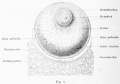
Fig. 1. Human egg from a mature follicle
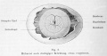


- Kollmann Atlas 1: Predevelopment | Ontogeny | Fetal membranes | Body shape | Systems and organs | Kollmann Atlas 1 | Kollmann Atlas 2 | Julius Kollmann
- Kollmann Atlas 2: Gastrointestinal | Respiratory | Urogenital | Cardiovascular | Neural | Integumentary | Smell | Vision | Hearing | Kollmann Atlas 1 | Kollmann Atlas 2 | Julius Kollmann
Glossary Links
- Glossary: A | B | C | D | E | F | G | H | I | J | K | L | M | N | O | P | Q | R | S | T | U | V | W | X | Y | Z | Numbers | Symbols | Term Link
Cite this page: Hill, M.A. (2026, September 19) Embryology Atlas of the Development of Man 1. Retrieved from https://embryology.med.unsw.edu.au/embryology/index.php/Atlas_of_the_Development_of_Man_1
- © Dr Mark Hill 2026, UNSW Embryology ISBN: 978 0 7334 2609 4 - UNSW CRICOS Provider Code No. 00098G
