Morula: Difference between revisions
mNo edit summary |
mNo edit summary |
||
| Line 1: | Line 1: | ||
==Introduction== | ==Introduction== | ||
[[File:Human embryo day 2.jpg|thumb|300px|Human morula (day 2)<ref name="PMID19924284"><pubmed>19924284</pubmed>| [http://www.ncbi.nlm.nih.gov/pmc/articles/PMC2773928 PMC2773928] | [http://www.plosone.org/article/info%3Adoi%2F10.1371%2Fjournal.pone.0007844 PLoS One]</ref>]] | [[File:Human embryo day 2.jpg|thumb|300px|Human morula (day 2)<ref name="PMID19924284"><pubmed>19924284</pubmed>| [http://www.ncbi.nlm.nih.gov/pmc/articles/PMC2773928 PMC2773928] | [http://www.plosone.org/article/info%3Adoi%2F10.1371%2Fjournal.pone.0007844 PLoS One]</ref>]] | ||
[[File:Human_embryo_day_3.jpg|thumb|300px|Human morula (day 3)<ref name="PMID19924284" />]] | |||
(Latin, ''morula'' = mulberry) An early stage in post-fertilization development when cells have rapidly mitotically divided to produce a solid mass of cells (12-15 cells) with a "mulberry" appearance. This stage is followed by formation of a cavity in this cellular mass [[blastocyst]] stage. | (Latin, ''morula'' = mulberry) An early stage in post-fertilization development when cells have rapidly mitotically divided to produce a solid mass of cells (12-15 cells) with a "mulberry" appearance. This stage is followed by formation of a cavity in this cellular mass [[blastocyst]] stage. | ||
| Line 12: | Line 13: | ||
== Some Recent Findings == | == Some Recent Findings == | ||
[[File: | [[File:Preimplantation blastomere biopsy.jpg|thumb|alt=ART Preimplantation blastomere biopsy|ART Preimplantation blastomere biopsy<ref name="PMID24783200"><pubmed>24783200</pubmed>| [http://www.ncbi.nlm.nih.gov/pmc/articles/PMC3982254 PMC3982254] | [http://www.hindawi.com/journals/bmri/2014/306505 Biomed Res Int.]</ref>]] | ||
{| | {| | ||
|-bgcolor="F5FAFF" | |-bgcolor="F5FAFF" | ||
Revision as of 10:41, 20 July 2015
Introduction


(Latin, morula = mulberry) An early stage in post-fertilization development when cells have rapidly mitotically divided to produce a solid mass of cells (12-15 cells) with a "mulberry" appearance. This stage is followed by formation of a cavity in this cellular mass blastocyst stage.
A key event prior to morula formation is "compaction", where the 8 cell embryo undergoes changes in cell morphology and cell-cell adhesion that initiates the formation of this solid ball of cells.
In humans, morula stage of development occurs during the first days of the first week following fertilization. This developmental stage is followed by formation of a cavity, the blastocoel, which defines formation of the blastocyst.
- Links: Carnegie stage 2 | Morula | Mitosis | Blastocyst | Fertilization | Week 1
Some Recent Findings

|
| More recent papers |
|---|
 This table allows an automated computer search of the external PubMed database using the listed "Search term" text link.
More? References | Discussion Page | Journal Searches | 2019 References | 2020 References Search term: Morula Development <pubmed limit=5>Morula Development</pubmed> |
Movies
| Morula Model |
| Page | Play |
Compaction
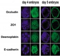
- E-cadherin mediated adhesion initiates at compaction at the 8-cell stage
- regulated post-translationally via protein kinase C and other signalling molecules
Blastomere Division

An in vitro study of human blastocyst development[6] showed that those blastomeres that initially divide quickly are more likely to develop to blastocyst stage.
A recent study in mice showed that there was no specific orientation of the mitotic spindle during cell division in the 8 to 16 cell stage transition.[5] This suggests no predetermined cleavage pattern (pre-patterned) at the 8 cell stage and only modulated by the extent of cell rounding up during mitosis. In other species, such as the worm C.elegans and ascidians, have specific patterns of spindle orientation from the zygote stage.
Model Human Morula Development
The following figure is from a recent study[4] using video and genetic analysis of in vitro human development during week 1 following fertilization.

- EGA - embryonic genome activation
- ESSP - embryonic stage–specific pattern, four unique embryonic stage–specific patterns (1-4)
- Links: Figure with legend
Morulas in Other Species
Mouse Morula
4 cell morula stage development
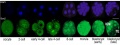
Sox2 expression
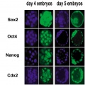
Early gene expression
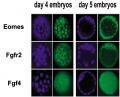
Early gene expression
Early gene expression
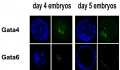
Early gene expression


- Links: Mouse Development
Sea Urchin Morula

Sea Urchin early embryo cleavage pattern (SDB Gallery Images)
- Links: Sea Urchin Development
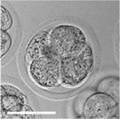
Bovine Morula

Bovine Morula[7]
- Image shows DNA staining (white) and f-actin filaments (orange) at day 4. Scale bars represent 100 µm.
- Pale staining round nuclei are at interphase.
- Arrow shows single nucleus at prophase.
- A single nucleus is seen at metaphase.
- Condensed bright nuclei are apoptotic.
- Links: Bovine Development | Mitosis
References
- ↑ 1.0 1.1 <pubmed>19924284</pubmed>| PMC2773928 | PLoS One
- ↑ <pubmed>24783200</pubmed>| PMC3982254 | Biomed Res Int.
- ↑ Galán A, Montaner D, Póo ME, Valbuena D, Ruiz V, Aguilar C, Dopazo J, Simón C. Functional genomics of 5- to 8-cell stage human embryos by blastomere single-cell cDNA analysis. PLoS One. 2010 Oct 26;5(10):e13615. PMID21049019 | PLoS One.
- ↑ 4.0 4.1 <pubmed>20890283</pubmed>| Nat Biotechnol.
- ↑ 5.0 5.1 <pubmed>19997595</pubmed>| PLoS One.
- ↑ <pubmed>11821286</pubmed>
- ↑ <pubmed>21811561</pubmed>| PLoS One.
Articles
<pubmed>19289087</pubmed> <pubmed>20157423</pubmed>
Search PubMed
Search Pubmed: morula development | blastomere development |
Glossary Links
- Glossary: A | B | C | D | E | F | G | H | I | J | K | L | M | N | O | P | Q | R | S | T | U | V | W | X | Y | Z | Numbers | Symbols | Term Link
Cite this page: Hill, M.A. (2024, June 27) Embryology Morula. Retrieved from https://embryology.med.unsw.edu.au/embryology/index.php/Morula
- © Dr Mark Hill 2024, UNSW Embryology ISBN: 978 0 7334 2609 4 - UNSW CRICOS Provider Code No. 00098G