Category:Liver: Difference between revisions
From Embryology
(Created page with 'This page lists UNSW Embryology content related to liver development.') |
mNo edit summary |
||
| Line 1: | Line 1: | ||
This | This {{Embryology}} category lists content related to liver development. | ||
Revision as of 10:56, 6 March 2015
This Embryology category lists content related to liver development.
Subcategories
This category has the following 2 subcategories, out of 2 total.
Pages in category 'Liver'
The following 66 pages are in this category, out of 66 total.
B
G
- Template:Gall bladder
- Template:Gall-bladder
- Template:Gallbladder
- Gastrointestinal Tract - Gall Bladder Development
- Gastrointestinal Tract - Gallbladder Development
- Gastrointestinal Tract - Gallbladder Histology
- Gastrointestinal Tract - Histology
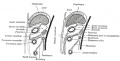
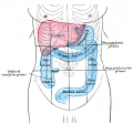
- Gastrointestinal Tract - Liver Development
- Gastrointestinal Tract - Liver Histology
- Gastrointestinal Tract - Pancreas Histology
L
P
- Paper - A Contribution to the Embryology of the Liver and Vascular System in Man
- Paper - A contribution to the morphology and development of the mammalian liver
- Paper - A contribution to the morphology and development of the mammalian liver (1908)
- Paper - A Note on the Development of the Septum Transversum and the Liver
- Paper - A note on the post-natal growth of the kidney, thyroid gland and liver (1924)
- Paper - A Study of the Structural Unit of the Liver
- Paper - Congenital Anomalies of the Liver (1929)
- Paper - Functions of the liver in the embryo
- Paper - Notes on the origin of the liver (1891)
- Paper - On the relation of the liver cells to the blood-vessels and lymphatics
- Paper - Retrogressive Changes in the Fetal Vessels and the Suspensory Ligament of the Liver
- Paper - The development of the lobule of the pig's liver (1919)
- Paper - The early morphogenesis and histogenesis of the liver in Sus scrofa domesticus, including notes on the morphogenesis of the ventral pancreas
- Paper - The embryogenesis of human bile capillaries and ducts
R
- Template:Ref-Bayon1912
- Template:Ref-Bloom1926
- Template:Ref-Bradley1908
- Template:Ref-Frazer1920
- Template:Ref-Gladstone1924b
- Template:Ref-Herring1906
- Template:Ref-Hilton1903
- Template:Ref-Ingalls1908
- Template:Ref-Jackson1909b
- Template:Ref-Johnson1919
- Template:Ref-Lewis1905c
- Template:Ref-MacMahon1929
- Template:Ref-Mall1901b
- Template:Ref-Meyer1914
- Template:Ref-Scammon1915
- Template:Ref-Scammon1916
- Template:Ref-Severn1968
- Template:Ref-Severn1971
- Template:Ref-Severn1972
- Template:Ref-Thompson1914
- Template:Ref-White1939
- Template:Ref-Zorn2008
- Template:Ribavirin
Media in category 'Liver'
The following 124 files are in this category, out of 124 total.
 Adult human liver cells.jpg 900 × 1,095; 245 KB
 Bailey264.jpg 881 × 562; 77 KB
 Bailey266.jpg 731 × 913; 178 KB
 Bailey270.jpg 736 × 448; 53 KB
 Bailey272.jpg 862 × 597; 120 KB
 Bailey273.jpg 560 × 522; 58 KB
 Bailey274.jpg 558 × 442; 32 KB
 Bailey275.jpg 485 × 483; 48 KB
 Bailey276.jpg 855 × 619; 126 KB
 Bailey277.jpg 896 × 480; 112 KB
 Bailey281.jpg 865 × 1,028; 211 KB
 Dog liver portosystemic shunts.jpg 800 × 264; 19 KB
 Embryo-1951-09-01-Slide-60 Scene11-5.jpg 1,440 × 900; 268 KB
 Embryo-1951-09-01-Slide-60 Scene11-6.jpg 1,440 × 900; 220 KB
 Embryo-1951-09-01-Slide-60 Scene11-7.jpg 1,440 × 900; 163 KB
 Embryo-1951-09-01-Slide-60 Scene11-8.jpg 1,440 × 900; 125 KB
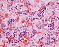
 Fetal liver erythroblasts 01.jpg 905 × 534; 69 KB
 Fetal liver weight growth graph.jpg 800 × 521; 34 KB
 Gray0475.jpg 2,042 × 1,363; 350 KB
 Gray0990.jpg 800 × 407; 60 KB
 Gray1223.png 537 × 500; 55 KB
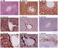
 Histology-fetal liver HEx100.jpg 1,280 × 1,024; 214 KB
 Histology-fetal liver HEx40.jpg 1,000 × 800; 281 KB
 HMB2011 Liver Histology 01.mp3 ; 1.08 MB
- HMB2011 Liver Histology 02.mp3 ; 870 KB
- HMB2011 Liver Histology 03.mp3 ; 757 KB
- HMB2011 Liver Histology 04.mp3 ; 638 KB
- HMB2011 Liver Histology 05.mp3 ; 1.04 MB
 Human liver week 9.jpg 1,200 × 991; 425 KB
 Ingalls1908 fig01.jpg 628 × 779; 35 KB
 Ingalls1908 fig02.jpg 628 × 779; 39 KB
 Ingalls1908 plate01.jpg 927 × 1,000; 204 KB
 Ingalls1908 plate02.jpg 988 × 1,000; 196 KB
 Keibel Mall 2 293.jpg 1,280 × 766; 304 KB
 Keibel Mall 2 295.jpg 1,000 × 920; 83 KB
 Keibel Mall 2 297-299.jpg 1,280 × 786; 135 KB
 Keibel Mall 2 297.jpg 354 × 359; 13 KB
 Keibel Mall 2 298.jpg 599 × 605; 40 KB
 Keibel Mall 2 299.jpg 733 × 1,034; 120 KB
 Keibel Mall 2 300-302.jpg 1,964 × 3,171; 814 KB
 Keibel Mall 2 300.jpg 1,279 × 627; 78 KB
 Keibel Mall 2 301.jpg 1,280 × 779; 140 KB
 Keibel Mall 2 302.jpg 1,278 × 803; 149 KB
 Keibel Mall 2 303-306.jpg 1,817 × 2,328; 520 KB
 Keibel Mall 2 303.jpg 659 × 833; 79 KB
 Keibel Mall 2 305.jpg 701 × 764; 77 KB
 Keibel Mall 2 306.jpg 569 × 832; 66 KB
 Keith1902 fig212.jpg 1,123 × 750; 139 KB
 Keith1902 fig213a.jpg 854 × 800; 166 KB
 Keith1902 fig213b.jpg 800 × 564; 64 KB
 Keith1902 fig214.jpg 750 × 549; 58 KB
 Keith1902 fig215.jpg 704 × 600; 56 KB
 Keith1902 fig216.jpg 1,000 × 771; 170 KB
 Keith1902 fig217.jpg 1,000 × 726; 144 KB
 Keith1902 fig218.jpg 800 × 475; 70 KB
 Keith1902 fig220.jpg 1,000 × 632; 116 KB
 Kollmann539.jpg 736 × 696; 132 KB
 Kollmann555.jpg 605 × 585; 61 KB
 Kollmann562.jpg 670 × 581; 68 KB
 Kollmann563.jpg 754 × 848; 143 KB
 Liver animated cartoon.gif 300 × 200; 239 KB
 Liver cholangiocyte tubulogenesis 01.jpg 800 × 236; 81 KB
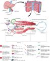
 Liver development signaling.jpg 600 × 467; 45 KB
 Liver hepatocyte from stem cell.png 600 × 444; 96 KB
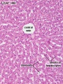 Liver histology 001.jpg 400 × 533; 94 KB
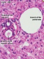 Liver histology 002.jpg 375 × 500; 54 KB
 Liver histology 003.jpg 375 × 500; 52 KB
 Liver histology 004.jpg 600 × 400; 70 KB
 Liver histology 005.jpg 800 × 664; 166 KB
 Liver histology 006.jpg 1,280 × 1,024; 664 KB
 Liver histology 007.jpg 1,280 × 1,024; 313 KB
 Liver histology 008.jpg 1,280 × 1,024; 214 KB
 Liver histology 009.jpg 1,280 × 1,024; 373 KB
 Liver histology 101.jpg 1,280 × 1,024; 410 KB
 Liver histology 102.jpg 1,280 × 1,024; 475 KB
 Liver histology 103.jpg 1,280 × 1,024; 330 KB
 Liver histology EM01.jpg 1,028 × 708; 141 KB
 Liver histology EM02.jpg 1,028 × 707; 154 KB
 Liver plasmodium infection cartoon.jpg 1,000 × 450; 78 KB
 Liver polyploidy 01.jpg 800 × 619; 119 KB
 Liver SEM01.jpg 2,000 × 1,333; 350 KB
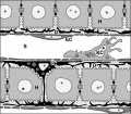
 Liver sinusoidal endothelial cell fenestrations.jpg 926 × 474; 184 KB
 Liver structure cartoon.jpg 1,000 × 451; 78 KB
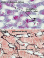 Liver- Kupffer cell and reticular fibre.jpg 600 × 800; 49 KB
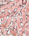 Liver-reticular fibre.jpg 700 × 875; 77 KB
 Liver-sinusiod cartoon.jpg 600 × 523; 51 KB
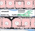 Liver-sinusoid colour cartoon.jpg 600 × 523; 64 KB
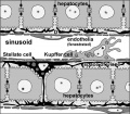 Liver-sinusoid-label cartoon.jpg 600 × 523; 58 KB
 Mall1906-fig06.jpg 600 × 716; 111 KB
 Mall1906-fig07.jpg 503 × 753; 102 KB
 Mall1906-fig08.jpg 726 × 753; 75 KB
 Mall1906-fig09.jpg 452 × 744; 111 KB
 Mall1906-fig10.jpg 660 × 744; 71 KB
 Mall1906-fig11.jpg 681 × 735; 89 KB
 Mall1906-fig12.jpg 544 × 735; 68 KB
 Mall1906-fig13.jpg 592 × 706; 129 KB
 Mall1906-fig14.jpg 673 × 699; 50 KB
 Mall1906-fig15.jpg 887 × 1,000; 115 KB
 Mall1906-fig16.jpg 708 × 890; 155 KB
 Mall1906-fig17.jpg 705 × 881; 98 KB
 Mall1906-fig18.jpg 933 × 584; 66 KB
 Mall1906-fig19.jpg 596 × 610; 102 KB
 Mall1906-fig20.jpg 768 × 1,000; 115 KB
 Mall1906-fig21.jpg 1,000 × 763; 72 KB
 Mall1906-fig22.jpg 694 × 880; 195 KB
 Mall1906-fig23.jpg 650 × 850; 191 KB
 Mall1906-fig24.jpg 577 × 850; 176 KB
 Mall1906-fig25.jpg 1,165 × 887; 153 KB
 Mall1906-fig26.jpg 766 × 639; 78 KB
 Mall1906-fig27.jpg 890 × 800; 168 KB
 Mall1906-fig28.jpg 1,000 × 803; 146 KB
 Mall1906-fig29.jpg 780 × 700; 59 KB
 Mall1906-fig30.jpg 845 × 650; 67 KB
 Mouse hematopoietic stem cell.gif 600 × 595; 40 KB
 Stage 22 image 131.jpg 1,000 × 668; 145 KB
 Stage 22 image 180.jpg 1,000 × 668; 145 KB
 Stage 22 image 181.jpg 1,000 × 653; 263 KB
 Stage 22 image 182.jpg 1,000 × 653; 211 KB
 Thompson1908 fig01.jpg 1,134 × 865; 295 KB
 Thompson1908 fig02.jpg 1,200 × 628; 142 KB
 Thompson1908 fig03.jpg 1,209 × 600; 121 KB
 Waterston07.jpg 562 × 655; 79 KB
 West05.jpg 457 × 759; 30 KB
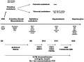
 Zorn2008 fig01.jpg 1,200 × 1,158; 110 KB


