File:Stage8 sem3.jpg: Difference between revisions
mNo edit summary |
mNo edit summary |
||
| (2 intermediate revisions by the same user not shown) | |||
| Line 1: | Line 1: | ||
==Scanning EM showing detail of the Nodal Cilia== | ==Scanning EM showing detail of the Nodal Cilia== | ||
Human embryo ([[Carnegie stage 8|Stage 8]], day 18) | Human embryo ([[Carnegie stage 8|Stage 8]], day 18) nodal cilia are the long cellular processes, the shorter processes visible on cells in the image are microvilli. | ||
Nodal cilia (microtubule-based motile cell structures) are thought to act to "stir" the overlying fluid layer at the time of gastrulation. This fluid contains secreted morphogens that are moved in one direction and therefore may have a role in establishing the initial embryo left/right axis. | |||
:'''Links:''' [[Carnegie stage 8|Stage 8]] | [[Gastrulation]] | |||
Microvilli (microfilament-based non-motile cell structures) increase the cell membrane surface area for absorption and secretion. | |||
:'''Links:''' [[Carnegie stage 8|Stage 8]] | [[Gastrulation]] | [[Nodal Cilia Movie]] | [[:File:Cilium cartoon.jpg|Cilium cartoon]] | |||
{{SEM}} | {{SEM}} | ||
Latest revision as of 23:16, 14 December 2013
Scanning EM showing detail of the Nodal Cilia
Human embryo (Stage 8, day 18) nodal cilia are the long cellular processes, the shorter processes visible on cells in the image are microvilli.
Nodal cilia (microtubule-based motile cell structures) are thought to act to "stir" the overlying fluid layer at the time of gastrulation. This fluid contains secreted morphogens that are moved in one direction and therefore may have a role in establishing the initial embryo left/right axis.
Microvilli (microfilament-based non-motile cell structures) increase the cell membrane surface area for absorption and secretion.
- Links: Stage 8 | Gastrulation | Nodal Cilia Movie | Cilium cartoon
Image Source: Scanning electron micrographs of the Carnegie stages of the early human embryos are reproduced with the permission of Prof Kathy Sulik, from embryos collected by Dr. Vekemans and Tania Attié-Bitach. Images are for educational purposes only and cannot be reproduced electronically or in writing without permission.
Original file name:Stage8day18nodalcilia.jpg
File history
Yi efo/eka'e gwa ebo wo le nyangagi wuncin ye kamina wunga tinya nan
| Gwalagizhi | Nyangagi | Dimensions | User | Comment | |
|---|---|---|---|---|---|
| current | 08:59, 22 August 2009 | 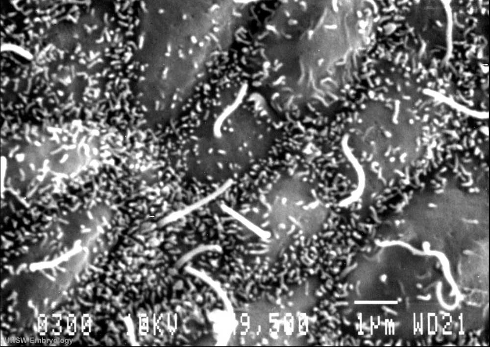 | 1,000 × 709 (91 KB) | S8600021 (talk | contribs) | Human embryo (Stage 8, day 18) Scanning EM showing detail of the nodal cilia. Original file name:Stage8day18nodalcilia.jpg {{Template:SEM}} |
You cannot overwrite this file.
File usage
The following 4 pages use this file: