File:Human uterine tube ciliated epithelium SEM.jpg: Difference between revisions
mNo edit summary |
|||
| Line 1: | Line 1: | ||
==Human Uterine Tube Ciliated Epithelium SEM== | ==Human Uterine Tube Ciliated Epithelium SEM== | ||
Uterine epithelial cilia are responsible for the initial movement of the ooycte and conceptus (zygote, morula, blastocyst). In humans, during the first week of development. | Uterine epithelial cilia are responsible for the initial movement of the ooycte and conceptus (zygote, morula, blastocyst). In humans, this is during the first week of development. | ||
Uterine epithelial microvilli are involved with the implantation process. | |||
The differences in size and shape of cilia and microvilli are well illustrated by scanning micrographs of the lumenal surface of the epithelium lining the mammalian oviduct. | The differences in size and shape of cilia and microvilli are well illustrated by scanning micrographs of the lumenal surface of the epithelium lining the mammalian oviduct. | ||
Revision as of 08:59, 5 May 2013
Human Uterine Tube Ciliated Epithelium SEM
Uterine epithelial cilia are responsible for the initial movement of the ooycte and conceptus (zygote, morula, blastocyst). In humans, this is during the first week of development.
Uterine epithelial microvilli are involved with the implantation process.
The differences in size and shape of cilia and microvilli are well illustrated by scanning micrographs of the lumenal surface of the epithelium lining the mammalian oviduct.
- The tufts of cilia associated with individual ciliated cells project several microns above the convex apices of nonciliated cells covered with short microvilli.
- The number of ciliated cells in this epithelium is under hormonal control by estrogens.
Cilia
|
Microvilli
|
- Links: Uterus Development | Week 1
Reference
Don W. Fawcett M.D. The Cell Chapter 13 (Cilia and Flagella) Figure 316.
Copyright
Attribution Non-Commercial; No Derivatives:This image is licensed under a Creative Commons Attribution, Non-Commercial, No Derivatives License.
Original file name: 11618.jpg http://www.cellimagelibrary.org/images/11618
NCBI Organism Classification: Homo sapiens
Cell Type: ciliated epithelial cell, oviduct epithielial cell
Cellular Component: cilium, microvillus
File history
Yi efo/eka'e gwa ebo wo le nyangagi wuncin ye kamina wunga tinya nan
| Gwalagizhi | Nyangagi | Dimensions | User | Comment | |
|---|---|---|---|---|---|
| current | 07:44, 26 April 2011 | 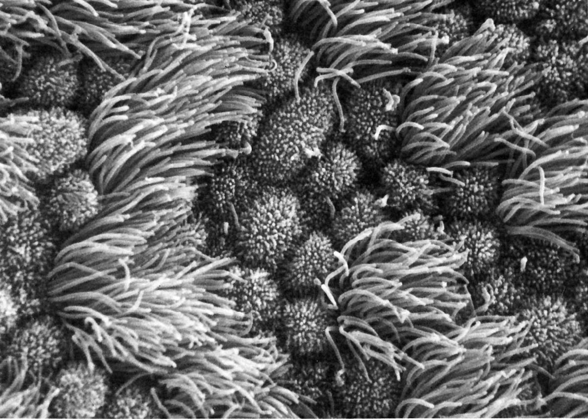 | 1,200 × 855 (248 KB) | S8600021 (talk | contribs) | ==Human uterine tube ciliated epithelium SEM== Figure 316 from Chapter 13 (Cilia and Flagella) of 'The Cell' by Don W. Fawcett M.D. The differences in size and shape of cilia and microvilli are well illustrated by scanning micrographs of the lumenal surf |
You cannot overwrite this file.
File usage
The following 4 pages use this file: