File:Hyaline cartilage 04.jpg: Difference between revisions
From Embryology
mNo edit summary |
mNo edit summary |
||
| Line 1: | Line 1: | ||
==Trachea | ==Respiratory Trachea - Layers== | ||
'''Mucosa''' - formed by epithelium and underlying lamina propria. | |||
* respiratory epithelium - (pseudostratified columnar and ciliated) ciliated cells, goblet cells, brush cells, endocrine cells, surfactant-producing cells (Clara cells), serous cells, basal cells, basement membrane. | |||
* lamina propria - loose connective tissue, many elastic fibres | |||
* elastic lamina - forming the border between the mucosa and submucosa is not visible in H&E stained slides. | |||
'''Submucosa''' - connective tissue and submucosal glands | |||
* submucosal gland - serous (dark) and mucous (light) parts have different staining appearance. | |||
'''Cartilage''' | |||
* perichondrium - surface of cartilage. | |||
* tracheal cartilage - hyaline cartilage, 16 to 20 C-shaped cartilages. | |||
* trachealis muscle - (smooth muscle) Not visible in this section, together with connective tissue fibres, join ends of the cartilages together. | |||
Revision as of 14:20, 10 March 2013
Respiratory Trachea - Layers
Mucosa - formed by epithelium and underlying lamina propria.
- respiratory epithelium - (pseudostratified columnar and ciliated) ciliated cells, goblet cells, brush cells, endocrine cells, surfactant-producing cells (Clara cells), serous cells, basal cells, basement membrane.
- lamina propria - loose connective tissue, many elastic fibres
- elastic lamina - forming the border between the mucosa and submucosa is not visible in H&E stained slides.
Submucosa - connective tissue and submucosal glands
- submucosal gland - serous (dark) and mucous (light) parts have different staining appearance.
Cartilage
- perichondrium - surface of cartilage.
- tracheal cartilage - hyaline cartilage, 16 to 20 C-shaped cartilages.
- trachealis muscle - (smooth muscle) Not visible in this section, together with connective tissue fibres, join ends of the cartilages together.
- Trachea Histology Links: Overview HE | Overview VG | Detail 1 HE Detail 2 HE | Respiratory Histology | Histology Stains | Histology
- Cartilage Histology: Developing | Hyaline HE | Hyaline VG | Hyaline HE | Hyaline VG | Elastic 1 | Elastic 2 | Fibrous - articular disc | Fibrous - intervertebral disc | Articular 1 | Articular 2
Links: Histology | Histology Stains | Blue Histology images copyright Lutz Slomianka 1998-2009. The literary and artistic works on the original Blue Histology website may be reproduced, adapted, published and distributed for non-commercial purposes. See also the page Histology Stains.
Cite this page: Hill, M.A. (2024, June 26) Embryology Hyaline cartilage 04.jpg. Retrieved from https://embryology.med.unsw.edu.au/embryology/index.php/File:Hyaline_cartilage_04.jpg
- © Dr Mark Hill 2024, UNSW Embryology ISBN: 978 0 7334 2609 4 - UNSW CRICOS Provider Code No. 00098G
File history
Yi efo/eka'e gwa ebo wo le nyangagi wuncin ye kamina wunga tinya nan
| Gwalagizhi | Nyangagi | Dimensions | User | Comment | |
|---|---|---|---|---|---|
| current | 23:24, 18 February 2013 | 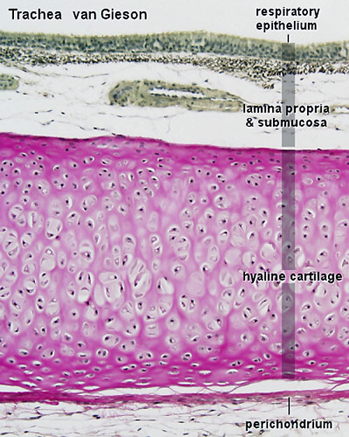 | 500 × 626 (101 KB) | Z8600021 (talk | contribs) | ==Trachea Hyaline Cartilage== {{Cartilage Histology}} {{Blue Histology}} |
You cannot overwrite this file.
File usage
The following 9 pages use this file: