Immune System Development: Difference between revisions
No edit summary |
|||
| Line 7: | Line 7: | ||
During prenatal development, maternal IgG antibodies are transferred from about week 13 (GA) across the placenta, from the maternal lacunae syncytiotrophoblast cell endosomes bind IgG through neonatal Fc receptors. | During prenatal development, maternal IgG antibodies are transferred from about week 13 (GA) across the placenta, from the maternal lacunae syncytiotrophoblast cell endosomes bind IgG through neonatal Fc receptors. | ||
{{Immune Links}} | |||
==Some Recent Findings== | ==Some Recent Findings== | ||
Revision as of 11:48, 15 February 2013
Introduction


Development of the immune system will also link to cardiovascular development notes (blood and vessel) and bone marrow development. Two organs which also relate to this system are the thymus and spleen, which have in the past been included in endocrine and gastrointestinal tract development respectively. There are now also movies showing lymphocyte (B and T cells) traffic within adult lymph nodes.
During prenatal development, maternal IgG antibodies are transferred from about week 13 (GA) across the placenta, from the maternal lacunae syncytiotrophoblast cell endosomes bind IgG through neonatal Fc receptors.
Some Recent Findings
|
Recent References | References
Spleen Development

|
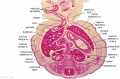
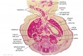
The human spleen arises in week 5 within the dorsal mesogastrium as proliferating mesenchyme overlying the dorsal pancreatic endoderm. Cells required for its hemopoietic function arise from the yolk sac wall and near dorsal aorta.
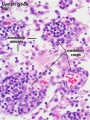
The spleen generates both red and white cells in the 2nd trimester. Note that many embryonic RBCs remain nucleated. |
| D4 Dorsal Mesogastrium (stage 13) |
- Links: Spleen Development
Thymus Development

|
The thymus has a key role in the development of an effective immune system as well as an endocrine function.
The thymus has two origins for the lymphoid thymocytes and the thymic epithelial cells. The thymic epithelium begins as two flask-shape endodermal diverticula that form from the third pharyngeal pouch and extend lateralward and backward into the surrounding mesoderm and neural crest-derived mesenchyme in front of the ventral aorta. |
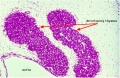
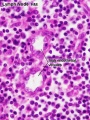
| D4 Developing Thymus (stage 22) |
- Links: Thymus Development
Lymph Node Development

- Links: Lymph Node Development
T Lymphocyte Development
A study of cord blood from 19 early second and third trimester fetuses (GA 18-36 weeks) and 16 term newborns (GA 37-42 weeks).[7]
- Percentage of lymphocytes in fetal white blood cells was 79.3%, reducing to 40% by term birth
- higher than that of adults.
- Mononuclear cells (cord blood mononuclear cells (CBMC)
- fetal mononuclear cells were unable to produce IL-2, IL-4 or IFN-gamma.
- spontaneously secreted IL-10, IL-6 and TNF-alpha in vitro.
- fail to respond to mitogen (PHA) or allogeneic stimulation in vitro.
- Stimulation with PHA up-regulated the production of IL-10, IL-6 and TNF-alpha substantially.
- CD3+ T cells in fetal (40.1%) and neonatal (42.4%)
- lower than that of men (59.6%) and pregnant women (53.6%).
- CD8+ T cells (9.5%)
- gamma delta - T cells (0.5%)
- NK cells (4.8%)
Other Organs
- Liver - The adult liver is a lymphoid organ with a predominantly innate immune system. NK cells are abundant in the normal liver (about one-third of intrahepatic lymphocytes), differs from other lymphoid organs and peripheral blood.
Maternal Antibodies
During prenatal development, maternal antibodies are transferred across the placenta to the fetus. Immunoglobulin G (IgG) is transferred across the syncytiotrophoblast cell layer is mediated by the Neonatal Fc receptor (FcRn). Once inside placental villi, immunoglobulins then need to enter fetal circulation by crossing the second cellular endothelial cell layer by an as yet unknown mechanism.
During postnatal development, maternal antibodies are transferred by maternal milk across the neonatal gastrointestinal tract epithelium.
- Links: Placenta Development | Milk
Additional Images
Developing Human Thymus (stage 22)
F1 Developing Human Spleen (stage 22)
F2 Developing Human Spleen (stage 22)
F3 Developing Human Spleen (stage 22)
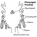
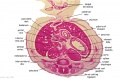
Embryonic origins of the endocrine organs of the neck


Histology
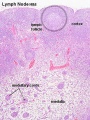
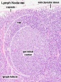
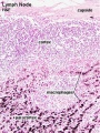
The images below are from adult immune Lymph Nodes.
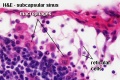
Lymph Node - Subcapsular sinus = marginal sinus
Rabbit Lymph Node
Rabbit Lymph Node
Lymph Node - medullary sinuses and medullary cords
Lymph Node - high endothelial venules
Lymph Node - macrophages


Immune Cells
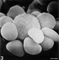
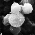
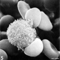
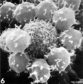
- Human natural killer cells (NK) - originate from CD34(+) hematopoietic progenitor cells.
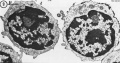
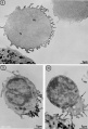
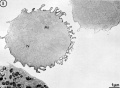
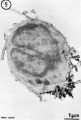
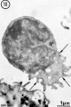
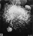
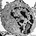
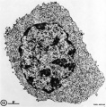
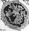
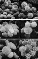
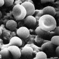
Adult Lymphocyte Histology
- Lymphocyte EM Images: T and B Lymphocytes 1 TEM | T and B Lymphocytes 2 TEM | T Lymphocyte SEM | B lymphocyte 1 TEM | B lymphocyte 2 TEM | B lymphocyte 3 TEM | Plasma Cell TEM | T2 Lymphocyte 1 TEM | T2 Lymphocyte 2 TEM | lymphocyte rosettes | T lymphocyte 1 | T lymphocyte 2 | T lymphocyte 3 | T lymphocyte 4 | T lymphocyte 5 | T lymphocyte 6 | B lymphocyte | B lymphocytes TEM | Immune System Development | Blood
T and B Lymphocytes TEM
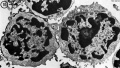
T and B Lymphocytes TEM
B lymphocytes TEM
B lymphocyte TEM
B lymphocyte TEM
B lymphocyte TEM
B lymphocyte SEM
Plasma Cell TEM
T2 Lymphocyte TEM
T2 Lymphocyte TEM
lymphocyte rosettes SEM
T lymphocyte SEM
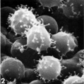
T lymphocyte SEM
T lymphocyte SEM
T lymphocyte SEM
T lymphocyte SEM
T lymphocyte SEM


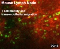
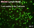
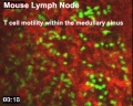
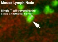
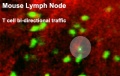
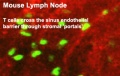
Adult Lymphocyte Motility Movies
|
|
|
|
|
|
| Transendothelial migration |
T cell zone | T cell zone | Sinus endothelial barrier |
Bi-directional traffic | Cross the sinus endothelial barrier |
| Quicktime | Flash | Quicktime | Flash | Quicktime | Flash | Quicktime | Flash | Quicktime | Flash | Quicktime | Flash |
References
- ↑ <pubmed>19138383</pubmed>| PMC2646268 | Genome Biology
- ↑ <pubmed>19906871</pubmed>
- ↑ <pubmed>19741595</pubmed>
- ↑ <pubmed>20231472</pubmed>
- ↑ <pubmed>19443732</pubmed>
- ↑ <pubmed>19255788</pubmed>
- ↑ <pubmed>12165087</pubmed>
Reviews
<pubmed>21071706</pubmed> <pubmed>18946678</pubmed> <pubmed>15804980</pubmed> <pubmed>15376314</pubmed> <pubmed>12669020</pubmed>
Articles
Search Pubmed
Search August 2010 "Immune System Development" All (90551) Review (15955) Free Full Text (28278)
Search Pubmed: Immune System Development | Embryo Immune System Development
Glossary Links
- Glossary: A | B | C | D | E | F | G | H | I | J | K | L | M | N | O | P | Q | R | S | T | U | V | W | X | Y | Z | Numbers | Symbols | Term Link
Cite this page: Hill, M.A. (2024, June 14) Embryology Immune System Development. Retrieved from https://embryology.med.unsw.edu.au/embryology/index.php/Immune_System_Development
- © Dr Mark Hill 2024, UNSW Embryology ISBN: 978 0 7334 2609 4 - UNSW CRICOS Provider Code No. 00098G