File:Buell08.jpg: Difference between revisions
(==Origin of the Pulmonary Vessels in the Chick - Fig. 8== {{Template:Historic Disclaimer}} A 36-somite chick of 60 hours' incubation, injected with ink and dissected by the paraffin method. Only the right half of the vascular tree is shown. This stage) |
|||
| (2 intermediate revisions by the same user not shown) | |||
| Line 1: | Line 1: | ||
==Origin of the Pulmonary Vessels in the | ==Fig. 8. Origin of the Pulmonary Vessels in the 36 somite chick== | ||
A 36-somite chick of 60 hours' incubation, injected with ink and dissected by the paraffin method. Only the | A 36-somite chick of 60 hours' incubation, injected with ink and dissected by the paraffin method. Only the | ||
right half of the vascular tree is shown. This stage is but slightly older than that in figure 7. The, | right half of the vascular tree is shown. This stage is but slightly older than that in [[:File:Buell08.jpg|figure 7]]. The, | ||
pulmonary arch is now complete but still retains a capillary appearance. The pulmonary artery | pulmonary arch is now complete but still retains a capillary appearance. The pulmonary artery | ||
can be recognized in the cephalic portion of the plexus. The right lobar tributary of the common vein | can be recognized in the cephalic portion of the plexus. The right lobar tributary of the common vein | ||
| Line 11: | Line 9: | ||
to show the opening of the common pulmonary vein into the sinus. | to show the opening of the common pulmonary vein into the sinus. | ||
{{Buell1922}} | |||
:'''Links:''' [[Book_-_Contributions_to_Embryology|Carnegie Institution of Washington - Contributions to Embryology]] | [[Chicken Development]] | [[Respiratory System Development]] | :'''Links:''' [[Book_-_Contributions_to_Embryology|Carnegie Institution of Washington - Contributions to Embryology]] | [[Chicken Development]] | [[Respiratory System Development]] | ||
{{Historic Disclaimer}} | |||
[[Category:Cardiovascular]] [[Category:Historic Embryology]] [[Category:Chicken]] [[Category:Respiratory]] | [[Category:Cardiovascular]] [[Category:Historic Embryology]] [[Category:Chicken]] [[Category:Respiratory]] | ||
Latest revision as of 07:42, 24 January 2013
Fig. 8. Origin of the Pulmonary Vessels in the 36 somite chick
A 36-somite chick of 60 hours' incubation, injected with ink and dissected by the paraffin method. Only the right half of the vascular tree is shown. This stage is but slightly older than that in figure 7. The, pulmonary arch is now complete but still retains a capillary appearance. The pulmonary artery can be recognized in the cephalic portion of the plexus. The right lobar tributary of the common vein is formed and is connected with its corresponding artery on the dorsal surface of the lung-bud. The cranial tributary of the common vein is plainly seen. The wall of the sinus venosus has been removed to show the opening of the common pulmonary vein into the sinus.
- 1922 Chicken Pulmonary: Fig 1 | Fig 2 | Fig 3 | Fig 4 | Fig 5 | Fig 6 | Fig 7 | Fig 8 | Fig 9 | Fig 10 | Plate 1 | Plate 2 | Carnegie No.66 | Chicken Development | Respiratory
- Links: Carnegie Institution of Washington - Contributions to Embryology | Chicken Development | Respiratory System Development
| Historic Disclaimer - information about historic embryology pages |
|---|
 |
File history
Yi efo/eka'e gwa ebo wo le nyangagi wuncin ye kamina wunga tinya nan
| Gwalagizhi | Nyangagi | Dimensions | User | Comment | |
|---|---|---|---|---|---|
| current | 16:23, 23 January 2013 | 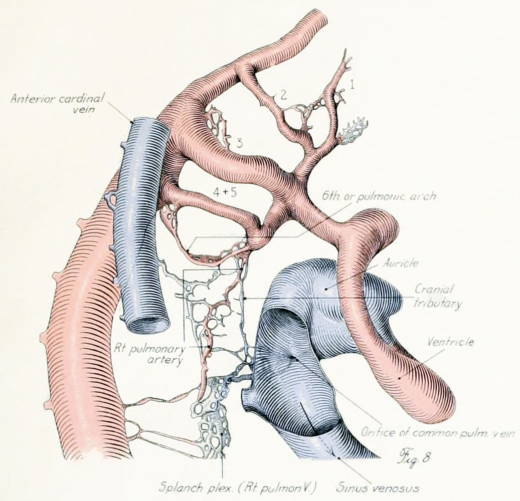 | 1,037 × 1,000 (176 KB) | Z8600021 (talk | contribs) | JP2 figure scan |
| 12:24, 29 March 2011 |  | 522 × 596 (62 KB) | S8600021 (talk | contribs) | ==Origin of the Pulmonary Vessels in the Chick - Fig. 8== {{Template:Historic Disclaimer}} A 36-somite chick of 60 hours' incubation, injected with ink and dissected by the paraffin method. Only the right half of the vascular tree is shown. This stage |
You cannot overwrite this file.
File usage
The following page uses this file: