Yolk Sac Development
| Embryology - 24 Jul 2026 |
|---|
| Google Translate - select your language from the list shown below (this will open a new external page) |
|
العربية | català | 中文 | 中國傳統的 | français | Deutsche | עִברִית | हिंदी | bahasa Indonesia | italiano | 日本語 | 한국어 | မြန်မာ | Pilipino | Polskie | português | ਪੰਜਾਬੀ ਦੇ | Română | русский | Español | Swahili | Svensk | ไทย | Türkçe | اردو | ייִדיש | Tiếng Việt These external translations are automated and may not be accurate. (More? About Translations) |
Introduction

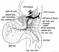
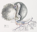
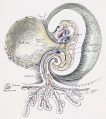
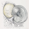
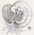
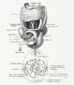
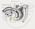
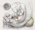
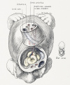
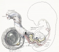
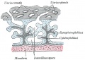
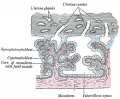
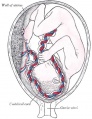
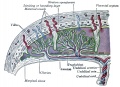
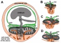
The yolk sac is an early extra-embryonic membrane which is endoderm origin and covered with extra-embryonic mesoderm. Yolk sac lies outside the embryo connected by a yolk stalk (vitelline duct, omphalomesenteric duct) to the midgut with which it forms a continuous connection. The endodermal lining is continuous with the endoderm of the gastrointestinal tract. The extra-embryonic mesoderm differentiates to form both blood and blood vessels of the vitelline system.
In reptiles and birds, the yolk sac has a function associated with nutrition. In mammals the yolk sac acts as a source of primordial germ cells and blood cells.
Note that in early human development (week 2) a transient structure called the "primitive yolk sac" forms from the hypoblast layer, this is an entirely different structure.
The yolk stalk normally degenerates around the time the midgut herniation return to the peritoneal cavity and the anterior body wall closes (week 8). Failure of complete degeneration of this structure can lead to a common intestinal abnormality, Meckel's diverticulum. (More? Meckel's diverticulum, Johann Meckel)
| Coelom Links: Introduction | Lecture - Week 3 Development | Lecture - Mesoderm Development | Placenta - Membranes | Category:Coelomic Cavity |
| Historic Embryology - Coelomic Cavity |
|---|
| 1891 peritoneal | 1897 human coelom | 1910 Coelom and Diaphragm | 1924 serous |
Some Recent Findings

|
| More recent papers |
|---|
 This table allows an automated computer search of the external PubMed database using the listed "Search term" text link.
More? References | Discussion Page | Journal Searches | 2019 References | 2020 References Search term: Yolk Sac Development | Vitelline Duct | Meckel's Diverticulum |
| Older papers |
|---|
| These papers originally appeared in the Some Recent Findings table, but as that list grew in length have now been shuffled down to this collapsible table.
See also the Discussion Page for other references listed by year and References on this current page.
|
Movies
| <html5media height="540" width="390">File:Week3_folding.mp4</html5media> | <html5media height="420" width="400">File:Amnion 001.mp4</html5media> | ||||||
|
|
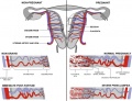
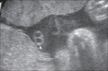
Development Overview
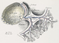
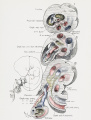
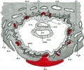
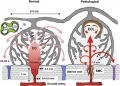
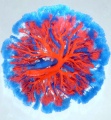
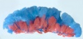
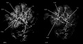
Yolk Sac Blood Vessels

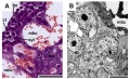
Yolk sac blood vessels and Notch model[6]
Abnormalities
Meckel's Diverticulum
ICD-11 LB15.0 Meckel diverticulum


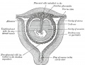
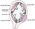
This gastrointestinal tract abnormality is a very common (incidence of 1–2% in the general population) and results from improper closure and absorption of the omphalomesenteric duct (vitelline duct) in development. This transient developmental duct connects the yolk to the primitive gastrointestinal tract.
In addition to Meckel's diverticulum there are a range of other vitelline duct abnormalities, which depend on the degree from a completely patent duct at the umbilicus to lesser remnants (cysts, fibrous cords connecting umbilicus to distal ileum, granulation tissue at umbilicus, or umbilical hernias).
- Links: GIT Abnormalities - Meckel's Diverticulum | OMIM - Meckel's Diverticulum | Pubmed - Meckel's Diverticulum | Pubmed - omphalomesenteric duct | Pubmed - vitelline duct
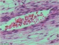
Yolk Sac Carcinoma
A yolk sac carcinoma (endodermal sinus tumor[7]) is a form of germ cell tumour.
References
- ↑ Too HC, Shibata M, Yayota M, Darras VM & Iwasawa A. (2017). Expression of thyroid hormone regulator genes in the yolk sac membrane of the developing chicken embryo. J. Reprod. Dev. , 63, 463-472. PMID: 28652559 DOI.
- ↑ Dong D, Reece EA, Lin X, Wu Y, AriasVillela N & Yang P. (2016). New development of the yolk sac theory in diabetic embryopathy: molecular mechanism and link to structural birth defects. Am. J. Obstet. Gynecol. , 214, 192-202. PMID: 26432466 DOI.
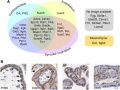
- ↑ Frame JM, Fegan KH, Conway SJ, McGrath KE & Palis J. (2016). Definitive Hematopoiesis in the Yolk Sac Emerges from Wnt-Responsive Hemogenic Endothelium Independently of Circulation and Arterial Identity. Stem Cells , 34, 431-44. PMID: 26418893 DOI.
- ↑ Ariza L, Carmona R, Cañete A, Cano E & Muñoz-Chápuli R. (2016). Coelomic epithelium-derived cells in visceral morphogenesis. Dev. Dyn. , 245, 307-22. PMID: 26638186 DOI.
- ↑ Renda MC, Giambona A, Fecarotta E, Leto F, Makrydimas G, Renda D, Damiani G, Jakil MC, Picciotto F, Piazza A, Valtieri M & Maggio A. (2010). Embryo-fetal erythroid megaloblasts in the human coelomic cavity. J. Cell. Physiol. , 225, 385-9. PMID: 20533375 DOI.
- ↑ Copeland JN, Feng Y, Neradugomma NK, Fields PE & Vivian JL. (2011). Notch signaling regulates remodeling and vessel diameter in the extraembryonic yolk sac. BMC Dev. Biol. , 11, 12. PMID: 21352545 DOI.
- ↑ Damjanov I. (1980). Animal model of human disease: yolk sac carcinoma (endodermal sinus tumor). Am. J. Pathol. , 98, 569-72. PMID: 6986787
Reviews
Yamane T. (2018). Mouse Yolk Sac Hematopoiesis. Front Cell Dev Biol , 6, 80. PMID: 30079337 DOI.
Palis J, Malik J, McGrath KE & Kingsley PD. (2010). Primitive erythropoiesis in the mammalian embryo. Int. J. Dev. Biol. , 54, 1011-8. PMID: 20711979 DOI.
Tavian M & Péault B. (2005). Embryonic development of the human hematopoietic system. Int. J. Dev. Biol. , 49, 243-50. PMID: 15906238 DOI.
Arendt D & Nübler-Jung K. (1999). Rearranging gastrulation in the name of yolk: evolution of gastrulation in yolk-rich amniote eggs. Mech. Dev. , 81, 3-22. PMID: 10330481
Auerbach R, Huang H & Lu L. (1996). Hematopoietic stem cells in the mouse embryonic yolk sac. Stem Cells , 14, 269-80. PMID: 8724693 DOI.
Articles
Funayama N, Sato Y, Matsumoto K, Ogura T & Takahashi Y. (1999). Coelom formation: binary decision of the lateral plate mesoderm is controlled by the ectoderm. Development , 126, 4129-38. PMID: 10457021
Search PubMed
Search Pubmed: Coelomic Cavity Development | pericardial cavity development | pleural cavity development | peritoneal cavity development
Additional Images
Historic
| Historic Disclaimer - information about historic embryology pages |
|---|
|
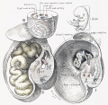
Keith A. Human Embryology and Morphology. (1902) London: Edward Arnold.
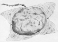
Yolk sac

Cullen TS. Embryology, anatomy, and diseases of the umbilicus together with diseases of the urachus. (1916) W. B. Saunders Company, Philadelphia And London.
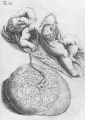
1 Human embryo 0.7 mm
2 Human embryo 1.7 mm
3 Human embryo 2.5 mm
4 Human embryo 3.5 mm
5 Human embryo 5 mm
6 Human embryo 7 mm
7 Human embryo 7 mm
8 Human embryo 10 mm
9 Human embryo 12.5 mm
10 Human embryo 10 mm
11 Human embryo 23 mm
12 Human embryo 3 cm
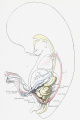
13 Human embryo 4.5 cm sagittal

14 Human Embryo 4.5 cm


Placenta and Fetus
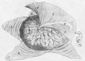
Placenta Fetal Side
Placenta Maternal Side

Fetal membrane and placenta cartoon
Placenta spiral artery conversion
Uterine and placental vasculature
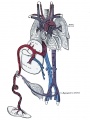
Fetal circulation overview
Placental trophospongium

Placenta anchoring villi
Fetal blood

Placental cord cross-section
Placenta_abnormalities
Mouse placenta E16.5
Mouse placenta E16.5
Human placenta viewed from the fetal side
Cord with one artery and one vein
Placenta gene expression


Terms
yolk stalk, vitelline duct, omphalomesenteric duct
Glossary Links
- Glossary: A | B | C | D | E | F | G | H | I | J | K | L | M | N | O | P | Q | R | S | T | U | V | W | X | Y | Z | Numbers | Symbols | Term Link
Cite this page: Hill, M.A. (2026, July 24) Embryology Yolk Sac Development. Retrieved from https://embryology.med.unsw.edu.au/embryology/index.php/Yolk_Sac_Development
- © Dr Mark Hill 2026, UNSW Embryology ISBN: 978 0 7334 2609 4 - UNSW CRICOS Provider Code No. 00098G