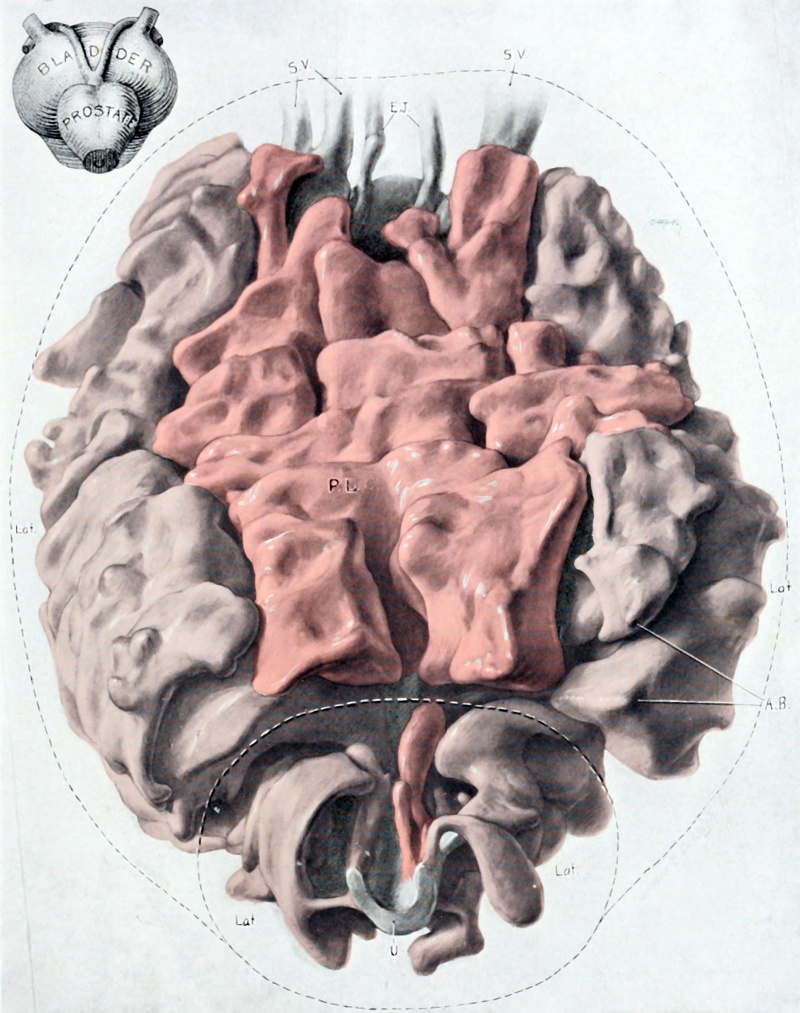
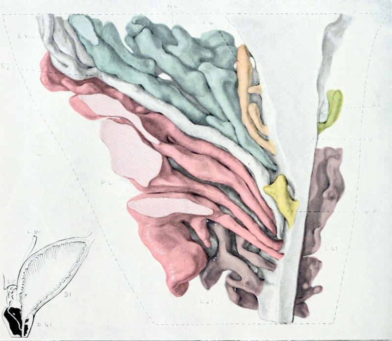
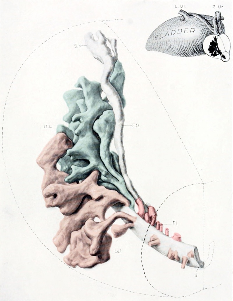
Paper - The development of the human prostate gland with reference to the development of other structures at the neck of the urinary bladder (1912)
| Embryology - 27 Apr 2024 |
|---|
| Google Translate - select your language from the list shown below (this will open a new external page) |
|
العربية | català | 中文 | 中國傳統的 | français | Deutsche | עִברִית | हिंदी | bahasa Indonesia | italiano | 日本語 | 한국어 | မြန်မာ | Pilipino | Polskie | português | ਪੰਜਾਬੀ ਦੇ | Română | русский | Español | Swahili | Svensk | ไทย | Türkçe | اردو | ייִדיש | Tiếng Việt These external translations are automated and may not be accurate. (More? About Translations) |
Lowsley OS. The development of the human prostate gland with reference to the development of other structures at the neck of the urinary bladder. (1912) Amer. J Anat. 13(3): 299-346.
| Online Editor |
|---|
| This historic 1912 paper by Watson described human human prostate gland and anatomically close structure development.
Watson EM. The development of the seminal vesicles in man. (1918) Amer. J Anat. 24(4): 395 - 439.
See also recent articles: Allgeier SH, Vezina CM, Lin TM, Moore RW, Silverstone AE, Mukai M, Gavalchin J, Cooke PS & Peterson RE. (2009). Estrogen signaling is not required for prostatic bud patterning or for its disruption by 2,3,7,8-tetrachlorodibenzo-p-dioxin. Toxicol. Appl. Pharmacol. , 239, 80-6. PMID: 19523480 DOI. Allgeier SH, Lin TM, Vezina CM, Moore RW, Fritz WA, Chiu SY, Zhang C & Peterson RE. (2008). WNT5A selectively inhibits mouse ventral prostate development. Dev. Biol. , 324, 10-7. PMID: 18804104 DOI. See also recent reviews: Meeks JJ & Schaeffer EM. (2011). Genetic regulation of prostate development. J. Androl. , 32, 210-7. PMID: 20930191 DOI. Cai Y. (2008). Participation of caudal müllerian mesenchyma in prostate development. J. Urol. , 180, 1898-903. PMID: 18801537 DOI. Thomson AA. (2008). Mesenchymal mechanisms in prostate organogenesis. Differentiation , 76, 587-98. PMID: 18752494 DOI. Cunha GR. (2008). Mesenchymal-epithelial interactions: past, present, and future. Differentiation , 76, 578-86. PMID: 18557761 DOI. |
| Historic Disclaimer - information about historic embryology pages |
|---|
| Pages where the terms "Historic" (textbooks, papers, people, recommendations) appear on this site, and sections within pages where this disclaimer appears, indicate that the content and scientific understanding are specific to the time of publication. This means that while some scientific descriptions are still accurate, the terminology and interpretation of the developmental mechanisms reflect the understanding at the time of original publication and those of the preceding periods, these terms, interpretations and recommendations may not reflect our current scientific understanding. (More? Embryology History | Historic Embryology Papers) |
The Development of the Human Prostate Gland with Reference to the Development of Other Structures at the Neck of the Urinary Bladder
Oswald S. Lowsley
From the Anatomical Laboratory, Johns Hopkins University
Eleven Figures (Three Color Plates)
Introduction
A review of the literature on the embryology of the prostate gland and other structures at the neck of the human bladder discloses a great diversity of findings. Much has been written about the middle lobe of the prostate and there seem to be two views very firmly held with regard to its development, one best expressed by Griffiths and utilized by Tandler and Zuckerkandl[1] to the effect that the middle lobe is an independent structure which may sometimes be lacking; the other supported by Pallin, Jores and others who believe that the middle lobe is always formed by ingrowths from the lateral lobes.
Griffiths[2] concludes from his studies: (1) That the middle lobe may be either present or absent at the time of puberty and in adult life before enlargement takes place. (2) That this lobe is independent, having glands of its own which open on parts of the hinder wall of the prostatic urethra. (3) That this region develops separately from the part of the urethra just mentioned in the same way as the lateral lobes do from the part of the urethra on each side of the verum montanum, and it is not the result of an extension back of gland tissue from the lateral lobes into the interval between the vasa deferentia beneath the neck of the bladder.
Griffiths[3] states in another paper that there can be no enlargement of the third lobe unless there were gland tubules originally there. Any one of the lobes may enlarge without involvement of the others. This author states that enlargement does not take place in that part of the prostate behind the urethra and anterior to the verum montanum (posterior lobe).
Mansell Moullin[4] states that the prostate is not an urinary organ but that the point of origin of the prostatic glands has simply been displaced in the course of racial development from the lining of the Wolffian ducts to that structure, into which they open. The question of the median lobe, according to this author, depends upon the extent to which this displacement has occurred in each individual. So long as the glands are restricted to the prostatic sinus there is no medium portion. In some instances a greater or smaller number are displaced towards the bladder and they not infrequently occupy the middle line. Usually they remain on the posterior wall of the urethra and form a more or less conspicuous median lobe. Exceptionally they make their appearance upon the anterior wall as well.
Keibel[5] does not agree with this opinion and considers the prostate to be an urinary organ because its glands arise from the urethra above the openings of the Wolffian and Miillerian ducts.
Evatt[6] in a study of a 12 cm. (crown-heel measurement) foetus which he considered to be three and one-half months of age, by means of a wax model described the middle lobe to be made up of branches of the two largest prostatic ducts which come together in the midline behind and these, with two smaller ducts immediately above them, form a centrally placed lobe above the level of the point of entrance of the genital cord. This he considers to be the middle lobe and consists of ducts derived from both sides of the prostate and cannot therefore be regarded as an azygos structure.
Gustaf Pallin[7] summarizes the results of his study of human embryos aged three and four months by means of wax models as follows :
The prostate gland is deposited in male embryos in the third month by separation of the solid longitudinal folds on the outer side of the epithelial wall of the urethra. Three groups of prostatic tissue are distinguished, (1) Cranialwards from the genital cords lying dorsally, (2) Caudalwards from the genital cords lying dorsally,. (3) Ventral.
Both of the first groups go out from the prostatic furrows. From the cranial the main mass of the base of the prostate will be composed; the third lobe seems not to be composed of independent glands but ramifications of the cranial glands can grow into the midline and then these become gland parts. The caudal dorsal structure forms the lateral and hind part of the side lobes. The ventral group at first occupies the greater part of the forward urethral wall. The number of its glands becomes reduced in the fourth month and it appears then in the midline as a forward lobe. In certain cases the reduction of this lobe amounts to complete atrophy.
This article is very extensively quoted and accompanied as it is by drawings from wax models and very accurate descriptions has influenced many workers.
Jores[8] states that the middle lobe can be considered only as a glandular commissure connecting the two lateral lobes and not as an independent structure.
The posterior lobe is generally referred to in the literature as originating from ingrowths from the lateral lobes.
The number of tubules of the various parts of the prostate emptying their secretions into the prostatic urethra is usually stated in text-books to be between twenty and thirty, while the proportion of glandular tissue compared to the interglandular stroma is quoted by different authors to be from one-third or one half (Kasuyoshi Nakasima) to five-sixths (Walker). The later writers seem to be agreed that the prostate begins to develop at about the third month of intra-uterine life. Kolliker thought that it was not present until the fourth month and Mihalkovics placed the fifth month as its beginning. The smooth muscle of the gland, according to the latter and also Tourneux, began to develop at the middle of the fifth month. Pallin, on the other hand, found the musculature to be developing at the fourth month.
Albarran[9] describes a group of glands under the neck of the bladder which open into the urethra and whose tubules lie between the mucosa and the musculature. He calls this the subcervical glandular group and states that it varies greatly in different individuals and may be entirely lacking in some.
The bladder, trigonum vesicae, and internal sphincter have received a great deal of attention by writers on the anatomy of this region.
J. Griffiths[10] believed the trigonum vesicae to be composed only of the innermost bands of muscular bundles of the bladder wall, while the outermost longitudinal bundles pass on to the neck of the bladder.
W. Waldeyer[11] called attention to the following facts in regard to the trigonum vesicae: (1) There is a separate development of its musculature which is continuous with the musculature of the ureters and the prostatic urethra. (2) There is an absence of a submucosa over the trigone. (3) It has a smooth, firm, thick layered mucous membrane.
Versari[12] concludes from his studies that normally the musculature of the trigonum vesicae is made up of (a) the trigonal portion of the internal sphincter, (b) part of the muscular layer of the ureters, and (c) the muscle bundles of their sheaths. In adults there are present in the trigonal region bundles which come from the muscular layer of the bladder.
Walker[13] agrees with the above findings in part. He observes that from the ureter on each side a thick band of muscle passes down towards the urethra. These bands converge and unite so that this longitudinal muscle flows over the margin of the urethral opening in a continuous sheet. In the center of the triangle formed by these bands of muscle the fibers appear to interlace indiscriminately.
Delbet [14] declares the trigonum vesicae to be an appendage of the urethral walls. Congenital absence of a ureter shows the trigonum to be lacking on that side. Passavant has described a case in which the trigone was entirely separate from the bladder wall.
In regard to the internal sphincter of the human bladder Versari [15] concludes from his investigations that (1) The smooth muscle sphincter of the urinary bladder of man constitutes a structure by itself, which develops independently of the middle (circular) layer of the bladder, the circular muscle layer of the urethra and the musculature of the ureters. (2) The sphincter is made up of an urethral and a trigonal portion, and it is the urethral portion only which assumes the form of a ring surrounding the initial part of the urethra. The first groups of the fibers of the sphincter arranged in bundles correspond to the anterior arch of the urethral portion; from there immediately follow those of the urethral portion of the posterior arch, and these last are apparently those of the trigonal portion. The posterior arch of muscle extends little by little, with new bundles either upwards to occupy part of the trigonal area or downwards along the posterior wall of the urethra, so that it comes to have an extent much greater than the anterior. On the other hand, the older view held by Krause, Hyrtl, Gegenbauer and others is that the sphincter is a continuation downward of the circular musculature of the bladder.
The seminal vesicles begin to develop at about the third month (McMurrich).[16] Pallin found that the ejaculatory ducts show no suggestion of smooth muscle at the sixth month. The colliculus seminalis of man is, according to D. Berry Hart,[17] the analogue of the hymen and lower one-third of the vagina, the Mullerian ducts not being represented in the adult human male except by the hydatid of Morgagni and some rudiments near the testes. Primrose[18] believes that the uterus masculinus must be looked upon as the homologue of the series of structures formed in the female by the fused portions of the Mullerian ducts. Minot[19] states that in the male the Mullerian ducts remain rudimentary and their middle portions usually abort leaving the upper fimbriated ends to develop into so-called hydatids of Morgagni, and the lower or caudal ends to unite within the genital cord to form the so-called uterus masculinus, a rudimentary representation of the female uterus and vagina.
The prostates used in this investigation were obtained from Dr. Mall's collection of human fetuses which were preserved in alcohol or in formalin (4 per cent). They were imbedded in paraffine and cut. in series, being stained with haematoxylin and eosin or Mallory's stain. The measurements taken are crownrump, and the ages of embryos are estimated according to the table in Keibel-Mall's Manual of Embryology.[20] Before taking up the discussion of the various specimens it seems best to state the terminology to be used. The various parts of the prostate gland will be referred to as follows: (1) The middle lobe, or that part of the gland which is situated between the bladder and the ejaculatory ducts under the floor of the urethra (prespermatic and post urethral). (2) Lateral lobes, or those parts of the gland which arise from the prostatic furrows and the lateral walls of the urethra and extend laterally and posteriorly from that structure. (3) Posterior lobe, or that part of the prostate which lies dorsal to the ejaculatory ducts above their entrance into the urethra and dorsal to the urethra below this point (post spermatic and post urethral). This is the part of the prostate which is felt per rectum. (4) Ventral lobe, or that part of the organ formed by glands arising from the anterior or ventral wall of the prostatic urethra.
Fetus 5 cm long
- These measurements are all crown-rump (CRL) and not crown-heel.
{ten weeks)
A study of the bladder and prostatic portion of the urethra of a human male fetus two and one-half months of age shows several interesting features. The bladder at the trigonal region has about the same circumference as it has over the rest of its area, being at thjs stage a tubular structure which narrows down gradually as it approaches its orifice and nowhere is there a sharply outlined portion which will later become the vesical orifice. The prostatic portion of this tubular structure is marked only by the change in its shape.
The organs all seem to be composed of embryonic connective tissue which is similar throughout. The walls of the bladder are uniform in size everywhere except in the region of the trigone which is nearly twice as thick as any other portion. The connective tissue strands can be traced from the ureters out into the trigone and the latter structure is quite definitely superimposed upon the bladder wall. The increased thickness of the base of the vesical wall extends throughout the trigonal region but is most marked at the beginning or interureteral region.
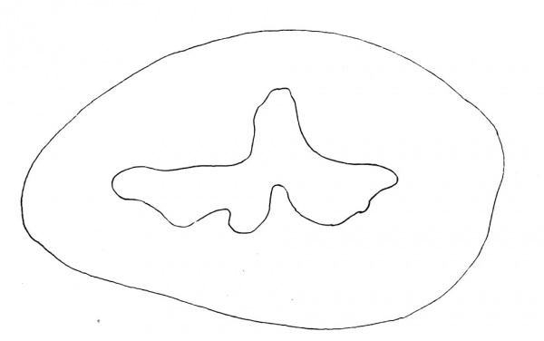
By following this gradually narrowing tube down there is noticed a change in shape so that there is a slight notch formed in the ventral wall of the urethra and a projection into the lumen of the posterior wall so that this structure takes on the shape of a very widely spread inverted V. This marks the beginning of the verum montanum. Further down there are noticed two little notches on the floor of the urethra, one on each side of the verum montanum. The one on the right is more pronounced than that on the left. The outer or lateral portions of the lumen which will later become the prostatic furrows point in a horizontal direction and at this period of development show no tendency to be directed downward (fig. 1).
In no portion of the prostatic urethra is there any thickening of tissue or outgrowth of epithelial cells indicating the development of prostatic gland tissue. There is no specific arrangement of tissue planes and it is not possible to pick out the exact site of the internal sphincter. Below the verum montanum the urethra becomes more or less star-shaped, indicating a collapsed circular tube.
Fetus 7.5 cm long
- These measurements are all crown-rump (CRL) and not crown-heel.
(thirteen weeks)
There is considerable change noted in the appearance of the bladder at this stage. Throughout its entire area the wall composing the base is thicker than at any other portion of its circumference, and the nearer one approaches the trigonal region the greater is the thickness. The musculature of the bladder is distinctly made out as deeply staining tissue composed of circular, interlacing and longitudinal strands which are easily differentiated from the connective tissue elements forming the major portion of the bladder wall. The strands of muscular tissue aie much larger and more abundant at the base or inferior portion than at any other. In the region just superior to the trigone where the bas-fond will later develop, the inferior wall is three times as thick as it is at any other part of the circumference. The mucous membrane is gathered in folds on the inferior interior surface of the organ throughout its length, while elsewhere it is smooth.
Fig. 1 5 cm. human fetus two and three-fourths months. Prostatic portion of urethra.
The trigonum vesicae is about five times as thick as the remaining' portion of the vesical wall. The muscular strands composing it are much finer in texture than those found elsewhere and many of them can be traced between the two ureters and out into other portions of the trigone.
There is a very sudden narrowing of the vesical walls at the site of the developing internal sphincter and the lumen of the bladder changes from an oval to a triangular shape and then into a rather narrow horizontal slit with a vertical slit connecting with its anterior wall as shown in fig. 2. Considerably below this it becomes triangular in shape again.
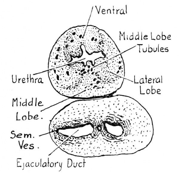
An examination of the urethra of the thirteen weeks old fetus from the bladder outwards reveals the fact that two large solid evaginations extend posteriorly from its floor. Outward or caudally from these buds other larger evaginations have developed forming tubules, in some of which, lumina are present. In others the lumina are poorly developed, and in still others are solid (fig. 2). These structures, 12 in number, which are without question developing prostatic tubules, are separated by a considerable space from the main mass of prostatic tissue and occur directty on the floor of the urethra in a position that is universally accorded to the middle lobe, i.e., between the bladder and the entrance of the ejaculatory ducts, and extend posteriorly to occupy a position under the floor of the urethra and between the bladder and ejaculatory ducts.
Fig. 2 7.5 cm. human fetus. Three months. X 20.
The tubules of the lateral lobes arise from the sides of the urethra and from the bottom and in some cases a little to the inside of the depressions at the sides of the urethra, which are commonly called prostatic furrows. These structures are in nearly every case larger than the ones making up the middle lobe. They are thirty-nine in number, twenty-six of which are arranged definitely in pairs and three of which are unpaired send branches forward.
In this series no glandular tissue is noted in the region dorsal to the ejaculatory ducts. At the lower or caudal end of the prostate the tubules become centrally located near the midline and here we have structures which later grow back dorsally to the ejaculatory ducts and become the posterior lobe of the prostate. Beginning evaginations and tubules occur throughout the prostatic urethra on its roof or ventral wall. The general direction of the growth in this region as well as in the others is bladderwards with the exception of the outermost of the lateral lobes and the posterior lobe which send a few branches caudalward. The .tubules of the ventral lobe are twelve in number, eight of which are paired, the other four being directly in the middle line. One of the latter tubules is quite long and presents a very definite lumen as all of the larger ones do; those smaller in size are nearly all solid epithelial outgrowths.
There are no glandular outgrowths from the urethra below the apex of the prostate and there are no signs of glandular growth in the subcervical region where Albarran's tubules are found in later stages.
The vasa deferentia appear under the bladder at the entrance of the ureters as two small tubes surrounded by a thick layer of developing muscle and connective tissue. As they descend they approach each other and behind the middle of the trigonum vesicae they are enveloped in the same tissue, being bound very firmly together, and between them is noticed a very small lumen surrounded by rather delicate but distinct connective tissue layers, which is taken to be the unobliterated upper portion of the fused Miillerian ducts. The vasa deferentia and their enveloping tissue increase enormously in size as they descend, so that under the internal sphincter this structure is larger than the beginning of the urethra and its surrounding tissue. Under the main portion of the middle lobe thelumina of the vasa deferentia spread out laterally as shown in fig. 2 and form a lateral branch or bending which marks the first appearance of the seminal vesicles. Immediately below this point at the beginning of the ejaculatory ducts, the lumina constrict, and the surrounding tissue is less abundant. The structures surrounding the ejaculatory ducts which have to this point remained separate now intermingle with the muscular walls of the urethra and firmly bind the two structures together.
At this point the utriculus prostaticus which has been very small begins to enlarge and finally becomes larger than the two ejaculatory ducts which decrease in size as they approach the urethral lumen.
In the progress of the two ejaculatory ducts and utriculus prostaticus toward the lumen of the urethra, the tissues surrounding the two structures are closely bound together, but the mass of tissue around the ejaculatory ducts and utriculus prostaticus maintains its identity and by its further progress pushes the flattened floor of the urethra up into a mound-like projection transforming its triangular lumen into an inverted semilunar shaped structure and forming the verum montanum, the main tissue of which is thus derived from the walls of the ejaculatory ducts.
Fetus 8 cm long
(thirteen weeks. No. 54 - in Dr. Mall's collection)
This fetus shows exactly the same distribution of elements as the one just described. The number of tubules is reduced, probably due to the fact that the sections were three times as thick as the ones discussed above and this might have obscured some of them.
The middle lobe region gives rise to seven glands. Twentyseven tubules are found in the two prostatic furrows which will form the lateral lobes and six others are located on the floor of the urethra caudalward from the entrance of the ejaculatory ducts which represent the posterior lobe of the prostate. These latter send branches caudalward or anteriorly as well as cranialward or posteriorly, and none of them extend as far back as the ejaculatory ducts.
On the ventral wall of the urethra are located thirteen structures composing the anterior lobe.
The subcervical glands of Albarran were not found.
Fetus 12.5 cm long
(sixteen weeks)
The bladder of the fetus studied here was contracted so that a direct comparison with the younger stages which were distended, is not possible. However, it is very evident that there has been an enormous development of smooth muscle between the last specimen described and this one. Throughout the whole vesical wall the muscle bundles making up the three coats of the bladder stand out very clearly. The mucous membrane and the connective tissue underlying it are thrown into thick folds extending into the lumen of the bladder and almost obliterating it. In this specimen as in those previously described the trigonum vesicae is very much thicker than any other part of the bladder wall. Muscle fibers are seen in large number extending from the ureteral walls and forming a thick layer which seems to be superimposed upon the true vesical wall as described by Poirier and Charpy.
The folds in the bladder mucosa disappear from the trigonal region, although they persist in other portions of the vesical lumen, until the region located at about the middle of the trigonum vesicae is reached where it is smooth and regular. In this same region the trigonal portion of the vesical wall is no thicker than any other portion, although lower down just before the commencement of the internal sphincter the trigonal portion is about twice as thick as any other part of the bladder wall.
Just below the internal sphincter which is quite well developed at this age there are noted eight very slight finger-like evaginations from the floor of the urethra. These structures extend only a short distance into the submucous tissue and will form the subcervical group of glands decribed by Albarran.
A very short distance below the lower part of the internal sphincter there are seen numerous gland tubules which are situated near the periphery of the greatly thickened urethral wall. These ends of prostatic tubules are arranged in four groups which are rather widely separated from one another by the stroma of the urethral wall. These groups of gland tubules are located one in each lateral wall, one between the floor of the urethra and ejaculatory ducts, and one in the anterior wall. Surrounding each tubule is noted a slight differentiation of tissue from that composing the wall of the urethra, which in this stage shows equal thickening on all sides, and there is nolas yet any bulging, although the posterior wall is enlarged as it was in the 13 weeks fetus due to the presence of the ejaculatory ducts and the utriculus prostaticus with their surrounding tissue layers.
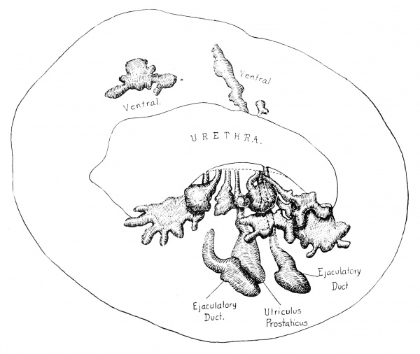
Careful identification of the various groups of tubules mentioned above and tracing their course from section to section reveals the fact that even at this early stage these structures have many branches which communicate with the urethra by means of one rather small duct. In one case there are three extensively branching tubules whose ducts join and empty into the urethra through a common duct (fig. 3).
In this series of sections the tubules are distinctly divided into five different groups as follows:
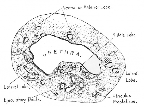
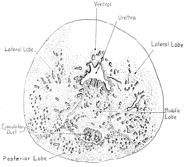
The middle lobe is composed of. ten extensively branching tubules which are separated from the two lateral lobes by a rather thick layer of connective tissue. The branches of these tubules which are situated in the posterior wall of the prostatic urethra between the ejaculatory ducts and the bladder join and communicate with the urethra by means of ten ducts whose mouths are situated upon the floor of the urethra a considerable distance bladderward from the openings of the ejaculatory ducts and are grouped together in a very characteristic manner, being surrounded and bound together by connective tissue and developing smooth muscle fibers (fig. 4). The location of these tubules, their course as graphically shown in fig. 3 by means of a composite drawing which represents the change in location of the various groups of tubules and not the individual branches, and the grouping of the duct openings of these tubules on the floor of the urethra as they communicate with it, demonstrate clearly that this structure is an independent part of the prostate gland.
Fig. 3 Composite drawing showing course of tubules of the middle lobe of prostate; 12.5 cm. human fetus. Four months. X 30.
The two lateral lobes are composed of tubules which are larger in size and branch more extensively than those of the middle lobe. The units composing these lobes grow posteriorly as well as laterally and occupy a region to the outside of the middle lobe and the ejaculatory ducts. In this specimen there are forty-six tubules composing the lateral lobes, the ducts of which communicate with the urethra not only at its sides but also in the depressions on each side of the verum montanum commonly referred to as the prostatic furrows.
In the posterior wall of the urethra outerward from the entrance of the ejaculatory ducts and widely separated from all of the other lobes of the prostate, are found four large branching tubules which form the posterior lobe of the prostate. The branches of these tubules are not at any place in close touch with the lateral lobe tubules which extend this low in the urethral wall and there seems to be a definite layer of connective and muscular tissue forming around the component parts of this lobe.
Extending along the ventral or anterior wall of the prostatic urethra are observed the tubules forming the anterior lobe. Fourteen ducts open into the urethra in this specimen. Most of the tubules are quite small. Only two are as large and extensively branching as those of the other lobes already described.
Below the outermost tubule of the posterior lobe the urethra changes its shape from the inverted semilunar type shown in the drawings to a stellate shape. Extending from the walls on all sides are small finger-like evaginations. Some of these are simple folds in the urethral mucosa but others are developing tubules, some of which have extended quite deeply into the submucous tissue and a few have small branches. These are considered to be the developing urethral glands. While they are quite numerous just below the apex of the prostate, below that they are not found at all.
The seminal vesicles in this series are composed of very tortuous tubes which have a thick muscular layer surrounding them and communicate by means of one small duct or opening into each vasa deferentia under the internal sphincter.
The two ejaculatory ducts bound together as described above become attached to the wall of the urethra below the point just mentioned and gradually become more deeply implanted in the thick posterior urethral wall. The utriculus prostaticus is entirely obliterated above but appears between the ejaculatory ducts in the middle of the prostatic urethra. These three structures bound together by connective and muscular tissue in a very characteristic way approach the urethra on a gradual slant maintaining the globe-like appearance shown in fig. 4, pushing the floor of the urethra up into its lumen, forming the verum montanum and causing the urethra to assume an inverted semilunar appearance. The ejaculatory ducts run parallel to the floor of the urethra for a considerable distance and then ascend vertically to empty on the sides of the verum montanum. The utriculus prostaticus in this series is very irregular in shape, being throughout most of its length in the shape of an inverted Y and giving the appearance of two tubes which have fused at their upper pole and not at the lower. Its opening into the urethra is below those of the ejaculatory ducts and there is a blind end extending a short distance down in the verum montanum under the floor of the urethra.
Fig. 4 12.5 cm. human fetus. Four months. Showing rather definite separation of the middle lobe from the lateral lobes.
Fetus 12 cm long
(sixteen weeks)
A series of sections cut in the longitudinal direction through the bladder and prostatic urethra of a fetus four months of age shows the arrangement of the various structures in a very striking manner. The smooth muscle of the bladder wall stains very deeply and is bound in place by the lighter staining connective tissue. The mucous membrane of the bladder and the submucous tissue is thrown into folds and finger-like projections into the vesical lumen except over the trigonum vesicae where it is tightly adherent to the muscle forming this structure. The trigonum vesicae is made up of muscular fibers which are finer in texture, more tightly bound together, and have lighter staining characteristics than the rather heavy, loose, deeply staining muscle bundles of the bladder wall proper upon which it is superimposed. The trigonal tissue extends down through the vesical neck and becomes lost among the muscle fibers of the prostatic urethra. The sphincter of the bladder appears as a large oval mass of circular fibers which surround the neck of the bladder and are quite distinct from the surrounding structures. There is a sharp line of differentiation around its lower part, while above some fibers from the trigone and vesical wall mingle with the fibers of the sphincter attaching it intimately to them.
The distribution of the tubules of the prostate gland is similar to that just described in the cross sections of the bladder and prostatic urethra of a fetus at this age. On the floor of the urethra just below the vesical sphincter several small evaginations represent the developing glands of Albarran. The middle lobe of the prostate is composed of several large tubules with a number of branches which extend back posterior to the sphincter and anterior to the ejaculatory ducts. The lateral lobes are somewhat larger than the middle lobe, being composed of numerous tubules with many large branches. The posterior lobe tubules are seen extending from the floor of the urethra below or outerward from the entrance of the ejaculatory ducts to a position behind them. On the roof or ventral wall of the urethra are found the tubules which make up the ventral lobe of the prostate. In this specimen they are not nearly so numerous or so large as in the series of cross sections just described.
Fetus 16.5 cm long
(twenty weeks)
The bladder and prostate of a fetus five months of age cut in cross sections show the musculature of the bladder wall and the superimposed trigonum vesicae much more clearly than any of the previous specimens described. A striking thing is the great thickness of the vesical sphincter whose many layers of circular fibers form a, mass about as thick as the bladder wall at its base. At its upper and also outer borders the muscle fibers of the bladder wall intermingle with it, thereby strengthening the vesical neck to a considerable degree. The vesical orifice in this specimen seems to be tightly closed. The muscular layers surrounding the tubules of the prostate gland whose earliest differentiation from the rest of the wall of the prostatic urethra was noted in the sixteen weeks fetus, have now become quite thick and the main mass of fibers are arranged circularly, although there is noted a certain amount of muscular tissue extending longitudinally as described by Walker.[21] The same distribution of the prostatic gland tubules is noted in this series of sections that has already been described in detail in the prostate of a fetus sixteen weeks old.
Fetus 16 cm long
(twenty weeks)
Sections of the bladder and prostate of a fetus five months old cut in a longitudinal direction show particularly well the arrangement of the structures at the vesical neck. The muscle bundles of the bladder wall are well developed and arranged in the characteristic manner already described. The bladder mucosa which elsewhere is thrown into folds is bound tightly to the surface of the trigone which is superimposed upon the vesical wall and is composed of muscle bundles made up of very fine fibers which do not stain so deeply as the muscle fibers elsewhere. The internal sphincter is sharply marked off and does not seem to be proportionally so large as the one observed in the twenty weeks fetus cut in cross sections.
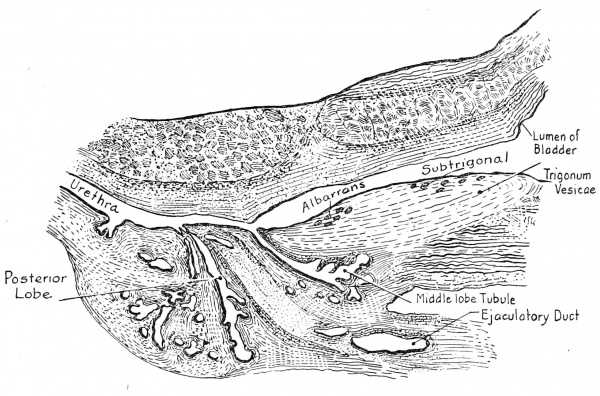
Just under the mucosa of the trigonum vesicae is seen a small collection of simple tubules which are taken to be the subtrigonal glands (fig. 5). They are few in number and are very delicate in structure.
Fig. 5 16 cm. human fetus. Prostate. X 15.
Below the internal sphincter another collection of glands is seen developing from the floor of the urethra. These have not as yet grown deeply into the wall of the prostatic urethra. They are identified as the subcervical glands of Albarran and in this series are very few in number and very slight as regards their architecture.
Extending from the floor of the urethra backward and downward is seen a tubule of the middle lobe of the prostate with numerous branches which extends into the region ascribed to the middle lobe and is posturethral and prespermatic. The ejaculatory duct is observed passing up into the verum montanum. Below its entrance there are several tubules of the posterior lobe with many branches and whose ducts communicate with the floor of the outer portion of the prostatic urethra. The section which shows this arrangement of structures is reproduced diagrammatically in fig. 5. It was cut a little to one side of the middle line as shown by the presence of part of the ejaculatory duct and its muscular wall, and shows quite conclusively that tubules of the middle and posterior lobes arise independently from the urethra and in this instance could not be construed as extending in towards the middle line from the lateral lobes.
Other sections of this series show the tubules developing from the prostatic furrows and sides of the urethra to form the lateral lobes. Others extending from the ventral or anterior wall make up the anterior lobe of the organ. In this specimen these latter are quite numerous and are extensively branched.
Fetus 19 cm long
(twenty-two weeks)
The bladder of this specimen is relaxed and the lumen almost filled with the loose folds of mucosa pulled into it by the contraction of the muscles of the wall, which are very large in this case compared to the specimens previously described. The three muscular layers are clearly made out and rather larger fibers than those heretofore described are seen passing from the wall of the entering ureters out upon the vesical wall, forming the trigonum vesicae. The upper border of this structure is about twice as thick as the bladder wall and more compactly arranged. Lower down the trigone is less thick and at its middle is not as thick as the bladder wall proper. The mucosa covering the trigonum vesicae is tightly bound to it so that in relaxation of the bladder the mucosa of the rest of that structure may be thrown into many large folds but the trigonal mucosa does not so arrange itself.
Extending parallel with the long axis of the lumen of the bladder just outside of the mucosa of the roof or ventral wall of that structure is seen a body which arises by two small branches and ends blindly in the vesical wall. It extends from the upper border to the middle of the trigonum vesicae and at its upper end the branches become indistinguishable from muscle bundles.
Throughout its lower portion after the two branches join it is surrounded by a thick layer of circularly arranged fibers enclosing this other structure, which in some places seems to be a lumen filled with degenerated epithelial cells and at others a space filled with bundles of fibers undergoing degeneration. This structure is about ten times as large as any of the blood vessels seen in the section and does not bear any resemblance to tissue seen in this or other series studied.
There are five small tubules seen developing from the floor of the bladder and extending down into the lower part of the trigone. These are the subtrigonal glands.
The circular fibers composing the sphincter at the vesical neck are quite thick in this case and are intimately connected with the muscle bundles of the bladder proper, while the trigonal fibers although they are very few in number down here are superimposed on the fibers composing the sphincter. This arrangement persists until the upper portion of the prostatic urethra is reached where they become lost in the musculature of the urethral wall.
Just below the vesical sphincter there are found eleven evaginations from the floor of the urethra which are lined with very fine cylindrical epithelium and which in no case branch. These tubules are recognized as the subcervical glands of Albarran and their direction of growth seems to be upward toward the bladder and in no instance do any of them extend deeply into the musculature of the urethra.
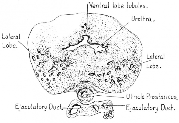
In the region usually occupied by the middle lobe tubules of the developing prostate gland, there is in this series an entire absence of such tubules (fig. • 6) . Very careful study fails to reveal any glandular tissue, except Albanian's tubules described above, developing from the floor of the urethra between the entrance of the ejaculatory ducts and the cervix of the bladder. Extending from each of the lateral lobes is seen a large branch from the tubule nearest the middle line, which, if it should continue to grow and send out additional branches, would ultimately form a bridge of tissue extending from one lateral lobe to the other and binding them intimately together. This is the condition which Pallin believes to exist in all prostate glands.
The two lateral lobes are composed of 42 distinct tubules which have numerous and large branches. These structures all extend laterally, posteriorly, and towards the bladder, their uppermost branches being found under the lower end of the vesical sphincter. The muscular layers around the tubules, the development of which has already been referred to, are very prominent in this specimen, particularly where the tubules are beginning to push out laterally and posteriorly to form the bulging lateral lobes.
Fig. 6 19 cm. human fetus. Five and one-half months.
Below the entrance of the ejaculatory ducts and utriculus prostaticus there are observed ten large branching tubules which extend back behind the structures mentioned above and between and behind the lateral lobe tubules. These tubules compose the posterior lobe of the prostate and make up the main mass of its apex, being distinctly separated from the two lateral lobes by a considerable area of tissue. The posterior lobe does not extend very far up behind the ejaculatory ducts in this specimen on account of the huge size of the utriculus prostaticus which occupies the space into which the tubules of this portion of the gland usuallv extend.
The anterior lobe consists of seven tubules which are very much smaller than those making up the other lobes and very few branches are sent off. There seems to be a shrinking into insignificance of these tubules as far as a comparison with the size of similar structures in the younger fetuses is concerned.
The seminal vesicles are very tortuous in this series. The ejaculatory ducts seem to be of normal size but in their course through the wall of the urethra they are separated by an utriculus prostaticus, the upper solid end of which is seen in fig. 6. This structure is a little larger in size than the ejaculatory ducts in most of the specimens studied, but in this case it is about twenty times as large an an ejaculatory duct and at least three times as large as the lumen of the urethra. It is as usual contained within the same muscular sheath as the ejaculatory ducts as shown in fig. 4 but here the muscular coat is very much thinned out. The utricle extends obliquely through the urethral wall accompanied by the ejaculatory ducts until one part of its circumference is quite close to the floor of the urethra, where a small part about the size of a normal utricle separates from the main part of the organ and runs along under the floor of the urethra for some distance below the openings of the ejaculatory ducts. It then opens in the midline on the crest of the verummontanum. The larger part of the lumen runs forward on the same plane that it occupied above and stops rather sharply in a blind end. There have not been seen either prostatic or ejaculatory ducts opening into the utriculus prostaticus of this or any other specimen studied.
The ejaculatory ducts are observed to run obliquely through the prostate until they approach quite close to the urethra in the crest of the verum montanum; then they run parallel with the axis of the urethra for a considerable distance (300/x in this case) , ultimately opening on a slant into the prostatic urethra on each side of the verum montanum, so that any pressure within the urethra would tend to close them very effectually and in a manner similar to that observed in the ureters in cases of distention of the bladder.
The verum montanum is formed in this case as already described by the ingrowth of the utriculus prostaticus and ejaculatory ducts,
Their development under the floor of the urethra pushes it up into the lumen causing the characteristic semilunar shape of the prostatic urethra. The fact that the lumen of the prostatic urethra below the entrance of the ejaculatory ducts is also crescentic in shape is due to the continuance of the muscular fibers that form the coats surrounding the utricle and ejaculatory ducts under the mucosa of the urethra where they finally become lost in the muscle of that structure.
Fetus 27 cm long
(thirty weeks)
Inspection of the bladder of this specimen cut in cross section corroborates the findings already described, the only difference being that the muscle bundles seem to be larger in this than in any of those previously observed. The trigone is superimposed in the characteristic way and its submucous blood vessels, which in all cases are much more numerous than those of any other portion of the viscus, are greatly increased in number and size in this case. The sphincter of the bladder is thicker and composed of heavier muscle fibers than the ones that have been described. Outside of the sphincter are seen the muscle bundles of the vesical wall which become blended with and are lost in the circular fibers forming that structure.
The subtrigonal glands are present in very small numbers, only four being detected. They extend through the submucous structures and only a very short distance into the musculature of the trigonum vesicae. Their architecture is very slight and there seems to be no particular differentiation of connective tissue or muscle fibers around them. They resemble the urethral glands of Littre more than any other tissue and probably are a continuation upward of those structures.
The subcervical glands are present in this series, being nine in number. They are all quite small and no branches are noted in any of them. In addition to these nine tubules on the floor of the urethra there are six found on its roof or ventral wall. These tubules are similar in extent and architecture to the subcervical group and are apparently not a continuation upward of the ventral lobe of the prostate as they are widely separated from that structure and are entirely different in their character from prostatic tubules. All of these gland tubules extend from the urethra upwards or towards the bladder under the mucosa, and in no case do they extend deeply into the muscular tissue. They push out into the submucous tissue between the numerous blood vessels in this region and are not surrounded by a specially arranged musculature. The lumina of these tubules are about three times the size of the blood vessels found here, but are much smaller than the prostatic gland tubules.
The middle lobe is made up of eleven- tubules, whose blind ends extend backward and upward under the sphincter of the bladder, each of which has a larger number of branches and communicates with the urethra through quite small ducts which open upon its floor some distance above the openings of the ejaculatory ducts and utriculus prostaticus. In the specimen studied previously there has been a wide and distinct separation of the middle lobe from the two lateral lobes. This distinctiveness is well shown in the fetus 16 weeks old (fig. 4). In this specimen it is easy to distinguish the middle lobe from its location, the direction in which the tubules extend, and the fact that its ducts communicate with the urethra at the same point that ducts of middle lobe tubules have in younger series. On the other hand, the layer of tissue separating the middle from the lateral lobes is not very extensive except at the base. There is no semblance of a special capsule and in some places the tubules of the middle lobe are side by side with the tubules of the lateral lobe, but in no place is there found an intermingling of the branches of one lobe with those of another. At the base of the prostate, up under the vesical sphincter where the prostatic tubules nave grown farthest away from their point of origin, the separation between the middle lobe and the two lateral lobes is quite wide and there is a great deal of connective and muscular tissue between them. In no case has it been possible to find any difference between the architecture of the tubules of the different lobes with the exception that those of the middle lobe are usually not quite so large as the lateral lobe tubules. Finer histological studies of the glandular epithelium have not always been possible, as some of the tissue is quite old and contraction of the epithelium has taken place.
The two lateral lobes are made up of thirty-six tubules which extend posteriorly and up under the internal sphincter as far as do those of the middle lobe. At the sides of the lower part of the sphincter there is a distinct bulging due to the further development of the lateral lobe tubules, which with the middle lobe form the base of the gland. This posterior and lateral bulging is noted throughout the entire prostatic region, although the larger number of tubules are massed in the base of the gland. There are a number of tubules whose ducts open into the sides of the urethra and which send branches up towards the ventral wall. These structures have many branches but they are much smaller than either the rest of the lateral or middle lobe tubules and their musculature is very slight compared to that of the other prostatic tubules. In the apex of the gland the tubules of the lateral lobe have a few branches which run downward or outward. Everywhere else the general direction of growth is towards the bladder.
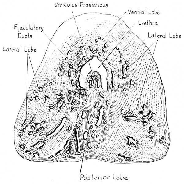
The glandular tissue making up the posterior lobe is found at the point where the ejaculatory ducts extend vertically towards the urethra. It is between and behind the lateral lobes and this part of the gland is distinctly separated from the lateral lobes by a thick layer of connective tissue (fig. 7). This lobe in the series under consideration is made up of nine branching tubules of large size whose ducts communicate with the urethra on its floor or posterior wall outerward from the openings of the ejaculatory ducts and at no point do any of the branches of these tubules intermingle with the tubules of the lateral lobes, being in all places separated from them by a definite layer of connective tissue. In the apex of the gland some of these tubules send branches forward or outward but elsewhere they extend in a bladderward direction.
The comparative decrease in size of the tubules of the anterior lobe noted in the prostate of the twenty-two weeks old fetus is also striking in this series. The number of tubules making up the anterior lobe noted in the fetuses younger than twenty-two weeks was about twice as great as that noted in the specimens twenty two weeks and older. In this prostate there are eight small branching tubules communicating with the anterior urethral wall. They are surrounded by muscular layers similar to the other prostatic tubules and are quite limited in their extent and widely separated from the lateral lobes.
Below the apex of the prostate there are noted a large number of glands, most of which are simple tubules, extending into the submucous tissue their ducts opening into the urethra. They are found on all sides of its lumen but most of them open on the ventral wall. They extend barely to the muscular bundles and none of them are found within the musculature.
Fig. 7 27 cm. human fetus. Seven and one-half months. X 14.
The seminal vesicles have grown back behind the base of the bladder to a point opposite the middle of the trigonum vesicae. They are made up of thick walled tortuous connected tubes which communicate with the vasa deferentia behind the lower part of the sphincter of the bladder rather deeply in the base of the prostate gland.
The ejaculatory ducts surrounded by their thick muscular wall and imbedded in the prostate traverse its posterior portion for a considerable distance in a plane horizontal with the floor of the urethra. A little below the point where the lowest of the middle lobe tubules opens out upon the floor of the urethra the ejaculatory ducts turn sharply and mount almost perpendicularly towards the urethra and with their thick coats and the utriculus prostaticus form the verum montanum. By referring to fig. 4 it is seen that the relative sizes of the urethra, the verum montanum and its contained structures are much smaller in the older specimen when compared with the size of the prostate gland. The ejaculatory ducts, which are greatly narrowed here, run for a considerable distance in the verum montanum parallel with the axis of the urethra and finally open on the sides of the verum montanum. The course of the ducts in this compressible structure and their lateral openings which are very small in size, are the features which make total occlusion of these important structures possible in case of distention of the prostatic urethra from any cause.
The utriculus prostaticus is about the size of an ejaculatory duct in this fetus. Its uppermost end begins at the point where the ejaculatory ducts assume their position in the verum montanum and extends outward in the apex of that structure opening in the middle line near the apex of the prostatic urethra. Outerward from the mouths of the ejaculatory ducts the utricle is surrounded by a thick muscular wall which includes a continuation of the fibers which surrounded the ejaculatory ducts. Below the mouth of the utricle the muscular tissue continues for a short distance only.
New-born infant 35 cm
The bladder and prostate of an infant who had died a few days after birth were cut in cross section and mounted in the usual way, being stained with haematoxylin and eosin. A very thorough study has been made of the prostate of this baby, which has in eluded drawings, diagrams, and the construction of a wax model, pictures of which are reproduced in this article.
The bladder was distended upon fixation and the natural curve at its neck mechanically straightened, so it is possible to get a good idea of the thickness of its walls at various points. At the place where the ureters join the vesical wall and begin their oblique course through to its lumen, there is an increase in the size of the base to twice that observed elsewhere in its circumference. This increased thickness of the base is maintained throughout the trigonal region, being most pronounced between the ureters where the interureteric ridge is made up of fibers extending from one ureteral wall mingling with those of the other. The trigone in this as in the previously described cases is made up of muscular fibers which originate in the walls of the ureters and pass over the musculature proper of the bladder, becoming lost among the fibers of the urethra. Just below the interureteric ridge the wall of the base of the bladder becomes comparatively smaller, being very little larger than the rest of the wall. As the sphincter is approached the entire vesical wall increases greatly in size, the base always being considerably larger than any other part and the trigonal fibers are always distinguishable from the tissue of the vesical wall proper. The mucosa over the trigone is much more richly supplied with blood vessels than any other portion.
The internal sphincter is made up of circular fibers which are very numerous at its upper part. It is larger at the floor of the vesical cervix, and while many of the fibers pass entirely around the orifice of the urethra the majority of them mingle with the muscle bundles of the bladder on its ventral aspect which approach much closer to the lumen of the orifice of the urethra here than in the posterior wall. At the outermost portion of the sphincter there is a great decrease in the number of fibers composing it and it is about equal in size at all points of its circumference. In its ventral portion a number of small longitudinally disposed fibers are seen. The lower part of the sphincter ends considerably outerward from the point where posterior lobe tubules have extended backward behind the two ejaculatory ducts.
In the mucosa at about the middle of the trigonum vesicae are found nine very delicately constructed tubules which are recognized as the subtrigonal glands. Most of them are simple tubules that extend down to the muscle but a few of them have one or two very small branches and these extend for a short distance into the musculature of the bladder wall. The blind ends of these tubules are a little closer to the base of the trigone than the mouths of their tubules.
Commencing at about the middle of the vesical sphincter and extending down to its lower border there are found on the floor of the orifice the tubules forming the glands of Albarran. There are nineteen of these structures in this series, most of which are simple tubular glands lined with very small columnar epithelium and do not extend very far beneath the mucosa. In a few cases there are several branches springing from a tubule and these in many instances extend a short distance into the musculature of the sphincter. None of these tubules are found in the ventral mucosa of the orifice but there are some very small evaginations in that region which may later develop into tubules of the same sort. The blood vessels in the mucosa of this region are quite numerous and large. The tubules of Albarran's group are not surrounded by any differentiated tissue as are the tubules of the prostate but seem to be merely imbedded in the submucous structures. They open for the most part near the middle line on the floor of the urethra but a few open in the angular depressions at the sides of the urethra which marks the beginning of the prostatic furrows.
The middle lobe tubules have extended up behind the sphincter to its uppermost border. The end of the tubule that has extended the highest is situated in the middle line just above the ampulla of the vas and its branches are surrounded by rather dense muscular tissue. The whole mass lies imbedded in the loose connective tissue beneath the bladder musculature. Lower down in the series these branches are reinforced by others of a like nature and a very short distance below become connected with the musculature surrounding the urethra. The tissues which envelop the middle lobe tubules are thicker in this specimen than in any of the younger ones observed being in places as dense as the walls of the ejaculatory ducts. A few of the most lateral ward of the middle lobe tubules have branches which extend to the sides for short distances but in no place is there noted an intermingling with lateral lobe tubules. There are no branches of the lateral lobes extending into the middle lobe region and the ducts of the nine large branching tubules which form the latter structure pour their secretions through mouths which empty upon the urethral floor bladderward from the openings of the ejaculatory ducts. An interesting thing noted in all of the prostates studied but more particularly in this one is the fact that an enormous number of branches join together to form one tubule which empties into the urethra through a duct which is no larger in diameter than one of its smallest branches. The ducts of the middle lobe tubules just before their entrance into the urethra are quite widely separated from other parts of the prostate, thereby retaining their embryological characteristic of an independent origin.
The uppermost ends of the left lateral lobe tubules have extended back under the bladder and are found contained in their thick muscular envelope adherent to the sides of the seminal vesicles. The right lateral lobe tubules are found to have extended back only to a point above the opening of the seminal vesicle into the ejaculatory duct. Lower down where the prostate is broadest the branches of the lateral lobe tubules are exceedingly numerous. By their great development they cause the base of the prostate to bulge laterally and posteriorly. The two lobes are made up of thirty-four tubules and are separated posteriorly by the ejaculatory ducts, the middle lobe and the posterior lobe. Anteriorly in this series of sections the branches of the lateral lobes approach each other very closely, especially in lower part of the gland at its apex. The outermost or caudalward portion of the lateral lobes, represented by two large gland tubules, send branches in a caudalward direction and this fact is of interest surgically because in nearly every successful enucleation these forward branches must be cut with curved scissors in order to free the lobes from the capsule as they seem to be particularly adherent. The posterior borders of these lobes are separated from the posterior lobe by a rather dense layer of connective tissue which also separates it from the middle lobe and ejaculatory ducts (fig. 8). An increase in the amount of connective tissue at this point would be of great interest surgically in cases of perineal prostatectomy, because the incision must be made entirely through it, otherwise the enucleation procedure would lead the instrument to the capsule instead of to the inside of that structure and hence could not be completed.
Fig. 8 36 cm. (new-born) baby prostate X 6, camera lucida.
The posterior lobe is made up of eleven glands, some of which have extended toward the bladder until they are almost as far back as the ends of the middle lobe tubules. Their course follows rather closely the dorsal aspect of the ejaculatory ducts until the latter structures ascend almost vertically towards the urethra, immediately caudalward to which there is a small area free from glandular elements. The ducts of posterior lobe tubules enter the floor of the urethra caudal wards from the entrance of the ejaculatory ducts. The posterior lobe is the part of the prostate that is palpated per rectum and presents in the middle of its posterior surface a slight depression which is ordinarily termed the median furrow. This depression in the specimen under discussion is more pronounced in the region of the apex and gradually becomes shallow at the middle of the gland and at the base assumes a rounded contour. At the apex two of the posterior lobe tubules send branches forward which appear outside of the muscular walls of the urethra, which at this stage are quite well developed.
The ventral lobe is composed of two very small tubules with just a few branches whose ducts open upon the ventral wall of the prostatic urethra at about its middle part. This lobe in the newborn has atrophied to. almost complete insignificance (fig. 10).
Just at the apex of the prostate and a little below that point there are noticed numerous gland tubules contained in the submucous tissue of the urethra which are easily differentiated from prostatic tubules, being much smaller in size and lacking the muscular coats of the latter. In most instances these tubules have several branches, but none of them are at all extensive as are the tubules of the prostate. The entire structures are contained within the muscular walls of the urethra and their ducts open into it on all sides. These tubules are considered to be the glands of Littre, and while very numerous just below the apex of the prostate are very few in number lower down in the urethra.
In the new-born the seminal vesicles have extended back under the bladder almost to the base of the trigone. Its uppermost portion consists of five lumina on each side with walls almost as thick as those of the vasa deferentia. These lumina all communicate lower down and connect with the ejaculatory ducts just below the point where they become imbedded in the musculature of the prostate gland. The ampullae of the vasa deferentia are easily distinguishable in this specimen and are marked by a considerable widening and great increase in the size of the lumen.
As the ejaculatory ducts become more deeply imbedded in the prostate they become situated in more immediate contact with one another until their musculature becomes intermingled as shown in fig. 8, and while each contains its own walls intact they are both contained within the same bundle. In their progress through the prostate they take the course already described in previous specimens and which is shown in fig. 10. In this as in the other specimens studied the verum montanum is observed to be composed of the ejaculatory ducts, utriculus prostaticus and their walls. The ejaculatory ducts run for a shorter distance in the verum montanum in this than in the prostates previously discussed, finally opening on the lateral walls of that structure in the characteristic way already described.
The utriculus prostaticus is found in the tip of the verum montanum extending only a short distance before it opens into the urethra in the midline, and in this case has a very large wide opening above the mouths of the ejaculatory ducts which is not the usual arrangement, as in all of the other specimens studied its mouth has been below those of the ejaculatory ducts. There have not been found in this or in any of the other prostates studied either an ejaculatory duct or a prostatic tubule opening into the utriculus prostaticus.
Discussion
1. The bladder of a fetus ten weeks old consists of a cylindrical tube composed of embryonic connective tissue. Near its base it is joined by the two ureters which pass through its wall and fibers from which are superimposed upon the bladder wall to form the trigonum vesicae. There is no tissue resembling muscle present and the future site of the vesical sphincter is not distinguishable except by the change in the size and shape of the lumen.
At the thirteenth week the muscular development has begun and is observed as circular interlacing and longitudinal strands of tissue that take on a deeper pink color than does the connective tissue which still forms the major portion of the bladder wall. The entire base of the bladder is thicker than any other portion of its circumference while the superimposed trigonum vesicae and the wall under it are five times as thick as the anterior wall. The muscle fibers observed in the trigonum are traced between the two ureters and down on the rest of the trigone. They are finer in texture than the muscle fibers in the bladder wall. The site of the internal sphincter is marked by a sudden narrowing in the size of the lumen and a few circularly arranged muscular fibers are made out.
By the sixteenth week there has been a very great development of the musculature of the bladder, so that the fibers are sharply defined from connective tissue. The muscle fibers forming the trigonum vesicae are easily traced out into that structure from the ureteral walls and the mucosa which is arranged in folds everywhere else in this specimen is attached tightly to the trigone and is smooth. The sphincter vesicae has become quite sharply outlined by many muscular fibers arranged circularly at the constricted lumen of the cervix.
At the twentieth week the muscular fibers have become very much more prominent but the most striking change is the enormous increase in the size of and the number of fibers forming the internal sphincter which seems to be tightly closing the vesical orifice. The longitudinal fibers of the bladder intermingle with the outermost of the fibers of the sphincter. The mucosa in this stage is smooth over the trigonum vesicae and folded elsewhere.
In the twenty-two weeks old fetus there is noted still greater increase in the size of the bladder musculature and a marked increase in the size of the interureteric bar. The trigone and the bladder wall under it do not show the great increase in size over the rest of the vesical wall observed in younger fetuses. The fibers forming the trigone are observed to be more tightly bound together than the fibers of the bladder wall and the mucosa is smooth over it. The sphincter is large and closely bound to the rest of bladder musculature.
Further increase in the size of muscles forming the vesical wall, trigonum and sphincter is noted at the thirtieth week and in the bladder of the new-born. The sphincter in the latter is larger at its upper margin than elsewhere and is very thick on its posterior quadrant. Many of the fibers appearing there do not entirely encircle the orifice but intermingle with longitudinally arranged fibers from the anterior surface of the bladder. At the lower or outermost part of the sphincter there are fewer fibers but they all entirely encircle the orifice.
2. There is found in some of the sections studied a small group of glands which open upon the mucosa at the middle and lower part of the trigonum vesicae and which barely extend into the underlying musculature. These glands are in nearly every case simple tubules of very delicate structure, no muscular or specialized tissue layers being found surrounding them. They are not found until the twentieth week but are observed in every fetus older than that and also in the new-born. These tubules form the subtrigonal group of glands and are in every case few in number, nine being the most found and four the fewest. They are apparently insignificant but occupy a strategic position as a very slight hypertrophy at this point might cause a considerable obstruction to urinary outflow. Cases have been observed by Dr. H. H. Young and Dr. J. T. Geraghty in which this group of tubules had become enlarged and a further growth had caused them to become almost free in the bladder lumen being connected to the original site by a small pedicle. Upon attempted urination this globular mass would- fall into the orifice of the urethra and blocking it would cause more obstruction than an enormous hypertrophied prostate. The structures are probably a continuation upward of urethral glands.
3. On the floor of the prostatic urethra of the fetus sixteen weeks of age at its commencement there are found eight small evaginations which are observed to extend only a short distance into the submucosa. These tubules are easily distinguishable from prostatic gland tubules being of very slight architectuie and lacking the muscular layers which surround the prostatic tubules. This group of tubules first described by Albarran is found in all of the specimens studied here older than sixteen weeks. In fetal life they have no branches at all but in the new-born a few very small branches were made out. In all cases these tubules grow back toward the bladder in the submucosa and never extend deeply into the musculature. In the new-born they have grown back within the sphincter. These tubules are found in one instance growing from the roof of the prostatic urethra. They are few in number, varying from eight to nineteen and are very similar in structure to the subtrigonal glands just described, except they are somewhat larger. The position of the sub-cervical glands of Albarran is even more strategic than that of the subtrigonal group. Growing back directly within the sphincter it is easy to see that a slight increase in size would form a very considerable obstruction to the passage of urine from the bladder.
4. The prostate gland begins to develop at the third month of fetal life. The tubules which compose it make their first appearance as solid epithelial outgrowths from five distinct parts of the prostatic urethra. These solid masses of deeply staining cells very soon become circularly arranged around lumen and branches are found very early. The five foci from which groups of prostatic tubules take their origin are located as follows: on the floor of the urethra between the neck of the bladder and the openings of the ejaculatory ducts and utriculus prostaticus, one in each prostatic furrow and on the sides of the urethra, on the floor of the urethra below the openings of the ejaculatory ducts and the utricle, and on the ventral or anterior wall of the prostatic urethra. The tubules originating from these five foci by their further growth and the development of stroma around them become the middle, right and left lateral, posterior, and anterior lobes respectively. In early fetal life they are widely separated from one another but in later stages the separation between the middle and two lateral lobes is not very great. There is not an intermingling of tubules in any of the specimens studied but in many places in the new-born the tubules of the middle lobe are observed side by side with those of the lateral lobes, there being no definite capsule separating them. The separation of the posterior lobe from the others is complete, as there is a rather dense layer of fibrous tissue between it and the lateral lobes. The anterior lobe is widely separated from the two lateral lobes.
The first appearance of muscular fibers developing around the tubules of the prostate is found at the sixteenth week at which time a slight differentiation in staining properties is noticed. At the twenty-second week the muscular layers are quite well developed and are particularly noted where some of the lateral lobe tubules have extended away from the base of the prostate. The musculature continues to become thicker and thicker until at birth some of the tubules are surrounded by very dense muscular layers.
In every case but one in this study the middle lobe of the prostate develops independently from tubules which originate from the floor of the urethra near the middle line between the bladder and the entrance of the ejaculatory ducts. The tubules composing this lobe are separated from the lateral lobes by a considerable area of tissue free from glandular elements, the younger the embryo the greater the separation. In later development while the tubules and their branches maintain their characteristic position, there does not seem to be any definite formation of a fibrous capsule separating the middle from the lateral lobes. The ducts of the largest of the middle lobe tubules which were originally near the middle line are pushed laterally by the further development of the structures in the verum montanum so that they open rather high up on its sides and not in the middle line, as that part is occupied by the utriculus prostaticus and the ejaculatory ducts. Rarely (once in our experience) there may be an absence of independent middle lobe tubules, in which case branches from the lateral lobes are seen approaching the middle line in the regionordinarily occupied by the middle lobe, and if the growth continues this region will be occupied by a glandular commissure such as Jores declares all middle lobes to be. In most cases the middle lobe is made up of nine or ten large tubules, the number in five cases varying from seven to twelve, the average being ten.
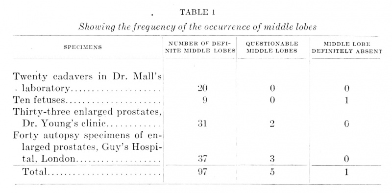
As one of our embryonic series showed an absence of tubules in the middle lobe region it was deemed advisable to determine the percentage of prostates that have a middle lobe.
Dissecting room and autopsy subjects to the number of twenty were examined and all demonstrated definite prostatic tissue in the middle lobe region. Ten sets of serial sections of embryonic tissue demonstrated only one case without independent middle lobe tubules. Thirty-three autopsy cases of enlarged prostate in the Johns Hopkins Hospital Genito-Urinary Museum were examined, with the result that in thirty-one, definite middle lobes were identified. The existence of middle lobes in the other two cases could not be determined, as the specimens were not sectioned. Two of the specimens examined had enlarged anterior lobes. Forty autopsy specimens of prostate gland enlargement were examined in the pathological museum at Guy's Hospital, London, thirty-seven of them showed the existence of glandular substance in the middle lobe region, while in three cases there was no macroscopic evidence of such a structure.
Table 1 Showing the frequency of the occurrence of middle lobes
Reference to table 1 shows that there is a definite absence of glandular tissue in the middle lobe region in one specimen out of one hundred and three studied, and five specimens in which it was not determined whether there was glandular tissue there or not.
The lateral lobes are composed of tubules which are greater in size and number than those of any of the other lobes. They originate from the right and left prostatic furrows and the lower parts of the lateral walls and extend backward and outward forming the main part of the base of the prostate. In the younger specimens these lobes are widely separated from each other and the remaining lobes but the older the fetus studied the less is the separation between the branches of the lateral and middle lobes, although there is in all cases a definite separation observed where the ducts communicate with the urethra. In the region of the apex some of the lateral lobe tubules send branches forward, at all other parts of the gland the direction of the growth of tubules is back towards the bladder.
In one specimen there was observed a growth of branches from lateral lobe tubules into the region usually occupied by the middle lobe, the latter structure in this case being absent.
In the prostate of the new-born near the apex the lateral lobes have two extensively branching tubules which extend up into the anterior commissure and practically form a glandular commissure in this region.
The lateral lobes are in most cases very well separated from each other anteriorly a considerable area of stroma being present, in which are found the anterior lobe tubules. Mesially the ejaculatory ducts, urethra and middle lobe are interposed and there is a definite plane of connective tissue which sharply separates their posterior branches from the posterior lobe.
The number of tubules forming the two lateral lobes vary from twenty-seven to forty-six, the average number being thirty-seven.
The posterior lobe is made up of tubules which begin to develop with the other prostatic tubules at the third month. They are found on the floor of the urethra below the openings of the ejaculatory ducts and their direction of growth is behind those structures back towards the bladder. They compose the main mass of the apex of the gland and the posterior lobe is that part of the gland which is palpated per rectum. Just anterior to this lobe is found in the older stages a stroma of connective tissue free from tubules which separates it from the posterior parts of the lateral lobes and from the ejaculatory ducts. The tubules of this part of the prostate send a few branches forward in the region of the apex as do the most anteriorly arranged branches of the lateral lobes. They are quite large with numerous branches but are not very numerous, the smallest -number being four, the greatest eleven, and the average in six specimens is eight.
This lobe is present in all fetuses here studied and in the newborn. It is an independent structure developing from tubules which are separated from those composing the other lobes and being divided from them by a definite capsule which is laterally connected with the capsule of the gland.
The posterior lobe is of considerable importance for several reasons. In doing Young's operation for perineal prostatectomy operators have found it absolutely necessary to make their two parallel incisions quite deep so that they go completely through the connective tissue layer separating the posterior from the two lateral lobes. In case the incision is only made through the capsule of the gland into the posterior lobe, an attempted enucleation leads the operator's instrument or finger laterally into the outer capsule again where the anterior capsule of the posterior lobe becomes lost in it and enucleation of the real offenders in hypertrophy, lateral and middle lobes, is not possible until the incision is made into the capsule containing them.
Recent studies by Dr. John T. Geraghty and Dr. Montague L. Boyd on the pathology of the prostate gland have confirmed the facts (1) that hypertrophy rarely or never occurs in this lobe, (2) that primary carcinoma of the prostate usually arises in it. This knowledge coupled with the embryological fact that the tubules forming this structure arise independently from a localized area in the urethra and remain independent throughout, increasing enormously in size in the normal adult prostate, suggests the possibility that it may have a different function from the other parts of the gland.
The tubules forming the anterior or ventral lobe begin to develop at the same time as do those of the other lobes. They are large and have numerous branches at first, but in the sixteenth week they are slightly smaller than the tubules of the other lobes. At the twenty-second week these tubules have decreased in size and number and very few branches are noted. There seems to have been a shrinking into insignificance of the anterior lobe after the sixteenth week, but the tubules persist until birth, at which time there are found two very small tubules. We have evidence of the fact that the anterior lobe may persist throughout life in that among the ninety-three specimens of the adult prostate examined two were found with hyper trophied anterior lobes. The average number of anterior lobe tubules in the first half of fetal life is thirteen while that of the last half is six. Two tubules in the new-born were the fewest found, fourteen in the sixteen weeks old fetus being the greatest number.
In most descriptions of the prostatic urethra it is stated that there are from twenty to thirty duct openings upon its floor. My studies have convinced me that this number is far too low. The results of microscopic studies of the number of tubules composing the prostate have been arranged in the form of a table.
Table 2 Showing number of tubules of each lobe opening into prostatic urethra
| Table showing number of tubules of each Prostate Lobe opening into prostatic urethra, the number of Albarran' s tubules, and the number of subtrigonal tubules. | |||||||
| Size of Fetus Crown-Rump |
Middle Lobe |
Lateral Lobes |
Posterior Lobe |
Anterior Lobe |
Total No. of Prostatic Tubules |
Subcervical Glands of Albarran |
Subtrigonal Glands |
|---|---|---|---|---|---|---|---|
| 7.5 | 12 | 39 | 11 | 12 | 74 | 0 | 0 |
| 8 | 7 | 27 | 6 | 13 | 53 | 0 | 0 |
| 12.5 | 10 | 46 | 4 | 14 | 74 | 8 | 0 |
| 19 | 0 | 42 | 10 | 7 | 59 | 11 | 5 |
| 27 | 11 | 36 | 9 | 8 | 64 | 9 | 4 |
| 36 | 9 | 34 | 11 | 2 | 56 | 19 | 9 |
| Averages | 10 | 37 | 8 | 9 | 63 | 12 | 6 |
| The averages are taken from the specimens in which the structure is present in case of middle lobe and the groups of Albarran and the subtrigonal group. | |||||||
By referring to table 2 it is seen that in no case were there fewer than fifty-three prostatic ducts opening into the urethra, and in two specimens there were as many as seventy-four, the average for six specimens studied microscopically in series being sixtythree, including one specimen in which the middle lobe was entirely lacking. The number of middle lobe tubules vary from seven to twelve, the average in five prostates being ten. The lateral lobe tubules vary in number from twenty-seven to fortysix, the average in six specimens being thirty-seven. The posterior lobes show a variation of from four to eleven, eight being the average number of tubules in the six specimens recorded. The anterior lobes present a very interesting variation. It is seen that up until and including the sixteenth week the tubules composing the anterior lobe are quite large and numerous, but after that time there is a decided decrease in the number, and, as has already been stated, the size and branches of the individual tubules. The greatest change in this structure is noted in the new-born in which the anterior lobe is made up of two insignificant tubules situated on the anterior part of the gland at its middle as shown in fig. 10.
The subcervical glands of Albarran are seen to occur in the specimen sixteen weeks of age and in all of the older ones. Their number varies from eight to nineteen, the average in the four series being twelve.
The subtrigonal glands are not found until the twenty-second week and they are in all cases few in number, varying from four to nine, the average in three specimens being six.
5. The vasa' deferentia descend behind the posterior surface of the bladder, each one being situated near the ureter. Lower down they gradually approach one another and in the earlier stages become contiguous behind the middle of the trigonum vesicae. In their descent they increase enormously in size, so that under the internal sphincter of the bladder they with their enveloping tissue are larger than the commencement of the urethra with its surrounding tissue. At this point the lumen of the vasa deferentia have widened considerably denoting the first appearance of the ampullae. These structures remain comparatively large until the sixteenth week after which they become relatively very much smaller.
The seminal vesicles originate in the thirteenth week. They appear first as an evagination lateralward from each vasa deferens being covered by the same tissue that envelops the latter structures. They grow backwards and laterally consisting of a main part which is convoluted and from which rather numerous, short, convoluted branches grow out as described by Pallin.[22] In the further development of these structures the opening into the ejaculatory ducts becomes comparatively smaller and its component parts become larger and more tortuous. In the thirty weeks old fetus they are found back under the trigonum vesicae at about its middle point and communicate with the vasa deferentia deeply in the base of the prostate to form the ejaculatory ducts. At birth the ends of the seminal vesicles have extended back almost as far as the base of the trigonum vesicae and four branches from the main lumen of the organ are made out. Its walls are made up of fibrous and muscular strands and are almost as thick as those of the ejaculatory ducts.
The ejaculatory ducts are the continuations of the vasa deferentia below the entrance of the seminal vesicles. They are like the vasa deferentia very large in size in the younger fetuses but gradually become comparatively smaller in size as the other structures grow larger. They pass obliquely through the posterior wall of the prostatic urethra accompanied by the utricle and the surrounding stroma layers the outer fibers of which attach this cylindrical mass intimately to the urethral wall by intermingling with the tissue forming it. As these structures approach the lumen of the urethra its floor is pushed up into a mound and its lumen converted from a triangular into a semilunar shape. This mound, called the verum montanum, is made up entirely of the ejaculatory ducts, utricle and surrounding envelopes, and gradually disappears below the openings of these several structures into the urethra, the stroma cells becoming intermingled with those forming the urethral wall.
The ejaculatory ducts pass through the posterior wall of the urethra on a gradual slant in the younger fetuses but in older ones their rise through the prostate is a very sudden one until they are within a short distance from the lumen of the urethra, then they run along parallel to its axis for a considerable distance so that their course in the prostate is very much like that shown in fig. 10 in all of the older specimens. They open on the sides of the verum montanum in such a way that there is a small area of tissue over them which, if the slightest amount of pressure were exerted upon it, would close them most effectively. In fact their whole course in the verum montanum parallel with the axis of the urethra near its lumen makes for a closure of the duct in case of distention of the urethra.
6. The urticulus prostaticus in the thirteen weeks old fetus is observed as a very small lumen between the vasa deferentia. It descends between them, and just below the beginning of the ejaculatory ducts becomes much larger than either of these structures. It passes through the wall of the prostatic urethra in company with them and opens in the midline just below the openings of the ejaculatory ducts. In the sixteen weeks old fetus the utricle seems to be composed of two partially fused tubes. Its upper end is obliterated and nothing is seen of it until the point is reached where the ejaculatory ducts and their envelopes are entirely within the posterior wall of the prostatic urethra. It opens below the mouths of the ejaculatory ducts. The utricle begins between the ejaculatory ducts before they enter the prostate in the twenty-two weeks old fetus as shown in fig. 6. It is very much larger than those observed in other specimens and shows other peculiarities already described. In all of the specimens older than twenty-two weeks the utricle appears only in the tip of the verum montanum. It opens in the middle line and in nearly every case below the openings of the ejaculatory ducts. In none of the specimens studied has there been found a single case in which an ejaculatory duct for a prostatic tubule opened into the utriculus prostaticus. There is no evidence that its mouth is protected in any way from invasion of substances or organisms in the posterior urethra.
7. Below the apex of the prostate there is noted in some cases a large number of glands of Littre. They are quite large and some of them have branches. They do not extend into the muscular layers of the urethra. They become very few in number lower down in the urethra.
Conclusions
- The mucosa is always free from folds over the fetal trigonum vesicae. The musculature of the bladder wall, trigonum, and sphincter begins to develop at the thirteenth week and by the sixteenth week is very pronounced.
- The subtrigonal glands begin to develop at the twentieth week. They are found at all ages after that, are few in number and insignificant in appearance, their only importance being that they occupy a strategic position where a small pedunculated enlargement might cause great obstruction to urinary outflow.
- The subcervical glands of Albarran are constant after the age of sixteen weeks. They are similar in structure but are more numerous and larger than the sub trigonal tubules. Their importance also lies in their position. They originate from the floor of the urethra within and below the internal sphincter and grow back under the mucosa so that they lie directly within it. Hence a slight increase in their size would cause very grave obstruction to urinary outflow.
- The prostate gland originates from five independent groups of tubules which begin to develop at about the twelfth week as follows :
- The middle lobe is made up of nine or ten large branching tubules originating on the floor of the urethra between the bladder and the openings of the ejaculatory ducts. There may be an absence of the middle lobe in which case there may be an ingrowth of tubules from the lateral lobes to form a commissure beneath the urethra. Embryologically the middle lobe is an independent structure. Practically it makes no difference because it is not separated by a capsule from the lateral lobes. The middle lobe is rarely absent, being found definitely present in ninety-seven specimens examined, possibly lacking in five others and definitely absent in one.
- The right and left lateral lobe tubules originate in the prosstatic furrows and from the lateral walls of the urethra. They are composed of from twenty-seven to forty-six tubules which grow back to form the main part of the base of the prostate. They are well separated from each other by the anterior lobe and commissure, the urethra, the middle lobe and the ejaculatory ducts. Posteriorly they are separated from the posterior lobe by a fibrous capsule.
- The posterior lobe is an independent structure being made up of tubules which originate from the floor of the prostatic urethra below the openings of the ejaculatory ducts. They grow back behind the latter structures and are in no sense a glandular commissure as they are definitely separated from the other parts of the gland. The posterior lobe is the part of the prostate palpated per rectum and is an important consideration in the performance of perineal prostatectomy. Hypertrophy rarely or never occurs in it and primary carcinoma of the prostate rarely or never begins anywhere else (Boyd and Geraghty).
- The anterior lobe is fairly large until the sixteenth week after which time it becomes greatly decreased in size and in the number of its tubules. It was found in all of the microscopic specimens studied but shrinks into insignificance at the twentysecond week. There is evidence in the occasional finding of enlarged anterior lobes at autopsy that this structure may persist throughout life.[23] The number of openings of prostatic tubules into the urethra is usually said to be between twenty and thirty. My studies have convinced me that this number is too low as in the specimens here recorded the number of tubules opening into the urethra varies from fifty-three to seventy-four, the average being sixtythree.
- The vasa deferentia in early fetal life are comparatively speaking very large, being greater in size than the urethra at the thirteenth week. Their lumina broaden out behind the vesical sphincter at this time showing the earliest appearance of the ampullae. They become relatively decreased in size after the sixteenth week.
- The seminal vesicles begin to develop as lateral evaginations from the vasa deferentia at the thirteenth week. They grow backward and laterally becoming more or less tortuous and send off as many as four short tortuous branches. In the later stages they communicate through a very narrow duct with the vasa deferentia just within the base of the prostate gland.
- In the younger fetuses the ejaculatory ducts pass obliquely through the posterior wall of the prostatic urethra forming with their envelopes the verum montanum. In the older specimens their course through the prostate is not so regular. At the base they progress on a gradual slant until the middle of the gland is reached, where they rise quite sharply until they lie in the top of the verum montanum, after which their course lies parallel to the axis of the urethra for some distance. They open into the urethra on the sides of the verum montanum, their mouths being composed of a collapsible fold of tissue so that pressure within the prostatic urethra very effectively closes them.
- The fused Mullerian ducts may persist intact until the thirteenth week, after which time the lower end (utriculus prostaticus) which has become quite large in size and surrounded by a rather dense layer of stroma cells, contracts until after the twenty-second week when it is found only in the tip of the verum montanum and is relatively very small in size. It usually opens in the midline just below the ejaculatory duct openings and rarely if ever is there an ejaculatory duct or a prostatic tubule opening into it.
Description of a wax model of prostate of a new-born infant
The sections from which this model was constructed were cut 30 micromillimeters in thickness. Every fifth section was magnified twenty times and drawn by means of the projection apparatus in use at the Anatomical Laboratory of the Johns Hopkins University. These drawings were traced upon wax plates 3 mm. in thickness, the tubules and their branches first being identified microscopically with great accuracy. The wax plates were then cut in such a way that the bladder lumen and those of the prostatic tubules were left with bridges of wax between to preserve the exact contour. The wax plates were then piled, the axis of the bladder and urethral lumen and the lumen of the ejaculatory ducts being used as points upon which to build. The prostatic tubules are represented with their branches grouped, it being obviously impossible to represent every branch of each tubule. The various parts of the model are held together by means of pins and copper wire so that the exact position of the various structures represented is maintained. The various parts of the model are painted with several coats of different colored enamel to make clearer the different structures reproduced.
In conclusion I wish to express my thanks to Drs. Clark, Mall and Young for many suggestions which were of great value to me in this investigation.
References
- ↑ Tandler and Zuckerhandl, Folia Urologica, Bd. 5, 1911, p. 587, in their discussion of prostatic hypertrophy state that the middle lobe is anatomically constant and believe that it is morphologically and embryologically independent.
- ↑ Griffiths. Jour of Anatomy and Physiology, vol. 23, p. 374, 1889.
- ↑ Griffiths, Jour, of Anatomy and Physiology, London, vol. 24, p. 236, 1890.
- ↑ Mansell Moullin, Jour, of Anatomy and Physiology, vol. 29, 1895.
- ↑ Keibel, Archiv fur Anatomic und Physiologie, 1896.
- ↑ Evatt, Jour, of Anatomy and Physiology, vol. 43, p. 314, 1908.
- ↑ Gustaf Pallin, Archiv fur Anatomie und Physiologie, 1901.
- ↑ Jores, Virchow's Archiv fur pathologische Anatomie, Bd. 135, 1894, p. 224.
- ↑ Albarran, Maladies de la prostate, p. 526, 1902.
- ↑ J. Griffiths, Jour, of Anatomy and Physiology, 1891, p. 535.
- ↑ W. Waldeyer, Das Trigonum Vesicae, Sitzungsberichte der akadamie der Wissenschaften in Berlin, 1897, p. 732.
- ↑ Versari, Ric. d. Lab. d. Roma, vol. 13, 1907.
- ↑ Walker, Jour, of Anatomy and Physiology, vol. 40, p. 190, 190G.
- ↑ Poirier and Charpy: Traite d'anatomie humaine, vol. 5, p. 110.
- ↑ Versari, Ric. d. Lab. d. Roma, vol. 13, 1907.
- ↑ McMurrich JP. The Development Of The Human Body. (1914) P. Blakiston's Son & Co., Philadelphia, Pennsylvania.
- ↑ Hart, A contribution to the morphology of the human urino-genital tract, Jour, of Anatomy and Physiology, 1901, p. 330.
- ↑ Primrose, Jour, of Anatomy and Physiology, 1899, p. 64.
- ↑ Minot CS. Human Embryology. (1897) London: The Macmillan Company., p. 490.
- ↑ Keibel F. and Mall FP. Manual of Human Embryology I. (1910) J. B. Lippincott Company, Philadelphia., pp. 180-200.
- ↑ Walker, Johns Hopkins Hospital Bulletin, vol. 11, p. 246.
- ↑ Gustaf Pallin, Archiv fur Anatomie und Physiologie, 1901.
- ↑ Kuznitzky found a persistent ventral lobe in one out of every fifteen prostates.
Plates
The diagram in the corner of each plate shows the exact region of the prostate reproduced in the drawing
Plate 1
Fig. 9 Dorsal view of a wax model of the prostate of a new-born infant. X 14. Lat., lateral lobes; P.L., posterior lobe; E.J., ampullae of vasa deferential S. V., seminal vesicles; A.B., anterior branches of lateral and posterior lobes; U., urethra.
Plate 2
Fig. 10 Sagittal view of a wax model of the prostate of anew-born infant. X 14. Lo/., anterior branches of lateral lobes; P.L., posterior lobe; E.J., ejaculatory duct; S.V., seminal vesicle; A.L., anterior lobe tubule; U., urethra; U.P., utriculus prostaticus; A.G., subcervical glands of Albarran; M.L., middle lobe tubules; L.Ur., left ureter; Bl., bladder; P. 67., prostate gland.
Plate 3
Fig. 11 View of wax model of the prostate of a new-born infant with posterior and most of lateral lobe removed. X 14. Led., lateral lobe; E.D., ejaculatory duct; S.V., seminal vesicle; U., urethra; M.L., middle lobe tubule; P.L.. cut ducts of posterior lobe; L.Ur., left ureter; R.Ur., right ureter.
| Historic Disclaimer - information about historic embryology pages |
|---|
| Pages where the terms "Historic" (textbooks, papers, people, recommendations) appear on this site, and sections within pages where this disclaimer appears, indicate that the content and scientific understanding are specific to the time of publication. This means that while some scientific descriptions are still accurate, the terminology and interpretation of the developmental mechanisms reflect the understanding at the time of original publication and those of the preceding periods, these terms, interpretations and recommendations may not reflect our current scientific understanding. (More? Embryology History | Historic Embryology Papers) |
Cite this page: Hill, M.A. (2024, April 27) Embryology Paper - The development of the human prostate gland with reference to the development of other structures at the neck of the urinary bladder (1912). Retrieved from https://embryology.med.unsw.edu.au/embryology/index.php/Paper_-_The_development_of_the_human_prostate_gland_with_reference_to_the_development_of_other_structures_at_the_neck_of_the_urinary_bladder_(1912)
- © Dr Mark Hill 2024, UNSW Embryology ISBN: 978 0 7334 2609 4 - UNSW CRICOS Provider Code No. 00098G


