Paper - The development and ossification of the human clavicle
| Embryology - 30 Apr 2024 |
|---|
| Google Translate - select your language from the list shown below (this will open a new external page) |
|
العربية | català | 中文 | 中國傳統的 | français | Deutsche | עִברִית | हिंदी | bahasa Indonesia | italiano | 日本語 | 한국어 | မြန်မာ | Pilipino | Polskie | português | ਪੰਜਾਬੀ ਦੇ | Română | русский | Español | Swahili | Svensk | ไทย | Türkçe | اردو | ייִדיש | Tiếng Việt These external translations are automated and may not be accurate. (More? About Translations) |
Fawcett E. The development and ossification of the human clavicle. (1913) J Anat Physiol. 47(2): 225-34. PMID:17232952 | PMC1289013
| Historic Disclaimer - information about historic embryology pages |
|---|
| Pages where the terms "Historic" (textbooks, papers, people, recommendations) appear on this site, and sections within pages where this disclaimer appears, indicate that the content and scientific understanding are specific to the time of publication. This means that while some scientific descriptions are still accurate, the terminology and interpretation of the developmental mechanisms reflect the understanding at the time of original publication and those of the preceding periods, these terms, interpretations and recommendations may not reflect our current scientific understanding. (More? Embryology History | Historic Embryology Papers) |
The Development and Ossification of the Human Clavicle
By Professor FAWCETT, M.D., University of Bristol.
In an interesting communication to the Lancet of 19th November 1910, by Mr D. C. L. Fitzwilliams, on hereditary cranio-cleido dysostosis, the question of the mode of development of the human clavicle is reopened. Fitzwilliams defines hereditary cranio-cleido dysostosis as a condition which affects those bones which develop wholly or partly in membrane, and states for this reason it throws some light on the developmental origin of bones about whose morphology there still exists some doubt.
In this disease the clavicle is affected, and Fitzwilliams has given an elaborately constructed table of 60 cases, and made an analysis of this table which I produce here bodily.
| Left | Right | |
|---|---|---|
| Both clavicles absent, 6 cases | ||
| One clavicle alone absent | 2 | 0 |
| One bone alone defective | 1 | 3 |
| Sternal end alone represented | 23 | 27 |
| Acromial alone represented | 1 | 2 |
| Both portions present, but ununited | 14 | 17 |
| Both portions joined, but showing, by angling, notching, or arching, formation from two parts | 5 | 2 |
| Ligament prolonging inner end outwards | 13 | 11 |
Personal Observations
For some time I have been engaged on this question, and could have wished here to present a complete series of personal observations on the development of the clavicle from its first appearance, but that, I regret, I am unable to do from want of suitable material. Fortunately, there are comparatively recent observations of others which will here be used to supply the deficiencies.
In the account given of the morphogenesis of the upper limb in Keibel and Mall's Text-book of Embryology, reference is several times made to the clavicle. Thus, on p. 379 vol. i., the clavicle in an 11 mm. embryo is described as an ill-defined mass of condensed tissue which extends from the acromion about a third of the distance to the tip of the first rib. The coraco-clavicular ligament is partially differentiated. On p.380, vol.i., in an embryo of 14 mm., the clavicle is described as a rod composed of dense tissue. It extends from the acromnion to the tip of the first rib,where it is continued into the sternal anlage. It contains a small core of precartilaginous tissue. The acromio-clavicular ligament is distinct on p.381; in an embryo 20 mm. long the clavicle isstated to extend from the acromion tothesternum. It is composed of a peculiar kind of pre cartilaginous tissue. On p. 507 an admirable and most instructive figure, fig. 368, shows the clavicle and its relation to the sterno-mastoid and trapezius muscles. In this figure the clavicle presents a well-marked bend opposite the interval between the two muscles.
On p. 342 in the same work four figures are given of the chondro-skeleton of the thorax, and the clavicles are pictured there. From the appearances presented in these figures there would appear to be some discrepancy between Charlotte Muller's observations and those above cited. There is nothing, however, textually to indicate it.
On p.389, in a table of ossification from Franklin Mall's paper on results obtained by the Schultze method of clearing, and which appeared in the American Journal of Anatomy, vol. v., 1906, it is definitely stated that there are two centres of ossification in the shaft of the clavicle-a median and a lateral, which blend on the 45th day.
To Franklin Mall, then, belongs the credit of first having recognised two centres in the shaft of the clavicle. Fitzwilliams is correct in his surmise that the clavicle ossifies by two centres, and the evidence which I hope to bring forward as the result of examination of a considerable number of embryos at or about the critical stage, i.e.about the 17 to 19 mm period crown rump measurement, will amply confirm these expressions of opinion and deal with the various details of development in a more exhaustive manner.
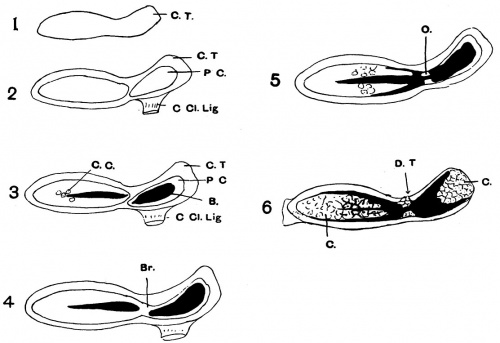
Accepting as correct the statements above quoted from Keibel and Mall, that the clavicle in its earliest stages is a condensed rod of connective tissue (see Scheme 1), placed caudal to the sterno-mastoid and trapezius muscles high up in the neck, about opposite the 4th cervical vertebra, we can con- tinue at the 15th mm. stage, of which I have representative material, and it shows that this condensed rod has undergone central changes. In the inner half there has appeared a mass of curious tissue, generally alluded to as a peculiar form of precartilage; the same holds good for the outer seg- ment. These two masses of pre cartilaginous tissue are separated from one another by the investing connective-tissue jacket (Scheme 2). No cartilage cells are to be recognized at this stage in either mass. At the 17mm. stage (and I owe the opportunity of examining such a stage to the kindness of Professor Arthur Robinson), ossification has commenced in the pre-cartilaginous tissue above mentioned; it is seen to commence at two separate spots--at the outer end of the inner segment, and the inner end of the outer segment; and it at first seems to involve the central part or core of each pre cartilaginous mass. At this stage a few cartilage cells can be distinguished in the inner segment, but none can be made out in the outer segment. The coraco-clavicular ligament is well developed,and it passes from the root of the coracoid process to the outer segment.
In al specimens which I have examined this is the invariable rule; the coraco-clavicular ligament always passes to the outer segment. The inner segment, then, has no direct connexion with the coracoid process. This is a point of very great importance, as it is dificult to see how Fitzwilliams' views on the morphology of the clavicle can, in the face of this fact, be maintained- but of this later.
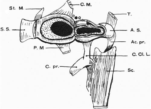
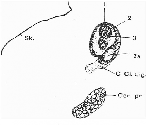
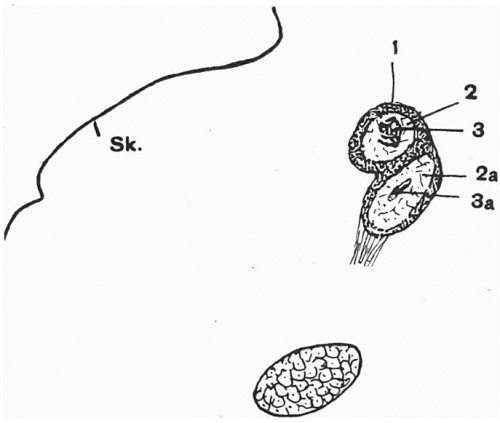
I have made a reconstruction model of the shoulder girdle of this embryo (fig. 1),and it shows how the inner segment is somewhat fusiformn in shape,whilst the outer segment is flattened. Each segment in cross section shows the following parts: most externally the connective-tissue jacket- periosteum or perichondrium; then a cylinder of precartilage, finally a centralmassofbone. Both masses of pre cartilage lie in a common jacket of perichondrium, and the bony masses in them are placed at right angles to one another, i.e.the inner mass when seen from the front is oval, whereas the outer mass is oval when seen from above. The inner precartilaginous mass slightly overrides above and in front by its outer extremity the outer mass of precartilage (figs. 2 and 3, and Scheme 3).
Fig.1. Drawing of a model of the right shoulder girdle of the 17mm. (Robinson) embryo, viewed from behind. The sternal segment (S.S.) has been cut coronally to expose the interior, and the acromial segment has been cut horizontally for the same purpose. Theblackareaineachis bone, the dotted area surrounding the bone is precartilage, and the area surrounding this is perichondrium. Above the junction of the two segments, two circles and a black dot are seen; the circles represent supraclavicular nerves, the black dot represents the cephalic vein. The scapula has been purposely shortened.
Ac. pr., acromion process; A. S., acromial segment of clavicle; C. Al., clei-do-mastoid; C. Cl. I., coraco-clavicular ligament; C. pr., coracoid process; T., trapezius muscle; P.M., pectoralis major; St. MI.,sterno-miastoid; S. S., sternal segment of clavicle.
Fig. 2. Sagittal section of left clavicle of 17mm. (Robinson) embryo.
Fig. 3. Sagittal section of leftclavicle of 17 mm. (Robinson) embryo. This is taken a little further to the left than the preceding one, and shows a bony core in the acromial segment (3a).
At a litle later stage the two precartilaginous masses fuse by their adjacent extremities. This fusion line is well seen in several of my specimens of about the 18-19 mm. stage, when horizontal sections of the clavicle show two bony masses separated by a non-stained precartilaginous interval. At this stage, too, chondrification has made a great advance in the inner segment,but cartilage cells cannot be recognized in the outer segment (fig.4 and Scheme 4).
Fig. 4. Semi-schematic drawing of semi-coronal section of right clavicle of an 18 mm embryo. All the parts shown save the subclavius muscle were shown in the section drawn, but the subelavitus was added from its appearance in several sections. Notice how the sternal segment overrides the acromial segment ofthe clavicle.
A.p,acromion process; B.1,bone of sternal segment; B.2, bone of acromial segment; Ct.b.,pre- cartilaginous bridge unossified and connecting acromial and sternal segments; C. Cl. Lig., coraco-clavicular ligament; C, coracoid process; Cl.M., cleido-mastoid muscle; C.Cl.L.,costo-clavicular ligament; R.1, first rib; S. M., subclavius muscle.
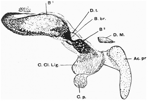
During the 19 mm. stage a further advance in ossification takes place. This brings about union by bone between the two independently ossified centres. This bony extension takes place in the bridge of pre cartilaginous tissue which unites the two main masses of precartilage. How precisely this bridge of bone is formed, whether by mutual outgrowth from the two bonymasses orfromone totheother,Iam notina position to state,nor doesitprobablymatter. The main point is that during the 19mm. stage a small bony bridge connecting the at first independent osific centres is developed (fig. 5 and Scheme 5).
Fig. 5. Semi-horizontal section of right clavicle of a 19 mm embryo.
B.', cartilage and bone of sternal segment; B.2, bone of acromial segment; B. br., bony bridge connecting sternal and acromial ossific centres; D.t.,pre cartilage which probably becomes the deltoid tubercle; D.M., clavicular part of deltoid muscle; Ac.pr.,acromion process; C.Cl.Lig.,coraco-clavicular ligament;C.p., coracoid process.
At the 27 mm. stage great advance in ossification has been made, and now for the first time cartilage is evident in the outer segment of the clavicle; the unossified precartilage has become definitely chondrified. The inner segment, too, has considerably enlarged, and cartilage is well marked initial stages of formation. The further inwards one traces itthe newer and younger the cartilage becomes, until we are led finally to ordinary connective tissue.
The bone now appears not only to be formed at the expense of the cartilage, but also from the investing perichondriunm, and it is always best developed where ossification commenced, and the appearance given is as if there were a bony cylinder solid in the middle but open at each end, the hollows at the extremities being filed with cartilage,a condition very much like that described by Gegenbaur.
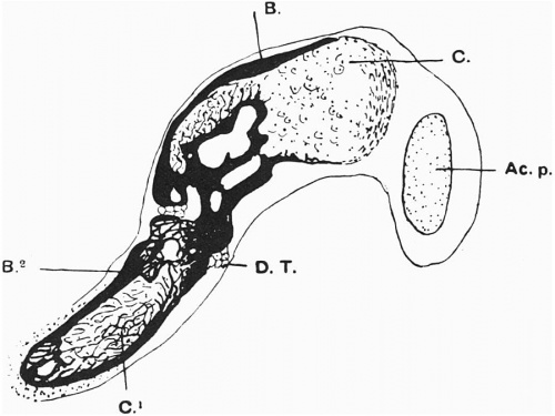
At one spot, however, there is not much bone developed, and that is at the site of junction of the two pre cartilaginous masses; the junctional bridge of precartilage has become converted into cartilage, and it projects forwards as a wedge-shaped mass between the two segments. I am inclined to think that it is this mass which ultimately gives rise to the deltoid tubercle (fig. 6 and Scheme 6).
Fig. 6. Horizontal section of left clavicle of 27 mm embryo.
B., bone of acromial segment; B.2, bone of sternal segment; C., cartilage of acromial segment; C.1,cartilageofsternalsegment; D.T.,cartilageremainingfrom precarti- laginous bridge which probably forms deltoid tubercle; Ac. p., acromion process.
Fig. 7. Schemes of right clavicle.
1.C.T.,connective-tissuerod. 2.C.T., connective tissue; P.C.,precartilage;C.Cl.Lig.,coraco.clavicular ligament. 3.C.C., cartilage cells in sternal segment; B.,bone. 4.Br., bridge formed by fusion of pre cartilage masses. 5.O.,ossification in pre cartilaginous bridge. 6.C.,cartilage in outer and inner segments; D.T.,chondrified bridge forming deltoid tubercle.
The stages through which the clavicle passes may be summarized thus
- At 11 mm., a condensed curved connective-tissue rod stretching from acromion inwards towards the first rib (Scheme 1).
- At 15 mm., an inner and an outer mass of precartilage is developed in the connective-tissue rod, the inner overriding above and in front the outer mass. The outer segment is at this time distinctly connected with the base of the coracoid process by the coraco- clavicular ligament (Scheme 2).
- At the 17 mm. stage, each precartilaginous mass undergoes in- dependent ossification, the bone forming a central core to the precartilage; cartilage cells are seen in inner segment (Scheme3).
- At 18-19 mm., the two precartilaginous masses fuse by their approximate ends (Scheme 4).
- At 19 mm., a bony bridge develops in the precartilaginous one (Scheme 5).
- At 24-27 mmn., both chondral and perichondrial bone are formed. Cartilage is now present in the hollow at the outer end of the outer segment, and the anlage of the deltoid tubercle is present (Scheme 6).
Consideration Of The Above Statements
If the statements brought forward here be correct-and they have only been arrived at after the examination of a considerable amount of material aided by the reconstruction method where necessary-then they confirm in the first place the statements of Mall (1906) based upon direct observation of Schultze cleared material, and the surmise of Fitzwilliams based on observations of the condition of the clavicle in hereditary cranio-cleido-dysostosis (1910). But it seems to me that the results which I have obtained are fatal to the morphological views of the later.
As has been previously stated, Fitzwilliams has argued that the inner segment of the clavicle must be the ancestral coracoid. In the first place, because he is of opinion that it develops in cartilage, whereas the outer end develops in membrane. Now, I hope I have conclusively shown that the two segments are at first identical, being formed of a peculiar precartilage. It is true that the inner segment shows cartilage cells at an earlier date than the outer segment,but it is only a question of time. By the 27 mm. stage the outer end of the clavicle is subtended by a huge mass of cartilage, which resembles in appearance in every way that of the inner end, though it has not arrived at the same degree of ossification. Tomy mind, there seems to be no more adequate reason for assigning a morphological value to this cartilage in the clavicle than to that which develops in certain of the face bones,e.g. the mandible.
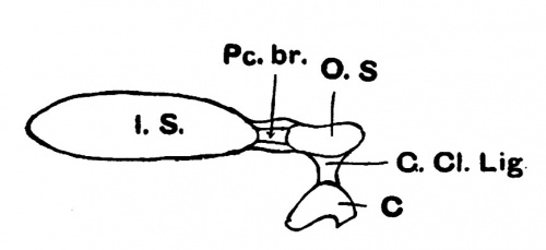
Fitzwilliams has further supported his argument by adducing the presence of a ligament prolonging the inner segment of the clavicle-in cases where the outer segment is apparently absent-to the root of the coracoid process. A part of this ligament he calls the coraco-clavicular ligament. But the evidence I have brought forward shows beyond doubt that the coraco-clavicular ligament is from the first connected with the outer segment of the clavicle, and with that only. The question, then, which arises is, How are we to explain this ligamentous prolongation of the inner end of the clavicle in an outward direction to the coracoid process on other than morphological grounds? That the ligament in question exists there can be no manner of doubt, as the author cited has himself personally seen it, and he has collected quite a number of cases which record its presence. It has occurred to me that the following suggestion may inpart, at al events, meet the difficulty. Let us assume that,owingtothenature of the disease, bone has not been laid down at the site usually laid down at the 17 to 19 mm. stages; the result will be that the inner segment of the clavicle will be prolonged outwards, first by its own outer unossified end, next by the unossified precartilaginous bridge and covering connective- tissue jacket, finally by that part of the outer segment which remains ligamentous through non-ossification and which has attached to it the coraco-clavicular ligament (fig.8).
Fig. 8. - Scheme to illustrate suggested explanation of the connexion of the sternal extremity of the clavicle with the coracoid process by ligament.
I. S., inner segment; Pc. br., precartilaginous bridge; 0. S., outer segment; C. Ci. Lig., coraco-clavicular ligament; C., coracoid process.
Such a hypothesis as this involves no morphological speculation, and it seems to account for the commonest anomaly met with save in one particular, and that is, why there should be no cartilaginous element developed representing the outer segment of the clavicle beyond the limits of the coraco-clavicular ligament. Itispossible that the answer to this problem may be found in the absence of the anterior fibres of the trapezius and deltoid, which I am inclined to think play a great part in regard to the development of the outer end of the clavicle. The relation between muscular attachment and the form, position, even the very existence of bones, is one which has not been made the subject of study, but is one which may be productive of the greatest results.
Jenkins has made a model of the shoulder girdle of an embryo of 16mm. in length. The model, which I have not seen, is stated to show the clavicle in two parts, both apparently cartilaginous. Jenkins believes that the inner mass forms the sternal end of the clavicle, whilst the outer mass disappears, the acronmial end being developed from separate mesodermal tissue. So far as I know, there is no evidence whatever for this view.
Cases of existence of the clavicle in two apparently separate masses may, I think, be explained on the ground of non-ossification of the pre- cartilaginous bridge connecting the sternal and acromial segments with one another during the19 mm. stage. The bony connexion is for along period normally only as light one.
In conclusion, let me say that I personally feel that we owe a debt of gratitude to Mr Fitzwilliams for the very able-way in which he has stated his views and marshalled his array of facts.
| Historic Disclaimer - information about historic embryology pages |
|---|
| Pages where the terms "Historic" (textbooks, papers, people, recommendations) appear on this site, and sections within pages where this disclaimer appears, indicate that the content and scientific understanding are specific to the time of publication. This means that while some scientific descriptions are still accurate, the terminology and interpretation of the developmental mechanisms reflect the understanding at the time of original publication and those of the preceding periods, these terms, interpretations and recommendations may not reflect our current scientific understanding. (More? Embryology History | Historic Embryology Papers) |
| Links and more recent papers |
|---|
|
<pubmed>14534796</pubmed> Search term: Clavicle Development <pubmed limit=5>Clavicle Development</pubmed> |
Cite this page: Hill, M.A. (2024, April 30) Embryology Paper - The development and ossification of the human clavicle. Retrieved from https://embryology.med.unsw.edu.au/embryology/index.php/Paper_-_The_development_and_ossification_of_the_human_clavicle
- © Dr Mark Hill 2024, UNSW Embryology ISBN: 978 0 7334 2609 4 - UNSW CRICOS Provider Code No. 00098G


