Paper - On the development and morphology of the human sphenoid bone
| Embryology - 1 May 2024 |
|---|
| Google Translate - select your language from the list shown below (this will open a new external page) |
|
العربية | català | 中文 | 中國傳統的 | français | Deutsche | עִברִית | हिंदी | bahasa Indonesia | italiano | 日本語 | 한국어 | မြန်မာ | Pilipino | Polskie | português | ਪੰਜਾਬੀ ਦੇ | Română | русский | Español | Swahili | Svensk | ไทย | Türkçe | اردو | ייִדיש | Tiếng Việt These external translations are automated and may not be accurate. (More? About Translations) |
Sutton JB. On the development and morphology of the human sphenoid bone. (1885) Proc. Zool. Soc. 308: 577-.
| Historic Disclaimer - information about historic embryology pages |
|---|
| Pages where the terms "Historic" (textbooks, papers, people, recommendations) appear on this site, and sections within pages where this disclaimer appears, indicate that the content and scientific understanding are specific to the time of publication. This means that while some scientific descriptions are still accurate, the terminology and interpretation of the developmental mechanisms reflect the understanding at the time of original publication and those of the preceding periods, these terms, interpretations and recommendations may not reflect our current scientific understanding. (More? Embryology History | Historic Embryology Papers) |
On the Development and Morphology of the Human Sphenoid Bone
By J. Bland Sutton, F.B.C.S.,
Lecturer on Comparative Anatomy at the Middlesex
Hospital Medical School. (Received May 25, 1885.) (Plate XXXV.)
Introduction
It has been truly remarked that the mode of ossification of the sphenoid bone is one of the most difficult questions in osteogenesis. We may go further and say that the morphological relations of the precursors of the sphenoid bone, the trabeculae cranii, present even greater difficulties. For some years past I have been, with the kind assistance of my pupils, gradually accumulating material for a thorough investigation of this interesting and important region of the skull. The result of the inquiry I propose to embody in this paper.
In order that the various constituent nuclei of the complex human sphenoid bone may be correctly comprehended, it is essential that the early stages of the human cbondro-cranium be briefly sketched.
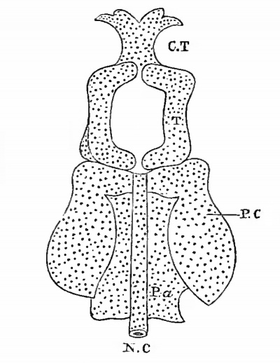
The embryological history of the cranial skeleton clearly shows that a uniform plan of construction underlies the skull in all Craniata ; and that it may be divided into a basi-cranial region, formed by two cartilaginous plates enclosing the notochord, known as the parachordals, which later on fuse and form a continuous platform known as the basilar plate. The anterior limit is marked off by the pituitary body. This basilar plate or notochordal region of the skull-base forms a floor for the hind and mid brain. The second portion is composed of a pair of bars, the trabeculae, embracing posteriorly the termination of the notochord, then separating to enclose a spaee, which is afterwards occupied by the pituitary body, they again come into contact, in most cases coalesce, and extend forward into the nasal region. This section of the primordial skull may be conveniently termed the basi-faeial region, the trabeculae forming a support for the fore brain (see fig. 1, p. 578).
The third element in the skull consists of the sense-capsules, auditory and olfactory ; the optic capsules as a rule remaiu distinct.
Lastly, the appendicular elements claim consideration ; they comprise the palato-pterygoid, meckelian, and hyoid cartilages, and the remaining branchial bars.
The portion which more immediately concerns us in this paper is the trabecular region.
It is now admitted unreservedly by those anatomists who have dealt with the morphology of the skull from the vantage point afforded by embryology, that the basi-cranial region - the portion extending from the margin of the foramen magnum to the summit of the dorsum sellse - is composed of the same dements as the vertebral column, hut differs from it in that it lies not passed through any stage of vertebration T , the notochordal portion with the parachordals representing the centrum, with the laminae which meet dorsally in man and mammals, over the developing brain in the occipital segment only.
Anteriorly the representatives of the laminae take on a very different disposition. During development the skull, whose long axis was originally a direct continuation of, and in the same plane as, the vertebral column, becomes at an early period bent, or, as it is usually described, flexed downwards. One of the important results of this flexion is the dissociation of the anterior portion of the lateral neural walls from the parts immediately adjacent. Eventually these dismembered portions of the neural walls coalesce around the down-bent brain and are recognized as the trabeculae cranii. This admirable explanation was first promulgated by Goctte (Entwicklunggeschichte der Unke, page 629) ; and this view has certainly much more to recommend it than the notion that the trabeculae are to be regarded as a pair of branchial arches.
Fig. 1. A diagram to represent the disposition of parts in tlie base of the primitive skull.
N.C, Notochord ; Pa, parachordals ; P*C, periotic capsules; T, trabeculae; C.T, ethmo-vomerinc region.
- 1 Ide Huxley, The Cranio-facial Apparatus of Petromyzon Journal Anatomy and Physiology, vol. x. p. 418,
After the trabeculae have coalesced, growth occurs in them in three directions:
- Laterally, to form the side walls of the skull anterior to the periotic capsules.
- Mesially, to fill up the floor of the pituitary fossa.
- Forwards, to form the ethmo-vomeriue aud fronto-nasal plates.
Although the trabeculae at a very early period form a floor to the pituitary fossa, yet this fossa is never completely shut off from the pharynx in the chondro-cranium ; a small opening persists for a very 'long time, the consideration of which leads to some very interesting conclusions, and which renders necessary the study of the early stages of the formation of the mouth and pharynx. It has been very satisfactorily proved that the buccal mucous membrane is derived from the epiblast ; the process by which this derivation occurs is usually described as a tucking-in of the epiblast ; but in reality it is a necessary outcome of the primary cranial flexure. Passing between the open arms of the trabeculae is a narrow tubular portion of the anterior primary encephalic vesicle, knowm as the infundibulum ; tins diverticulum from the primitive brain comes into contact with the buccal epiblast; the meeting point of the two structures is represented by the pituitary body. This disposition of the parts has long been known.
Whilst engaged working over the development of this complex region, I found that even at the mid period of intra-uterine life of the human embryo a narrow cavity may be detected passing from the pharynx through the basisphenoid, so as to come into close relation with the infundibulum.
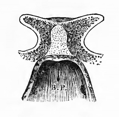
The point of communication with the pharynx is in the middle line in contact with the basisphenoid, the spot being indicated at birth by a recess in the mucous membrane known as the bursa pharyngea (see fig. 2, p. 580). After birth the canal suffers obliteration ; but a band of fibrous tissue, passing from the pituitary body to the pharynx, represents the original position of the canal. When the bone is macerated the fibrous tissue disappears, leaving a hole in the basisphenoid, termed by Landzert the canalis cranio-pharyngeus (Petersburger med. Zeitscbrift, Bd. xiv.). This cranio-pharyngeal canal may be detected in the floor of the sella turcica in very many mammals at birth.
Up to the present time I have been unable to assure myself that the low r er end of the infundibulum ever opens into the pharynx, but there is every probability that such is the case. However, the existence of this diverticulum of the first encephalic vesicle raises an exceedingly interesting question. It will be remembered that the central canal of the spinal cord at its caudal end is brought into relation in the early embryo of very many of the Yertebrata with the hind gut, by a narrow and, in most cases, very temporary passage known as the neurenteric canal, which passes around the caudal end of the notochord, but afterwards becomes obliterated.
Turning now to the cephalic end of the notochord, we find the central canal of the encephalic vesicles turning round the most anterior end of the notochord in the form of an infundibulum, to form a continuity with the buccal epiblast and come in very close relationship at first with the extreme end of the foregut ; the place where these highly interesting occurrences take place being for a long time represented by the cranio-pharyngeal canal. The inquiry needed to make the demonstration complete is, to determine positively whether the infundibulum is always a cul-de-sac, or at some period communicates with the cavity of the future pharynx.
Fig. 2. The posterior wall of the pharynx, showing the position of the Bursa pharyngea, B.P. From a foetus at the fifth month.
It is a very remarkable fact that the pituitary fossa and the space between the bladder and the rectum are common situations for those curious tumours known as teratomata; it will be interesting to ascertain the relation of the curious neurenteric passages to these morbid growths.
The ossification of the sphenoid must now be considered.
The alae arise from single nuclei in the cartilage forming the lateral walls of the skull immediately anterior to the periotic cartilage, commencing about the eighth week of intra-uterine life. They are the first centres of this important bone to make an appearance.
A little later two circular spots may be detected in the cartilage forming the floor of the pituitary fossa. They first become visible on the under surface of the bone, and are not affected when the perichondrium is removed : these are the paired basisphenoidal nuclei.
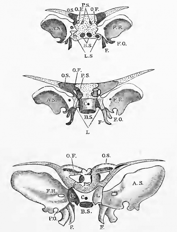
They are very quickly followed by two earthy spots in the lingulae, lying between the alae and basisphenoidal nuclei, as shown in fig. 3 A, which represents the disposition of these six nuclei, three on either side, viz. basi-, lingulae-, and alisphenoid centres.
The two for the basisphenoid become quickly confluent from below upwards, and the lingulae soon fuse with them and form a porous mass. The alae grow rapidly, but remain separated from the lingulae by a thin layer of cartilage until the first year after birth.
These six centres constitute with the internal pterygoid plate, which will be considered further on, the posterior portion of the sphenoid hone.
The details of the ossification of the presphenoid, or that portion in relation with the optic nerves, may be summarized as follows :
Fig. 3. A series of figures to show the disposition of the various nuclei of the human sphenoid bone.
The upper figure shows the relation of the centres to the cartilage; the latter in all cases is represented by dots.
In the middle figure the basisphenoiclal nuclei have coalesced, the orbitosphenoids have joined the presphenoids, and the internal pterygoids have joined the alisphenoids.
In the lowest figure the sphenoid bone is represented as at the eighth month of foetal life.
A.S, Alisphenoid; B.S, basisphenoid ; L , lingulae; F, pterygoids ; O.S , orbitosphenoids ; P.S, presphenoid ; O.F, optic foramen ; F.U, foramen rotundum; F.O, foramen ovale; <7, cranio-pliaryngeal canal.
The dorsum sellae at this date is cartilaginous, and therefore it is not represented in the figures. The foramen ovale, until some time after birth, is only a notch in the alisphenoid.
Ossific matter is deposited immediately external to the optic foramen, and extends rapidly outwards to form the orbitosphenoid. This occurs about the commencement of the third month.
Later a nucleus makes its appearance on the inner side of each optic foramen, on the deep aspect of the perichondrium : these are the presphenoidal centres. They attain some considerable size before involving the cartilage, and can during the first month of their existence be removed without in -any wav disturbing the subjacent cartilage. The orbitosphenoid quickly sends down two spurs around the optic nerve, which fuse with the presphenoid. The presphenoids in their turn send a thin shell of bone across the dorsal aspect of the cartilage to fuse with each other ; a small circular spot of cartilage long remains to indicate the point around which they united. It is long before these presphenoidal nuclei fuse below ; a large piece of cartilage, belonging to the ethmo-vomerine plate, separates them, even for some months after birth. Long before this occurs the presphenoids, bearing their allies the orbito-sphenoids, have fused with the basisphenoids ; the line of fusion being represented in afterlife by the ridge known as the olivary process.
Before dismissing the orbito-sphenoid, it is necessary to draw attention to one circumstance connected with it, of some interest. The lateral extension of the trabeculae to form the side-wall of the chrondro-cranium is in the later stages replaced almost entirely by the alisphenoids.
If the region of the side-wall of the skull known as the anterior lateral fontanelle in the foetus (in the adult it is called the pterion) be examined between the fourth and seventh months of intra-uterine life, it will be easily noted that the cartilaginous orbito-sphenoid extends into this fontanelle, so that for a considerable period it helps to form the side-wall of the skull. In man the permanent orbitosphenoid never extends so far outwards as its cartilaginous forerunner, leaving the space to be filled in by the epipteric bone. The details of the ossification of this region I have considered elsewhere.
There remains little to add concerning the later development of the orbito-sphenoid, except to note that eventually the orbitosphenoids of opposite sides send a thin lamella across that portion of the presphenoid which is anterior to the optic groove, thus excluding it from the cranial cavity.
The portions of the sphenoid previously considered are strictly cranial, but it receives an additional element from one of the appendages of the skull, viz. the palato-pterygoid bar.
In a paper published in the Proceedings of this Society for 1884, “On the Parasphenoid 55 &c., facts were adduced to show that the anterior portion of the palato-pterygoid cartilage in man became ossified to form the internal pterygoid plate ; the nucleus for this bone may be detected as early as the commencement of the third month of intra-uterine life. The length of time it may remain as a separate ossicle varies within wide limits. I have seen it distinct from the sphenoid as late as the fifth month ; but as a rule it will be found united with the under surface of the alisphenoid at the commencement of the fourth, so that it joins the alisphenoid before that hone
fuses with the corresponding lingula, with which element the internal pterygoid also has after birth an osseous union ; the space left between this triple union of ala, lingula, and pterygoid being occupied by the vidian or great superficial petrosal nerve, a nervous cord of no small morphological significance, as I have previously shown. The external pterygoid plate is simply an apophysis from the under surface of the alisphenoid, becoming conspicuous about the third month of intra-uteriue life. As a matter of convenience and ready reference, the dates of appearance of the individual centres and their fusion with one another are here given in a collected form.
At the eighth week the following centres appear quickly one after the other, in the following order :
- Alisphenoids.
- Basisphenoids.
- Lingulae sphenoidales.
- Internal pterygoids.
During the third month the ossific points fuse in the following order :
- Basispbenoidal nuclei coalesce, and the
- Lingulae join the basisphenoid.
At the third month the following centres appear :
- Orbito-sphenoid.
- Presphenoids.
At the fourth month the orbito-sphenoids fuse with the presphenoids and the internal pterygoids join the alisphenoids. At the seventh month the presphenoid and postsphenoid coalesce. At the eighth month the presphenoids fuse together.
During the first year after birth the alisphenoids bearing the internal pterygoids coalesce with the lingulae, and the so-called sphenoidal’ tubinals develop. The strip of cartilage which is prolonged from orbito-sphenoid to the anterior lateral fontanelle (the pterion) now disappears.
We must now deal with the morphology of the various centres of the sphenoid.
In the determination of the regions of the skulls in different types we are greatly assisted by the disposition of the cranial nerves, which in the majority of cases serve as fairly reliable guides.
The optic nerves always embrace the presphenoid, whilst the third division of the fifth cranial nerve usually quits the skull between the most anterior part of the periotic capsule and the alisphenoid, whilst the auditory nerve stands in very definite relationship with the various constituent nuclei of the periotic capsule, in those forms in which this cartilage undergoes ossification, so that as a rule we have no difficulty in distinguishing between the regions. When dealing with the individual ossific nuclei the case is very different, it being absolutely necessary to watch every stage of the development to avoid falling into the numerous pitfalls which abound on every side.
The presphenoid nuclei do not offer very much that is important, but the basisphenoid and its associated nuclei are in no small degree interesting.
Mr. Parker, in his very valuable paper, - On the Skull of the Common Fowl†(Phil. Trans. I 859), introduces us to two very remarkable bones which he names the basitemporals. I will describe them in Mr. Parker's own words : The subcranial region, which in the Frog is ossified by the basitemporal wings of the parasphenoid, is here supplied with a pair of distinct and large basitemporal bones which extend from near the median line, beneath the cochlea and so far outwards as to constitute a floor for the tympanic cavity ; their anterior limit is near the fore margin of the alisphenoid cartilage. These ossifications arise in a thick weft of fibrous tissue in the hinder part of the palate ; the matrix is abundant in the middle line, extending forwards to the bone next to be described. The Eustachian tubes run forwards and inwards above the anterior edge of these bones, and meet in the middle line beneath the pituitary fossa 1 . The Fowl's basitemporals are shown on Plate XXXV. fig. 1.
It is needful to explain what is here meant by the basitemporal wings of the parasphenoid.
Underlying the Frog's skull (as shown in Plate XXXY. fig. 2) is a dagger-shaped bone termed by Prof. Huxley the parasphenoid, but it is simply the representative of the vomer of the mammalian skull : the lateral portions marked L in the figure are what Mr. Parker refers to as the basitemporal portions. The morphological value which the latter writer places upon the bird's basitemporal is so singular that it is needful again to cpiote his own description, contained in a footnote in the “Fowl†paper: "From a careful comparison of these parts in the lower Mammalia with those of man, 1 feel satisfied that the bony lingulae in that class answer to the basitemporal rudiments of the parasphenoid†(p. 770). This comparison was first suggested to Mr. Parker by Prof. Huxley, who states in a footnote in one of his admirable ‘"Lectures’ that “ Mr. Parker agrees wdth my suggestion that the basitemporals of the Sauropsitla are the homologues of the lingula sphenoidales of Man †(p. 220).
My intention is now to proceed to show beyond all doubt that the “suggestion†and the “ agreement†are out of harmony with the facts of the case and opposed to the usual methods of morphological reasoning.
The lingulae of the Mammalian sphenoid have no relationship whatever with the basitemporals of birds.
The proof is as follows : The basitemporals, and no one doubts the facts, arise in membrane. It is a well-established truth that a bone preformed in cartilage cannot be homologous with one simply of membranous origin. The basitemporals are membrane-bones ; the lingulae are preceded by cartilage. On this ground alone the evidence of identity fails. On Plate XXXV. fig. 3, is represented the base of the skull of a young Ostrich ( Strut hio camelas) : two distinct osseous nuclei are seen lying on either side of the basisphenoid, between it and the alisphenoid ; they are developed in cartilage, thus in mode of ossification as in their relations they correspond to the lingulae of mammals. In the same specimen the basitemporal bones are present . The skull of this Ostrich is sufficient in itself to prove that the conclusions of Mr. Parker regarding the identity of the basitemporals of birds and the mammalian lingulae sphenoidales are based on erroneous premises.
It now behoves me, seeing that I challenge these views, to identify the lingulae, and explain the apparently anomalous condition of the basi temporals.
I shall address myself to the lingulae first. In the skull of certain fish there is a centre known as the sphenotic, which occupies the antero-external region of the periotic capsule, but the cartilage in which it arises is always of a composite character, being due to the confluence of proper cranial cartilage with that of the periotic cartilage. This centre is present in the Fowl in the very spot where the lingulae ought to be represented.
In the chondro-cranium of Man, the cochlear region of the periotic capsule comes into union with the lingulae of the sphenoid, and the remains of the uniting cartilage are familiar to students of human anatomy as the cartilage filling up the foramen lacerum medium. If the cartilaginous lingulae of the bird and man are homologous, and on that score there can be uo doubt, then the ossific nuclei which transform them into bone should certainly be considered homologous also. On these grounds my contention is, that the nuclei called sphenotic in the Fowl and Ostrich are the true morphological representatives of the human lingulae. It is now necessary to find out to what ossifications in the mammalian skull the basitemporals of the bird really correspond.
Turn from the Bird for a brief space, and inspect the hard palate of a Crocodile. From before backwards we find the following bones : — premaxilla, prepalatine portion of the maxilla, palate, and a bone usually marked pterygoid ; passing from the outer edge of this bone to the maxilla is a bony bar known to anatomists as the os transversum, the general relations of which can be readily seen by reference to Plate XXXV. fig. 4.
The anatomical relations of the hone marked pterygoid are important in the following particulars : they surround the posterior nares, it being due to their intervention that the nasal passages are prolonged posteriorly to such a marked extent in the Crocodile. Above, they have the Eustachian passages, and externally they support the os transversum. This latter bone ought to be really regarded as the pterygoid. In Man's skull, and it is most probably true of other mammals, the internal pterygoid arises as an ossification of the distal end of the palato-pterygoid cartilage. The bone in the Crocodile's hard palate marked pterygoid arises as a membrane-bone, and during its growth the outer end invades to a slight extent the middle portion of the palato-pterygoid cartilage, and thus cuts off the distal end of the chondral rod, which becomes the os transversum, really the internal pterygoid.
Even at the risk of being tedious I must make myself clear on this point. In Man a rod of hyaline cartilage stretches from, and is continuous with, the malleus at the eighth week of intra-uterine life ; it passes towards the anterior limit of the fronto-nasal plate. Later the rod becomes segmented as follows: — the distal end becomes internal pterygoid plate, the middle portion persists as the cartilaginous piece of the Eustachian tube, and the proximal portion degenerates into ligament. The details of the metamorphosis of this bar will be found in my paper on the Parasphenoids 99 &c. (Proc. Zool. Soc. November 1884).
In the Crocodile the corresponding bar of cartilage is continuous posteriorly with the huge quadrate, its middle portion is considerably infringed by the so-called pterygoids, whilst its distal end is continuous with the os transversum.
If the bones forming the posterior limit of the hard palate in Crocodiles are not to be regarded as pterygoids, to what do they correspond ? They are the homologues of the avian basitemporals. As is the case with the so-called pterygoids of the Crocodile, the bird’s basitemporals are preformed in membrane, they underlie the basisphenoid, and the Eustachian tubes lie above them ; but the bird in its aerial mode of life needs not a long tubular nasal passage, indeed its hard palate may be considered defective, and each basitemporal, instead of sending a process of bone to curve around the posterior nares, merely persist as a flat plate of bone which eventually becomes welded to the skull-base.
In birds the pterygoids take a different direction from that of the Crocodile’s os transversum : in the former case they converge anteriorly, and in some avian skulls actually come into contact at the spot where they join the palatines, whilst posteriorly they abut upon the quadrate bone ; this last fact is sufflcient to prevent any misinterpretation as to their nature. In the Crocodile the anterior ends of the pterygoids are carried outwards, until they rest on the maxillae, and the postpalatine bones (the so-called pterygoids), being wedged inbetween them, separate the pterygoids from the quadrate to such an extent as to disguise their real nature and make them appear as additional ossifications.
According to this view the pterygoid of Birds, the os transversum of Crocodiles, the transpalatine of the Snake, and the internal pterygoid of Mammals, including Man, arise in connection with the distal end of the palato-quadrate cartilage, and must therefore be regarded as homologous bones.
The so-called pterygoids of Snakes and of Crocodiles and the basitemporals of Birds agree in their mode of development and relationship to the main morphological landmarks of the skull; they must therefore be regarded as homologous ossifications, and as a matter of convenience it is proposed to name them postpalatines . Whether these ossifications are represented in the mammalian skull by the socalled sphenoidal tubinals, or by certain accessory ossicles which are developed in connection with the hinder end of the vomer in some tvpes (marsupials, hedgehog, &c.), or not represented at all, is a matter of very little importance. It would of course be very interesting to be able to determine whether the bones which prolong the hard palate in some of the Edentates, Myrmecojihaya for example, arise in the same manner as the bird’s basitemporals. My conviction, so far as I have been able to look into the question, is that they do, but until iny material is more abuudant the question, with me, remains sub jutiice.
Fig. 4 is an attempt to represent in a graphic manner the metamorphosis of the palato-pterygoid bar in a Bird, in a Crocodile, and in Man, so as to explain how it comes about that in a Bird the true pterygoids rest on the quadrate, but in the Crocodile and in Man the true pterygoids are separated by a piece of cartilage.
Fig. 4. A series of diagiams to illustrate the metamorphoses of the palato-pterygoid arch in Birds, Crocodiles, and in Man.
P = pterygoid ; C = cartilage; Q = the quadrate. In Man the malleus is the equivalent of the quadrate, and is represented thus : Q = M. M.C., Meckel's cartilage ; P.G. processus gracilis.
The upper row of figures represent the cartilaginous bars, arranged in order from left to right - Bird, Crocodile, and Man. The lower row represent the adult condition.
Lastly, in a preceding paper I endeavoured to dispose of the blade of the famous parasphenoid. On the present occasion I try to show that the view which would regard these basitemporals (postpalatines) as the homologucs of the lateral portions of the Frog's parasphenoid is against the weight of evidence. The investigation supports my view previously expressed, that the parasphenoid of the amphibian skull is represented in the highest mammals by the vomer , and by that bone alone .
Explanation Of Plate XXXV
Fig. 1. View of the base of the skull of a Chick at end of second week. P.P, Postpalatine (ba si -temporals of Parker).
2. Under view of the skull of a Frog, to show the general appearance and relation of the so-called parasphenoid, P. The lateral portions are marked L.
3. The sella turcica of a young Ostrich, Strut hio camelus , to show the lingulae,!/. A.S, Alisphenoid ; B.S, basi sphenoid ; F.Af t foramen magnum.
4. The hard palate of a Crocodile, to show the so-called pterygoid bones. P.P , The author's postpalatines; P.N, posterior nures ; P, opening of Eustachian tube.
(Figs. 3, 2, and 4 after Parker.)
Cite this page: Hill, M.A. (2024, May 1) Embryology Paper - On the development and morphology of the human sphenoid bone. Retrieved from https://embryology.med.unsw.edu.au/embryology/index.php/Paper_-_On_the_development_and_morphology_of_the_human_sphenoid_bone
- © Dr Mark Hill 2024, UNSW Embryology ISBN: 978 0 7334 2609 4 - UNSW CRICOS Provider Code No. 00098G


