Paper - Notes on the Wolffian body of higher mammals (1902)
| Embryology - 30 Apr 2024 |
|---|
| Google Translate - select your language from the list shown below (this will open a new external page) |
|
العربية | català | 中文 | 中國傳統的 | français | Deutsche | עִברִית | हिंदी | bahasa Indonesia | italiano | 日本語 | 한국어 | မြန်မာ | Pilipino | Polskie | português | ਪੰਜਾਬੀ ਦੇ | Română | русский | Español | Swahili | Svensk | ไทย | Türkçe | اردو | ייִדיש | Tiếng Việt These external translations are automated and may not be accurate. (More? About Translations) |
MacCallum JB. Notes on the Wolffian body of higher mammals. (1902) Amer. J Anat. 1(2): -259.
| Online Editor | ||
|---|---|---|
| This historic 1902 paper by John Bruce MacCallum describes the development of the mammalian Wolffian body or mesonephros, the intermediate embryonic kidney stage.
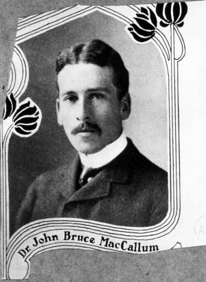
John Bruce MacCallum (1876-1906) "During his short years at Baltimore and in California, under Loeb, where he bravely fought a losing fight against tuberculosis, he did pioneer work of permanent value on the embryology and histology of the heart muscle and the intestines, and on the action of calcium and the salines. His letters give an introspective picture of his student days, his work, his summers in Muskoka, his travels—to Germany, Jamaica, Denver (where he practised briefly) and California, and in addition they have a literary, romantic, and psychological interest. He was also an artist, musician, story-writer, and poet; and the few poems included make one wish that room had been found for more." (Short Years, the Life and Letters of John Bruce MacCallum)
|
| Historic Disclaimer - information about historic embryology pages |
|---|
| Pages where the terms "Historic" (textbooks, papers, people, recommendations) appear on this site, and sections within pages where this disclaimer appears, indicate that the content and scientific understanding are specific to the time of publication. This means that while some scientific descriptions are still accurate, the terminology and interpretation of the developmental mechanisms reflect the understanding at the time of original publication and those of the preceding periods, these terms, interpretations and recommendations may not reflect our current scientific understanding. (More? Embryology History | Historic Embryology Papers) |
Notes on the Wolffian Body of Higher Mammals
By
John Bruce MacCallum, M. D.
From the Anatomical Laboratory, Johns Hopkins University.
With 17 Text Figures.
Introduction
In studying the Wolffian body of human and pigs’ embryos, certain facts were arrived at which will be set down in the following order:
- Methods of study and material.
- Tubular system of Wolffian body in human embryos.
- Tubular system of Wolffian body in pigs’ embryos with a description of a wax reconstruction showing the course of the tubules.
- Blood-vessels of Wolffian body of pig’s embryo.
- Relation of the tubular systems of the testis and the Wolffian body.
1. Methods of Study and Material
A large number of pigs’ embryos were studied, varying in length from 8 mm. to 200 mm. Since a fresh supply of these could be obtained at any time, it was not difiicult to make a considerable number of injections and preparations with various methods. The human embryos were available through the kindness of Professor Mall, and free use was made of the large collection which he possesses.
A very instructive method in the study of the tubular system consisted in the injection of the organ through the allantois with a colored solution. Various injection masses were made use of, but the most sa.tisfactory proved to be the saturated aqueous solution of Berlin blue, and the ordinary carmine gelatin mass. Gelatin in which cinnabar or lamp-black granules were suspended had the disadvantage of being less transparent. Double injections were also made with carmine gelatin for the tubules and Berlin blue for the blood-vessels. In the Wolffian body there is no difficulty in distinguishing veins from arteries with a general vascular injection. Double injections of the vessels, however, were also made to supplement the general ones.
In forcing fluid into the tubules of the Wolffian body it is necessary to inject either through the allantois or the cloaca. In small embryos it is easier to tie off the cloaca below the entrance of the Wolffian duct, and fill the allantois with the injection mass. Then by gently squeezing the allantois between the fingers the fluid can be forced slowly into the tubules of the Wolffian body. In older embryos the cannula can be placed in the cloaca, and the allantois tied off. Where possible this is the best method, because the soft gelatinous tissues around the cloaca make it difficult to close it in any way. It proved to be a most instructive thing to cause the injection mass to flow into the tubules slowly, and to watch with a lens its course. Injections with lamp—black agar and subsequent digestion with pepsin and hydrochloric acid were unsatisfactory; for the delicacy of the structures made it impossible to isolate complete tubules.
The ordinary methods of histological study were employed. The reconstruction was made by the Born wax-plate method.
2. Tubular System of Wolffian Body in Human Embryos
In all higher Amniota the first part of the urinogenital apparatus to make its appearance is the Wolffian duct. According to Hensen and v. Spee this tube is derived from the ectoblast; while His and Kowalewsky believe that it has its origin in the middle plate of the mesoblast. Remak, v. Kolliker, VValdeyer, and others trace its development from the lateral plate of the mesoblast. Romiti, Rensen, Dansky, and others find that it springs from the coelomic epithelium. Ac cording to Michalkovics
Fig. 1. Transverse section of human embryo CLXIV ' ' ' (3.5 mm.) in length. A. beginning of anterior part of It IS at first 3‘ Sohd mass urinary apparatus. W. D., Wolffian duct of Cells differentiated from the mesoblast. The canal is blind at both ends in the beginning, and becomes lined by an epithelium—like layer.
In a human embryo, CLXIV, 3.5 mm. in length, possessing 19 myotomes (probable age 2.5 weeks), the Wolffian duct is found in an early stage of its development. It is in close connection with the coelomic epithelium in many places; and at its anterior extremity is evidently a direct turning in of the lining of the coelom, Fig. 1 A. The duct consists of a rod of cells ending anteriorly in a depression or groove. This is shown on the left side of Fig. 1; while on the right side the duct is cut more posteriorly and shows already some indication of the formation of a lumen. Examined closely, the organ is found to consist of two parts, an anterior, and a posterior division. The anterior part begins opposite 3 the 6th myotome, and ends opposite the 9th.
The posterior part begins opposite the 10th myotome and extends throughout the rest of 4 the body to the level of the last myotome. 7 This is shown diagrammatically in Fig. 2.
The anterior part, A., is a simple rod—shaped mass of cells possessing a small lumen at the anterior end opening into the body cavity, as shown in Fig. 1. The posterior part, W. B., extends backward parallel with the last nine myotomes; and at 13 places on its course it is thickened to form rounded masses, from a few of which there project lateral outgrowths. These are the beginnings of the Wolffian tubules, W. T. At this stage there is no indication whatever of glomeruli.
Fig. 2. Diagrammatic reconstruction of The Slgnlficance of these two urinary appa at ' h e b 0 CLXIV. ' ‘ A.. anterior ryezilirsifi ailignziligtturg: ’l¥’.B.. Wo1f- parts of the unnary apparatus 13
tubules. The myotomes are not Clean one is tempted to consider the anterior mass of cells as in some way related to the pronephros of lower animals; and the posterior mass, as the developing Wolfiian body. The fact that the anterior mass possesses a lumen which opens into the body cavity would seem to support this idea, as this is the case with the pronephric tubules in lower vertebrates. Between the anterior and posterior parts there is a space of five or six sections, each 20 /1 thick.
- 1 The Roman numerals refer to embryos in the collection of human embryos belonging to Dr. Mall, in the Anatomical Laboratory of the Johns Hopkins University.
In a somewhat older embryo, LXXX (V. B. 4.5, N. B. 5.; probable age 3 weeks), a slightly more advanced stage in the development of the tubules is noticed. The urinary apparatus begins opposite the 7th myotome as a small. duct. This extends for 4 sections, each 20 /1 thick, and then ceases. During its course one small, blind, slightly curved, tubule is given ofl’. In the next four sections no trace of this duet can be made out. In the section following, however, another duct starts and is continued to the posterior end of the body. It seems at first probable that this interruption in the duct may be due to a misplacement of four sections in the series; but a close examination shows that this is quite impossible. Furthermore, the tubule which arises from the anterior duct proceeds from its ventral side and curves with its convexity ventralwards; while the tubules of the posterior duet all arise from the dorsal side of the duct with the convexity dorsalwards, as shown in Fig. 3.
There are no glomeruli opposite the anterior duct; while in the posterior part of Wolffian body proper, there is a glomerulus corresponding With each tubule. The tubules are curved as represented in Fig. 3 and number 15 on one side of the body and 17 on the other. There proved to be 1'?’ glomeruli on one side and 18 on the other. Considering the possibility of error in counting these by following them through a series of sections, it seems that the organ is a fairly symmetrical one; and
Fig. 8. Diagram — that there are approximately as many tubules as glomermatlc reconstruction or ur1naryan— uli, and an equal number of each on the two sides.
paratus In human
A very similar condition is met with in another embryo, LXXVI (V. B. 4.8, probable age 3 weeks). Here
also the urinary apparatus consists of an anterior and a posterior part. Opposite the 7th myotome the duct first appears. At the upper end of the 8th it ceases, and a new duct begins at the lower end of the 8th myotome. The latter duct extends backward to the posterior end of the body cavity. The tubules have a structure similar to that described in the preceding embryo. There is no difl*Terentiation into a secretory and a conducting part, Fig. 4. As nearly as can be determined there are 19 tubules in the Wolffian body of this embryo. The Malpighian bodies are not fully formed. A crescent—shaped bending of the end of the tubule is present with the concave side thickened, and the opposite side thinned out to a layer of flat cells. A small mass of capillaries is pushed into the concave side of this end structure, as shown in Fig. 4. The tubule is curved like the letter S. The lining epithelium of the Wolffian duct is not different in any essentials from that of the tubules.
V. B. and N. B. refer to the vertex-breech and the neck-breech measurement.
An older human embryo, II (V. B. 3, N. B. 7, probable age -1 weeks), shows a slight advance on this last stage. In the Wolffian body there are 30 tubules and 30 glomeruli. These correspond throughout with the greatest accuracy. There is no trace of the short anterior duct described in the preceding embryos. The of Wolffian tubules are S Shaped, corpuscle is shown in process of with a slight dilatation near the Malpighian body. Except for this there is no difierentiation into a secreting and a conducting region.
In an embryo, CLXIII (V. B. 9, N. B. 9, probable age 4% Weeks), this diiferentiation is well marked. The duct itself is lined by regularly arranged cells. The tubules near the duct possess a small lumen and are lined by small polygonal cells. In the region of the Malpighian body the lumen becomes considerably wider, and the cells lining the tubule are large and rich in protoplasm. This difference, which was first noticed by J. Miiller, is seen in a human embryo about 5 weeks old, CIX (V. B. 11, N. B. 10.5).
In embryo CXLIV (V. B. 14, N. B. 12, probable age 5% weeks), the Wolffian body possesses 27 tubules and approximately 25 glomeruli. Figure 5 is a longitudinal section of the Wolffian body taken from a sagittal series of the embryo, showing the relation of the organ to the testis and kidney. The close relation between the Wolffian body and the testis in which tubules are just beginning to develop, must be noted. That these tubules become connected with the Wolffian body tubules through the Malpighian bodies, and that their subsequent connection With the epididymis is thus established will be shown later. In this embryo the Mullerian duct can be seen only as a very short tube extending backward from the peritoneal cavity at the anterior end of the Wolffian body to end blindly posteriorly.
In a somewhat older embryo, CXXVIII (V. B. 20, N. B. 14, probable age 11 weeks), there are distinct signs of retrogression in the Wolffian body. There are 20 tubules on each side, while in younger embryos as many as 30 were observed. The most anterior tubules possess a somewhat widened lumen, and the most posterior show signs of obliteration. Tubules half filled with cells can be made out together with the remains of glomeruli. Into the anterior 8 or 9 Malpighian bodies there is a growth of the testis tubules. The Bowman’s capsule is broken through by the testis tubules and a connectionithus established between testis and epididymis. This will be described more in detail in pig’s embryo, where fresh material allowed of its more exact study.
In embryo LXXXVI (V. B. 30, N. B. 20), there are 9 tubules and 12 glomeruli in the left Wolffian body. It is evident in this specimen that the degeneration of tubules progresses from the posterior . end of the organ forwards.
In another embryo, LXXV (V- B- 30, N- 13- 20), there can be seen in the posterior half of the organ only vestiges of tubules. The anterior end is somewhat enlarged. The tubules here are considerable coiled, and the Malpighian bodies are in close connection with the tubules of the testis.
From the above notes, a rough idea of the course of development and metamorphosis undergone by the Wolffian body in man can he arrived at. The anterior tubules (pars sexualis) continue to increase in length and complexity to form the head of the epididymis in the male, and the parovarium in the female. The more posterior tubules (pars renalis) form in the male the paradidyinis or organ of Geraldé, and in the female the paroophoron. The Wolffian duet persists in the male as the tail of the epididymis and the vas deferens, and in the female as Gartner’s canal.
The Mullerian duct in the female forms the Fallopian tube and uterus. In the male the middle part disappears. The anterior portion gives rise to the hydatids of Morgagni, the posterior to Weber’s organ. When the whole tube persists it is called Rathke’s duct.
3. Tubular System of Wolffian Body in the Pig
The youngest pig’s embryo I was able to obtain was 8 mm. in length. At this stage the Wolffian body is fairly well formed. In Fig. 6 it is shown in transverse section. It is made up of a tubular and a glomerular part. The glomeruli are situated ventro-medially throughout nearly the whole length of the organ. At the posterior end they cease a short distance anterior to the hindmost Wolfiian tubules. They are directly connected with the aorta by a series of arteries which run across in a straight line through the dorso—medial portion of the gland, Fig. 6. The Wolffian duct runs in a slight ridge along the outer ventral border of the gland, and extends
F .6. T v secéfim ofmgfggfigg from the anterior end of the Wolffian body to the cloaca. In its course it is slightly curved with its concavity towards the median line. From it there proceed at right angles a number of tubules, each of which has a lumen considerably smaller than that of the duct. These have the course represented in Fig. 7. In this embryo there were 51 glorneruli and 42 tubules on the left side; 45 glomeruli and 40 tubules on the right side.
The Wolffian body reaches its greatest development when the embryo is about 40 mm. long. From this stage to the time
Fig. Diagmmmic reconstruction
. ' ' t t’ f W ltfian tub les and when it reaches a length of 90 mm. the filggtc E‘: pigs embry 38mm
gland remains in about the same condition. 1°"!!After this degeneration begins and changes take place which end in the almost complete disappearance of the organ.
In the Wolffian body, at the height of its development in pigs between 40 and 95 mm. in length, there can be recognized three main surfaces. The ventro-medial and dorso—medial sides are flattened by pressure from the sexual gland and the kidney respectively. The remaining lateral surface is rounded, as shown in Fig. 13. Three borders may be spoken of: the ventral border, which is caused by the ridge in which the Wolffian duct is situated; the medial border between the ventro- and dorso—medial surfaces, and the more or less rounded dorsal border. The blood-vessels enter and leave the gland at the medial border. The glomeruli are situated beneath the ventro-medial surface.
FIG. 8. Wax reconstruction of the Wolffian body of a pig’s embryo 80 mm. long. S, secretory portion of tubule; M, Malpighian bodies; D, dorsal border; W. D., Wolffian duct, anterior border.
An attempt was made to determine the course of the tubules by means of injections. Although by this method it was not possible to obtain specimens showing the entire course of any one tubule, some interesting facts were arrived at. By filling the allantois with colored fluid and keeping up a constant pressure on it, the injection could be followed with a lens. The fluid could be seen to run rapidly up the Wolffian duct almost to its anterior extremity. A moment later the fluid entered the tubules, beginning with the most posterior ones. In these it could be followed around the lateral surface of the gland to the dorsal border, where it plunged into the depths. A moment afterwards it entered the capsules of the glomeruli. This same process could be followed until the anterior end of the organ was reached. If pressure enough was exerted to fill the anterior tubules, the posterior ones became much dilated and overfilled. On the lateral surface the fluid at a certain point could be seen to run in opposite directions in the tubules on the surface, and in those just beneath these, which will be explained in the study of the entire course of the tubules. Partial injections were found to be very instructive. It was observed that some of the tubules branched soon after leaving the Wolffian duct. In examining thick sections of these injected specimens cleared in creosote, the tubules were seen sometimes to branch just before entering the glomeruli. In this way one tubule might be in connection with two or more glomeruli. Evidences of anastomosis and the formation of small networks of tubules were also made out. This was seen particularly in the region of the of the dorsal border.
To gain an exact idea of the course which the tubules take in the gland, a wax reconstruction was made according to the method of Born. This well-known method has been described in detail by Bardeen.“ Wax plates, 2 mm. thick, and a series of sections cut at 10 /1 were used. Every other section was reconstructed and controlled by the intervening ones. The magnification was thus 100 diameters. The model is represented in Fig. 8. The course of the tubule can be made out plainly from its beginning in the Malpighian body to its termination in the Wolffian duct. The Bowman’s capsule (to use a term usually employed in describing a similar structure in the permanent kidney) narrows down to a fine tube Which runs forwards towards the ventral border. Here it turns and follows the lateral surface of the gland to a short distance from the dorsal border, where it turns abruptly on itself, forming a large loop, and returns to the region of the anterior border. Here it becomes somewhat convoluted and then passes over to the region of the dorsal border, where it is again thrown into convolutions. From the dorsal border it proceeds around on the lateral surface of the gland to empty into the Wolffian duct. Certain differences in the calibre of the tubule are to be noted. The collecting tubule arising in the capsule of Bowman is small, and is lined by cubical epithelium. In the region of the lateral surface it passes into a tube many times larger, lined by large columnar epithelial cells containing granular protoplasm. These cells seem to be secretory in character. This large tube forms a complete loop, as shown in Fig. 8, and passes over in the region of the anterior border into a much smaller, somewhat convoluted segment of the tubule. This in turn runs across the surface of the Malpighian bodies, where it becomes again greater in diameter, to join with another convoluted segment in the region of the dorsal border. This whole middle part of the tubule has a much greater diameter than either the collecting tubule at the glomerulus end or that which empties into the Wolffian duct. The relative size of these various segments is shown in Fig. 8. Special names might be given to the different parts of the tubule, but until their significance is more definitely known this could be of little value. There is, however, a very distinct division into a secretory and a conducting part. In the two convoluted segments, anastomoses sometimes occur. It can readily be seen in examining the course of this tubule how fluid forced into it from the Wolffian duct could be seen on the lateral surface running in opposite directions. In comparing Figs. '7 and 8 the development of the tubule can be roughly traced. In Fig. 7 the large secretory loop S can already be recognized. The greatest increase in length thus takes place in the segment between this loop and the Wolfiian duct.
3 Bardeen: Johns Hopkins Hospital Bulletin, Aprll—May—June, 1901.
The epithelium lining the large secretory loop and the larger parts of the middle segment of the tubule is represented in Fig. 9, S. T. The cells are large, cylindrical, and somewhat rounded. The protoplasm is granular in the basal half of the cell and quite clear in the other half. The nucleus is oval and situated near the centre of the cell, usually at the edge of the granular half. The epithelium lining the collecting tubules, Fig. 9, C. T., is made up of cubical cells rich in granules throughout. The nuclei are round and stain deeply in haematoXylin. The lines of demarcation between these cells are not plainly visible, While in the secretory portions of the tubule each cell can be seen distinctly.
Evidences of degeneration can be observed in injecting‘ the Wolffian tubules
°f 10° W‘ 1-ength-' -Af ti”? on blood capmam stage the tubules sometimes ingec completely, while in other specimens the fluid runs only a short distance. In the male there is usually left a small uninjected region opposite the testis. The tubules injected anterior to this become the epididymis. At this stage also many of the tubules contain desquamated epithelial cells cast off into the lumen.
In pig’s embryos 120 mm. in length, the injection fluid cannot in any case be forced through the entire length of the tubules. Usually only an occasional tubule near the middle of the gland, and in the male the fine tubules at the extreme anterior end are filled with the colored solution. An injection of the gland in the female at this stage is shown in Fig. 10. It will be noticed that both the Wolffian and Mullerian ducts are injected. The organ is drawn from its lateral surface to show the extent to which the Wolffian tubules have been injected. The tubules are by no means so abundant as in younger embryos. The glomeruli are still present in considerable numbers. The interstitial tissue is relatively greater in amount than in earlier stages. Degenerating tubules in such a gland are shown in section in Fig. 11. In the upper tubule the lumen is seen to be partially filled with epithelial cells, while in the lower tubule the lumen is almost obliterated.
F .10.
In embryos 130 mm. long the tubules Mu1- - - - 1e,1;',‘,mdue°,f’;3'w_, wg§,7,1;§,, duo}; A" cannot be injected at all in the posterior part of the Wolffian body. In the female the fluid runs up in the Miillerian duct and flows into the body cavity. In the male there is a complete injection of the anterior (sexual) part of the organ, i. e., the epididymis. In pigs 140 mm. long it requires considerable pressure to force fluid into the Wolffian duct. On entering, however, it runs up to the anterior end and flows out as before into the tubules near the head of the testis. In emb;yosbl45 mm. lpng Eh: injection fluid iiillls gfité-31%-b§%*3§Z;§: a COI1S1 era e mass o u u es represen mg e , head of the epididymis. In the female it requires no mmllongl only very little pressure to cause the fluid to flow through the Miillerian duct to the body cavity. Here the Wolffian duct can no longer be injected.
4. Blood-Vessels of Wolffian Body of Embryo Pig
The arteries of the Wolffian body in an embryo 40-80 mm. in length arise from the lower half of the aorta. Their number Varies from five to eight. They run in a slant direction posteriorly across the lower part of the kidney and enter the Wolfiian body at its medial border. Entering the organ here they break up into branches which proceed to the glomeruli. The position of the latter in the organ has been described. It is shown again in Fig 12. Each arterial branch may supply one or more glomeruli; or, on the other hand, one glomerulus may receive several branches from one artery. The afierent arterial branches break up to form the large plexus of capillaries making up each glomerulus. No definite arrangement of these capillaries can be made out. The glomeruli are many times as large as those of the permanent kidney. From each glomerulus there arise two or more efferent arteries. These usually proceed from the side opposite the entry of the afferent vessels. As many as five of these are often seen in a thick section. They run out radially from the glomeruli and form networks of capfllaries around the Wolffian tubules. From these the veins arise, as shown in Fig. 12.
Fig. 12. Thick transverse section of Wolffian body of a pig’s embryo 4.5 mm. long. The blood vessels are injected. The arteries, veins and glomeruli can readily be distinguished.
Fig. 13. Ventral aspect of Wolffian body of a pig’s embryo 45 mm. long, showing the surface veins and the arteries entering the organ.
Fig. 14. Cross section of Wolffian body, kidney, and sexual gland, showing the relation of their veins.
Fig. 15. Three tubules from a thick section of the Wolffian body of embryo pig 45 mm. long, showing the capillary plexuses in the walls.
The veins of the Wolffian body arising from the capillary networks around the tubules are represented in Fig. 12. They gather together in two directions. A large number join to form veins which proceed towards the periphery of the organ, while the rest enter large veins which leave the Wolffian body by the hilus where the arteries entered. The surface veins are large branching vessels which run somewhat parallel with the tubules and divide the whole organ roughly into lobes. Their course on the ventral aspect of the organ is shown in Fig. 13. Arising from between the tubules they course over the surface and pass under the Wolffian duct. Figs. 12 and 13. On the ventro—medial surface of the organ they join together to form three or four large trunks, which enter a common vein at the medial border. From the dorsal region veins present a somewhat similar picture. They usually gather together into four large vessels, two of which drain the middle third of the gland, while the other two drain the anterior and posterior thirds. Running along the dorso-medial surface these venous trunks join with the veins from the ventro-medial surface and enter the inferior vena cava. In addition to these surface veins, there is a series of central veins which leave the Wolfiian body at the medial border. One of these veins is shown in Fig. 12. It is made up of the junction of several small branches derived from the capillary plexuses around the tubules. Passing down on the dorsal side of the glomeruli, it joins with the superficial veins to enter the common trunks.
Fig. 14 shows the relations of these three series of veins and their relation to the veins of the testis and kidney.
It is necessary to note particularly the disposition of the efferent arteries as shown in Fig. 12. As mentioned before, they pass out from the glomeruli in a radial direction. Each artery in this way occupies a territory of its own, and from all sides its small branches form capillary networks which collect to form the veins. This is a repetition of what has been noted in many organs, namely, the formation of blood vascu1ar units with an artery in the centre of each and veins at the periphery. In a transverse section, such is as represented in Fig. 12, six or seven units can be observed. The arterial end of each capillary plexus can be easily distinguished from the venous end by a. diflerence in structure just as the arteries and veins can without difficulty be recognized in a single injection of the Wolffian body. The venous end of this network is shown in Fig. 15. This figure is intended to represent a thick section of three tubules, the walls of which are seen obliquely. A fine plexus of irregular venous capillaries covers the walls and gives evidence of the remarkably rich blood supply of the organ.
5. Relation of the Tubular Systems of a Testis and Wolffian Body
An intimate relation between the tubular systems of these two organs
Fig. 16. Transverse section of Wolffian body, testis and part of embryo pig 95 mm.
is breaking into the. Mal i hian body ; '1‘. testis; K..kiduey; .,Wolfiian or!
W. D.. Wolffian duct; M. D.. Mti the kidney 0 long. 11.. tubules from tes
leriau duct.
are well developed, showing well-marked walls and a distinct lumen. In the centre of the gland and towards the hilus the tubules become very narrow and enter a mass of extremely fine tubules which run together to the hilus of the testis. These tubules are so small that it is difficult to observe a lumen in many of them. As shown in Fig. 16, the mass of tubules leaves the testis and passes directly over into the
was spoken of in describing the human organs. In pigs’ embryos the same relation can be made out with much greater distinctness, owing probably to the possibility of obtaining fresh material. In the pig the sexual gland is from the first very closely connected with the Woltfian body. It develops on the ventro-medial surface of the anterior part of the organ apparently from the cells covering this surface. Later on tubules develop in the testis, and the large characteristic sexual cells in the ovary. The exact method of development of these glands does not come within the scope of the present paper. It is sufficient to note that in a pig’s embryo 95 mm. long the testis tubules
Fig. 17. A portion of the preceding section with higher magnification. R., tubules from testis; . of tubule and Bowman’s capsule. The entrance of the testis tubules into this cavity can plainly be seen.
. glomerulus: 0.. cavity
Wolffian body through the neck of tissue which joins the two organs.
Here they come into contact with the Malpighian corpuscles, with the walls of which they seem to fuse, so that the tubules come to communicate with the cavities contained by Bowman’s capsules. This is shown in Figs. 16 and 17. It will readily be seen that a communication is thus established between the testis tubules and the Wolffian duct through the Malpighian bodies and tubules of the Wolffian body. The testis tubules break into only the anterior ten or twelve Malpighian bodies, so that the anterior tubules (10 or 12) are the only ones in connection with the testis. From these tubules the head of the epididymis must be formed. From the Wolffian duct the rest of the epididymis and the vas defferens arise. This mode of secondary communication between the testis and the Wolffian tubules is not unknown in the lower classes of vertebrates.
Cite this page: Hill, M.A. (2024, April 30) Embryology Paper - Notes on the Wolffian body of higher mammals (1902). Retrieved from https://embryology.med.unsw.edu.au/embryology/index.php/Paper_-_Notes_on_the_Wolffian_body_of_higher_mammals_(1902)
- © Dr Mark Hill 2024, UNSW Embryology ISBN: 978 0 7334 2609 4 - UNSW CRICOS Provider Code No. 00098G


