Paper - Development of the mammary gland
| Embryology - 27 Feb 2026 |
|---|
| Google Translate - select your language from the list shown below (this will open a new external page) |
|
العربية | català | 中文 | 中國傳統的 | français | Deutsche | עִברִית | हिंदी | bahasa Indonesia | italiano | 日本語 | 한국어 | မြန်မာ | Pilipino | Polskie | português | ਪੰਜਾਬੀ ਦੇ | Română | русский | Español | Swahili | Svensk | ไทย | Türkçe | اردو | ייִדיש | Tiếng Việt These external translations are automated and may not be accurate. (More? About Translations) |
Bowlby AA. Development of the mammary gland. (1882) Br Med J. 2(1145): 1143-5. PMID 20750403
| Historic Disclaimer - information about historic embryology pages |
|---|
| Pages where the terms "Historic" (textbooks, papers, people, recommendations) appear on this site, and sections within pages where this disclaimer appears, indicate that the content and scientific understanding are specific to the time of publication. This means that while some scientific descriptions are still accurate, the terminology and interpretation of the developmental mechanisms reflect the understanding at the time of original publication and those of the preceding periods, these terms, interpretations and recommendations may not reflect our current scientific understanding. (More? Embryology History | Historic Embryology Papers) |
Development of the Mammary Gland
By Anthony A. Bowlby, F.R.C.S., Curator of the Museum of St. Bartholomew's Hospital.
The manner of the development of the breast has never until recent times been the subject of much controversy; the accounts in all works which described it as occurring by a process of involution of the cells of the epiblast, and the subsequent hollowing out of the ingrowth into a series of tubes, from which the acini were developed, were generally accepted without hesitation.
The chief authority for this description was Kolliker, and, in his last edition of his Entwiclzluugsgesclzic/ire, he depicts the origin of the inamma as being visible at the fifth month as an ingrowth of epithelium, from which by the seventh month ten to fifteen processes are protruded. Langer, while agreeing partly with Kolliker, maintained that, “ the development of the mammary gland is bound up with the existence of a peculiar and inde endent body, in which the ducts develop without connection with t e groove in the skin.”
It was not, indeed, until Dr. Creighton, in his work on The Physiology and Pathology of the Breast, threw doubts on the accuracy of previous observers, and suggested that conclusions had been arrived at after too scanty investigation, that the possibility of the origin of the mammary gland to a great extent from mesoblast was seriously entertained. The reputation of the author, the manifest care expended on his researches, and the arguments adduced, all combined to bring into great prominence the original conclusions contained in this work.
It is, perhaps, on this account the more remarkable that, although four years have passed since the publication of Dr. Creighton’s book, no further investigations have been made public in this country with a view either to confirm or to refute the accuracy of his statements.
Yet, considering the importance to pathology, as well as to descriptive anatomy and: embryology, of an accurate knowledge of this subject, it appears to us that the matter ought not to be allowed to rest in its present unsettled state; and it is rather with a view of reawakening interest in it, than with any idea of settling the question offhand, that I have brought it under your notice.
The following is a brief statement of the opinions expressed by Dr. Creighton which are at variance with those of other authors. He maintains that the cpiblast is not the source from whence the acini are developed, and says, “ The contention of the first part of this chapter is that the acini, or secreting structure of the breast, develop from a matrix-tissue at numerous scattered points or centres; that the matrix-tissue or embryonic cells is the sairie from which the fat surrounding the inamma develops ; and that the mode of development of the acini is, for the individual cell, exactly the same process as in the development of the fat-lobnles. The mammary gland would thereiore be a further specialisation of fat-tissue, and a roduct of the mesoblast.”......“ The present description agrees wit that of Goodsir in the important point, that the development of acini is not by means of protru , of the ducts so as to form infundibula or recesses at many points along their course, but that it is an interstitial development from theenibryonic‘ tissue that surrounds the ducts.” (P e 99.)
As i ’ s the development of the due Dr. Creig ton writes : “ In the w p-shaped body of embryonic tissue in the groin of a foetal guinea- less than half grown, t with the nipple already formed, there are seen, extending backwards from the nipple, certain narrow tracts of ‘cells which are simply the embryonic eel s of the part closely packed together. These tracts of closely packed cells form at various points throughout the embryonic mass independently of each other, and no continuous extension of them can be traced rom the nipple.”
......“There is certainly no evidence of a process of growth downwards from the rete mucosum under the nipple.” ......“ The ducts are therefore held to be formed as aggregations of the embryonic cells of the matrix along certain predetermined lines; that preliminary point may be tal-'.en'a,s mtablished.”
"The conclusions of this inquiry are the following. many acini of the guinea-pig develo at many separate points in a matrix-tissue; the embryonic cells rom which they develop are of the same kind that give origin to the surrounding fat-tissue, and that the process of development of the mammary acini is step for step the same as that of the fat-lobules. 3. The ducts of the mamma develop from the same matrix-tissue by direct ag regation of the embryonic cells along predetermined lines; the facts develop in the individual guinea-pig before the acini, whereas, in the phylogeneticsiiccession, the ducts are a later acquisition; and this reversal of the order of acquisition of parts is in accordance with the principle stated by Mr. Herbert Spencer that, under certain circumstances, the direct mode of development tends to be substituted for the indirect.”
It is needless for me to summarise further Dr. Creighton’s opinions; they are expressed with admirable clearness; but it deserves to be remarked at the outset that the animals selected were mainly either kittens or guinea-pi , while the conclusions arrived at as a result of his observations, an of a consideration of the types of gland-develop ment in other animals, are equally applied to the human mamma (see pages 136 and 137); indeed, had it been otherwise, the work would ave lost much of its interest. My own investigations have been conducted entirely on the humansubject, the material at my dis posal consisted of thirty-four breasts taken from foetuses whose are varied
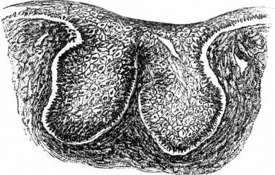
Fig. 1. Section of the mammary gland of a foetus in the fifth month, showing the ingrowth of the epithelium into the subjacent tissues.
Fig 2. Section of the mammary gland of a foetus at the beginning of the sixth month. The ingrowth is larger, and the bridge of epithelium yet intact.
from four months of intra-uterine life to one month-after birth. Sections were cut of, the whole-of these, and between zoo and 300 were mounted and examined; those ‘which’ I show you "to-day have been", selected as most suitable for my purpose, and most. of the conclusions I shall ask you to accept, may either be confirmed or negatived by. an examination of them. The age given must be taken as to a great extent approximated .
admits of some variation. I could not discover anything at the fourth month, but succeeded in doing so at the fifth (Fig. I) ; it consisted of a very slight thickening of the deeper cells of the epiblast, exactly similar to that which. may be noticed in a developing hair, or sweatgland. It was covered by a thick bridge of cuticle, and limited by an ingrowth of the cells forming the rete Malpighii. This ingrowth, at first simply of a round or flask shape, later on becomes bifurcated, and shows a tendency to become hollowed out at the point nearest to the horny layer of the epithelium. (Fig. 2.) “This hollowing out seems to be effected by a great increase in the size of the cells which are superficial to the rete Malpighii, so that their contiguous borders form a network, the meshes of which constantly tend to increase in diameter, the cell-wall finally giving way, and its substance shrivelling up. The horny layer of epithelium which is at first spread over the mass of cells then breaks away, and a hollowed out ingrowth, lined by the rete Malpighii, results.
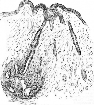
Before, however, this has occurred, processes of the epithelium have begun to grow downwards into the subjacent tissue, and by the seventh month have become tolerably numerous (Fig. 3). They in their turn are excavated in a manner similar to that described above, with the exception of some, which appear to be hollow at their commencement. During the last two months of intra-uterine life, the processes of epithelium penetrate still further into - the subjacent embryonic tissue ; they divide and subdivide, and at their extremities, and as direct outgrowths from their walls, thefimammary acini are formed. (Fig. 4.) During the month immediately subsequent to birth no material changes seem to occur; and, judging from the quietude of the gland, I think it may be safely assumed that,. until puberty, no further development takes place. I did not notice any material difference in the manner of the development in the two sexes, but the extent to which a gland had undergone developments in any given month was very variable. Now as, in all the 200 or 300 sectionsl examined, there was never any appearance of acini in the early months, as‘ the first rudiment of the gland was entirely epiblast, as the ductsccould be plainly seen to originate by a process of sprouting from the growing mass of epithelial cells; and, as the development of the acini was in every case strictly proportionate to, and in direct continuity with, the ingrowths of epiblastic tissue, I consider that the only conclusion, to be arrived .at is, that the acini as well as the ducts are developed from epiblast.
Fig. 3. Section of the mammary gland of a foetus aged seven months. The ingrowth is now hollowed, the bridge has broken away, and ducts are being developed from the epithelial ingrowth.
I shall now turn very shortly to the part played by- the subjacent mesoblastic tissues, and to some of the points on which I differ from Dr. Creighton. I have no hesitation in aflirming that, in the specimens I have examined, the mesoblast acts an entirely passive part. At the fifth month it consists almost (entirely of spindle-shaped cells and embryonic connective tissue, the former of which may be found collecting into groups where the fat is subsequently to be found.
Fig. 4. Section of a portion of the mammary gland of a foetus aged about seven months. Acini are being developed at the terminal extremity of one of the ducts.
There is no mapping out of the shape of the future gland in the mesoblast, there is no independent formation of acini; and. though I am quite prepared to admit the similarity which exists between the developing fat-tissue and the cells of the acini, it is merely a superficial resemblance, and, at any rate, in the human subject, the two cannot well be confounded.
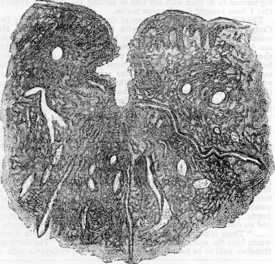
Fig. 5. Section of the mammary gland of a new-born child, showing the ducting from the nipple.
The description by Creighton of the development of the ducts in the guinea-pig, does not obtain in the human subject for, in this account, the ducts are said to first-make their appearance as small channels in the mesoblast; further, “there is certainly no evidence of a procc? loft growth downwardtsl from thekrete‘ trlnuccpsutrn undclr thfe ni pllg :” an a er on esame an or remar s: e uc sare ere ore e o be formed as aggregations of the embryonic cells of the matrix along predlcteilmingd lines; that preliminary point may be taken as esta listed. But, however much it may be established in the guinea-pig, I allege that there is before you ocular demonstration of a different method of formation in the human mamma. No attempt at the formation of a duct canbe discerned in the mesoblastic tissue, except in direct continuity with the epithelial ingrowth, and there is abundant evidence of a process of growth downwards from the rete mucosum under the nipple.
It will thus be seen that my observations are: in accordance with the views of Kolliker, as referred to above. The conclusions drawn from them may be briefly summarised as follows.
- The mammary gland is developed from epiblast.
- The acini do not appear until the ducts are formed by an ingrowth of the epithelial cells of the rete mucosum.
- The acini are developed from the epithelial cells forming the ducts.
- The mesoblast takes no part in the formation of the glandular structures.
| Historic Disclaimer - information about historic embryology pages |
|---|
| Pages where the terms "Historic" (textbooks, papers, people, recommendations) appear on this site, and sections within pages where this disclaimer appears, indicate that the content and scientific understanding are specific to the time of publication. This means that while some scientific descriptions are still accurate, the terminology and interpretation of the developmental mechanisms reflect the understanding at the time of original publication and those of the preceding periods, these terms, interpretations and recommendations may not reflect our current scientific understanding. (More? Embryology History | Historic Embryology Papers) |
Cite this page: Hill, M.A. (2026, February 27) Embryology Paper - Development of the mammary gland. Retrieved from https://embryology.med.unsw.edu.au/embryology/index.php/Paper_-_Development_of_the_mammary_gland
- © Dr Mark Hill 2026, UNSW Embryology ISBN: 978 0 7334 2609 4 - UNSW CRICOS Provider Code No. 00098G


