K12 - Communication
| Embryology - 28 Apr 2024 |
|---|
| Google Translate - select your language from the list shown below (this will open a new external page) |
|
العربية | català | 中文 | 中國傳統的 | français | Deutsche | עִברִית | हिंदी | bahasa Indonesia | italiano | 日本語 | 한국어 | မြန်မာ | Pilipino | Polskie | português | ਪੰਜਾਬੀ ਦੇ | Română | русский | Español | Swahili | Svensk | ไทย | Türkçe | اردو | ייִדיש | Tiếng Việt These external translations are automated and may not be accurate. (More? About Translations) |
Introduction
Biological communication occurs at many levels, from one cell communication with its neighbour in a tissue (paracrine), to signals released into the blood from one cell to signal to another cell or tissue (endocrine or hormone signaling). The signalling that occurs in the brain, spinal cord and other nervous tissues involves electrical (action potentials) signaling. All sensory information needs to be first converted into this form before the brain can interpret.
This page will introduce development of signaling in our special sensory nervous systems: the eyes for vision and the ears for hearing. Both systems convert signals in one medium (light or sound) into an electrical signal that our brain can understand.
Note that other animals may have additional sensory systems or sensory systems that allow highly specialised vision, hearing or touch.
Sound - Hearing
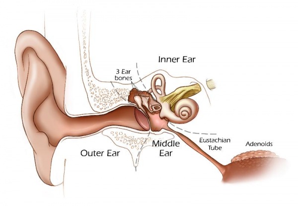
This cartoon shows the adult "ear" with the 3 main divisions (outer, middle, inner).
Outer Ear
|
Middle Ear
|
Inner Ear
|
Hearing Before Birth
Can a baby "hear" like we do before it is born?
External Ear Growth before Birth
What's so important about the outer ear?
| Month 3 - Fetus | Month 4 - Fetus | Month 5 - Fetus |
|---|---|---|
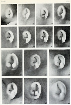
|
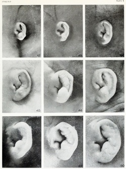
|
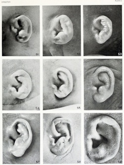
|
| About the Outer Ear shape and Position. |
|---|
|
The position and shape of the external ear can tell you about how the head grows before birth!
|
Testing a Baby's Hearing
If a baby cannot hear you talk they cannot easily learn to speak!
Much of your first learning comes from being able to copy sounds that you hear. The parents or doctors would only know that a baby could not hear if they were not developing speech correctly or meeting other developmental milestones.
- In the past - the tests were very simple, like turning their head to a bell or sound. They could also not be easily carried out on newborn infants.
- Today - the tests are computer-based, requiring no infant response and can be easily carried out on newborn infants.
| About Newborn Hearing Tests |
|---|
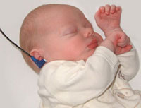
Whats an Automated Auditory Brainstem Response? (AABR) uses a computer generated stimulus (click) that is delivered through earphones and detected by scalp electrodes.
|
Light - Vision
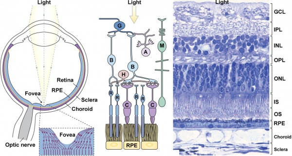
This cartoon[1] shows the eyeball (left), a cartoon of the retinal cell organisation (middle) and an actual slice of the human retina.
The Eye
|
Retinal Cell Organization
These are the names of the cells and neurones required to convert light into electrical signals. (detect, process and carry)
|
| Notice that light has to pass through all the other retina cell layers to the detection cell layer. Then when an electrical signal, be transferred back up through these neurons to the ganglion cells, that carry the signal to the brain in the optic nerve. | |
- ↑ <pubmed>20855501</pubmed>| PMC3101587 | JCB
Glossary Links
- Glossary: A | B | C | D | E | F | G | H | I | J | K | L | M | N | O | P | Q | R | S | T | U | V | W | X | Y | Z | Numbers | Symbols | Term Link
Cite this page: Hill, M.A. (2024, April 28) Embryology K12 - Communication. Retrieved from https://embryology.med.unsw.edu.au/embryology/index.php/K12_-_Communication
- © Dr Mark Hill 2024, UNSW Embryology ISBN: 978 0 7334 2609 4 - UNSW CRICOS Provider Code No. 00098G

