Introduction
The vernix caseosa consists of a bilayer formed by corneocytes embedded in a rich lipid matrix, formed by the sebaceous glands and epidermis, that is anchored to the fetal epithelium by lanugo hair.
Prenatally, this thick integumentary surface layer may, like the stratum corneum, protect the fetal skin from the watery amniotic environment and the enzymatic agents it contains. At birth, this slippery layer may aid the delivery process. The vernix has also many other known protective and immune as well as potential functions.[1] The constituent components and their concentrations appear to differ between individuals and the sexes.[2]
In late gestation, vernix from the fetal skin sheds as particles suspended within the amniotic fluid. Amniotic fluid is also continuously swallowed during this period and found within both the fetal gastrointestinal tract and respiratory tract.
Some Recent Findings
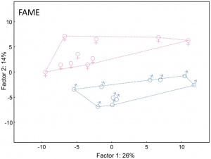
Newborn boys and girls differ in the lipid composition of vernix caseosa
[2]
- Review - Biology of the vernix caseosa[3] "The vernix caseosa is a complex membranous structure comprising 80% water, 10% protein, and 10% lipids including barrier lipids such as ceramides, free fatty acids, phospholipids and cholesterol, synthesized partly by fetal sebaceous glands during the last trimester of pregnancy in an antero-posterior and dorsoventral manner. Because of its lipid content, vernix is hydrophobic and protects the skin from excessive water exposure during the development of the stratum corneum. The vernix caseosa has various functions during fetal transition from an intrauterine to an extrauterine environment, including lubrication of the birth canal during parturition, barrier function to prevent water loss, temperature regulation, for innate immunity and for intestinal development. This review discusses the evidence supporting the prenatal and postnatal functions of vernix caseosa, along with its structure, composition, and physical and biological characteristics. Understanding the biology of the vernix may facilitate improved care of preterm infants immediately post-partum.}
- Applying a vernix caseosa based formulation accelerates skin barrier repair by modulating lipid biosynthesis[4] "Restoring the lipid homeostasis of the stratum corneum (SC) is a common strategy to enhance skin barrier function. Here, we used a ceramide containing vernix caseosa (VC)-based formulation and were able to accelerate barrier recovery in healthy volunteers. The recovery was examined over 16 days by monitoring trans-epidermal water loss (TEWL) after barrier disruption by tape-stripping. Four skin sites were used to examine the effects of both treatment and barrier recovery. After 16 days, samples were harvested at these sites to examine the SC ceramide composition and lipid organization. Changes in ceramide profiles were identified using principal component analysis. After barrier recovery, the untreated sites showed increased levels of ceramide subclass AS and ceramides with a 34 total carbon-atom chain length, while the mean ceramide chain length was reduced. These changes were diminished by treatment with the studied formulation, which concurrently increased the formulated ceramides. Correlations were observed between SC lipid composition, lipid organization, and TEWL, and changes in the ceramide subclass composition suggest changes in the ceramide biosynthesis. These results suggest that VC-based formulations enhance skin barrier recovery and are attractive candidates to treat skin disorders with impaired barrier properties."
- Skin barrier in the neonate[5] "The purpose of this review is to focus on determinants of skin barrier function in neonates at molecular and cellular levels. The skin barrier is critical in terms of water and gas exchanges during fetal life and undergoes rapid changes at birth, followed by a progressive maturation. Consequences of skin barrier disruption can be extremely detrimental or lethal, as shown in severe genetic epidermal defects. In this context, the fine-tuned rapid adaptation from a liquid to a gaseous milieu is not fully understood. The stratum corneum provides an air-liquid barrier, tight junctions in the granular layer provide a liquid-liquid barrier, aquaporins represent a plumbing system for water-glycerol as well as gas exchanges, and Langerhans cells are central to the immunological barrier. Acid mantle formation is essential for appropriate interaction between the skin and microbial symbionts. Temperature and pH regulate the key enzyme activities responsible for the integrity of the stratum corneum. Skin barrier permeability can be assessed noninvasively and simply with miniaturized devices measuring transepidermal water loss, where water flow is faster in cases of a damaged or functionally premature barrier. New avenues for therapeutic skin barrier research in neonates include a better delineation of the maturation of aquaporins in water balance and gas exchanges from fetal to neonatal life and a better understanding of the role of vernix caseosa, in particular, for the implantation of a healthy microbiote. Practical applications should be derived for caring for infant skin, particularly in fragile zones, such as the diaper area." neonatal
|
| More recent papers
|
|
This table allows an automated computer search of the external PubMed database using the listed "Search term" text link.
- This search now requires a manual link as the original PubMed extension has been disabled.
- The displayed list of references do not reflect any editorial selection of material based on content or relevance.
- References also appear on this list based upon the date of the actual page viewing.
References listed on the rest of the content page and the associated discussion page (listed under the publication year sub-headings) do include some editorial selection based upon both relevance and availability.
More? References | Discussion Page | Journal Searches | 2019 References | 2020 References
Search term: Vernix Caseosa | Vernix Development
|
| Older papers
|
| These papers originally appeared in the Some Recent Findings table, but as that list grew in length have now been shuffled down to this collapsible table.
See also the Discussion Page for other references listed by year and References on this current page.
- Newborn boys and girls differ in the lipid composition of vernix caseosa[2] "Vernix caseosa protects the skin of a human fetus during the last trimester of pregnancy and of a newborn after the delivery. Besides its cellular and proteinaceous components, an important constituent and functional agent is a complex lipid fraction, implicated in a multitude of salubrious effects of vernix caseosa. Little is known about how the chemical composition of vernix caseosa lipids is affected by various biological characteristics of the baby, such as the gestational age, birth weight, and, last but not least, the gender of the newborn. This study reports on the chemical variability of lipids contained in the vernix caseosa of twenty newborn girls and boys and shows that the quantitative patterns of the lipids are sex-specific. The specificity of lipids was investigated at the level of fatty acids in the total lipid extracts and intact lipids of several neutral lipid classes. Hydrocarbons, wax esters, cholesteryl esters, diol diesters and triacylglycerols were isolated using optimized semipreparative thin-layer chromatography, and the molecular species within each class were characterized using matrix-assisted laser desorption/ionization mass spectrometry. Statistical evaluation revealed significant quantitative sex-related differences in the lipid composition of vernix caseosa among the newborns, pronounced in the two lipid classes associated with the activity of sebaceous glands. Higher proportions of wax esters and triacylglycerols with longer hydrocarbon chains were observed in newborn girls."
|
Textbooks
- Human Embryology (2nd ed.) Larson Chapter 14 p443-455
- The Developing Human: Clinically Oriented Embryology (6th ed.) Moore and Persaud Chapter 20: P513-529
- Before We Are Born (5th ed.) Moore and Persaud Chapter 21: P481-496
- Essentials of Human Embryology Larson Chapter 14: P303-315
- Human Embryology, Fitzgerald and Fitzgerald
- Color Atlas of Clinical Embryology Moore Persaud and Shiota Chapter 15: p231-236
Integumentary Development Overview
4 weeks
- simple ectoderm epithelium over mesenchyme.
1-3 months
- ectoderm - germinative (basal) cell repeated division of generates stratified epithelium.
- mesoderm - differentiates into connective tissue and blood vessels.
- week 11 - (GA week 13) blood vessels visible in the early fetal skin, small blood vessels in the upper papillary region and larger vessels in the deep reticular dermis.[7]
|
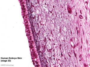
Integument Human Embryo (Week 8, Stage 22)
|
4 months
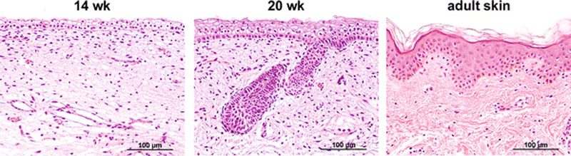
Fetal human integumentary histology[7](Weeks in figure are from LMP)
- Basal cell - proliferation generates folds in basement membrane.
- Neural crest cells - melanoblasts migrate into epithelium. These are the future melanocyte pigment cell of the skin.
- Embryonic connective tissue- differentiates into dermis, a loose ct layer over a dense ct layer. Beneath the dense ct layer is another loose ct layer that will form the subcutaneous layer.
- Ectoderm contributes to nails, hair follictles and glands.
- Nails form as thickening of ectoderm epidermis at the tips of fingers and toes. These form germinative cells of nail field.
- Cords of these cells extend into mesoderm forming epithelial columns. These form hair follicles, sebaceous and sweat glands.
5 months
- Hair growth initiated at base of cord, lateral outgrowths form associated sebaceous glands.
- Other cords elongate and coil to form sweat glands.
- Cords in mammary region branch as they elongate to form mammary glands. These glands will complete development in females at puberty. Functional maturity only occurs in late pregnancy.
Embryonic and Fetal Epidermis
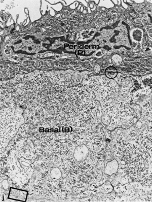
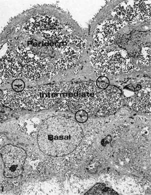
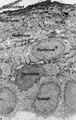
Electron Micrographs of the Developing Human Epidermis[8]
6 to 8 weeks (8-9 week EGA)
|
7 to 9 weeks (9-11 week EGA)
|
22 weeks (about 24 week EGA)
|
Functions
Vernix has many different known and potential functions.[1]
- a highly variable coating of the fetal skin
- develops cranio-caudally production coincides in utero with terminal differentiation of the epidermis and formation of the stratum corneum
- primarily composed of sebum, cells that have sloughed off the fetus's skin and shed lanugo hair
- can be absent in preterm infants
- dehydration and rehydration processes occur two to four times faster at 37 degrees celcius than at room temperature[9]
- towards term fragments of vernix can mix into the amniotic fluid resulting in (normal) turbidity
- fetal swallowing of amniotic fluid mixed with fragments of vernix can also occur
Components
- high water content (80%) largely compartmentalized within fetal corneocytes (cells forming the stratum corneum)
- saturated branched chain fatty acids (BCFA)
- squalene, a lipid produced by sebaceous glands, and also the biochemical precursor to steroids.
- cathelicidin LL-37, alpha-defensins, and LL-37 in neutrophils.[10]
- Lysozyme, lactoferrin, human neutrophil peptides 1-3, and secretory leukocyte protease inhibitor
Lanugo Hair
Lanugo hair is the first hair produced by the fetal hair follicles, appearing at about 5 months of gestation. The hair is very fine, soft, and usually unpigmented. Normally lost (shed) before birth, though sometimes present at birth.
- Links: hair
Abnormalities
Neonatal Aspiration Syndrome
Neonatal aspiration syndrome caused by vernix caseosa is very rare condition, caused by the infant oral cavity becoming filled with aggregates of vernix caseosa.[11]
Vernix Caseosa Peritonitis
Vernix caseosa peritonitis is a very rare post caesarean section inflammatory response occurring maternally after birth.[12]
References
- ↑ 1.0 1.1 Pickens WL, Warner RR, Boissy YL, Boissy RE & Hoath SB. (2000). Characterization of vernix caseosa: water content, morphology, and elemental analysis. J. Invest. Dermatol. , 115, 875-81. PMID: 11069626 DOI.
- ↑ 2.0 2.1 2.2 Míková R, Vrkoslav V, Hanus R, Háková E, Hábová Z, Doležal A, Plavka R, Coufal P & Cvačka J. (2014). Newborn boys and girls differ in the lipid composition of vernix caseosa. PLoS ONE , 9, e99173. PMID: 24911066 DOI.
- ↑ Nishijima K, Yoneda M, Hirai T, Takakuwa K & Enomoto T. (2019). Biology of the vernix caseosa: A review. J. Obstet. Gynaecol. Res. , 45, 2145-2149. PMID: 31507021 DOI.
- ↑ Boiten WA, Berkers T, Absalah S, van Smeden J, Lavrijsen APM & Bouwstra JA. (2018). Applying a vernix caseosa based formulation accelerates skin barrier repair by modulating lipid biosynthesis. J. Lipid Res. , 59, 250-260. PMID: 29217624 DOI.
- ↑ Taïeb A. (2018). Skin barrier in the neonate. Pediatr Dermatol , 35 Suppl 1, s5-s9. PMID: 29596733 DOI.
- ↑ Fuchs E. (2008). Skin stem cells: rising to the surface. J. Cell Biol. , 180, 273-84. PMID: 18209104 DOI.
- ↑ 7.0 7.1 Coolen NA, Schouten KC, Middelkoop E & Ulrich MM. (2010). Comparison between human fetal and adult skin. Arch. Dermatol. Res. , 302, 47-55. PMID: 19701759 DOI.
- ↑ Dale BA, Holbrook KA, Kimball JR, Hoff M & Sun TT. (1985). Expression of epidermal keratins and filaggrin during human fetal skin development. J. Cell Biol. , 101, 1257-69. PMID: 2413039
- ↑ Rissmann R, Groenink HW, Gooris GS, Oudshoorn MH, Hennink WE, Ponec M & Bouwstra JA. (2008). Temperature-induced changes in structural and physicochemical properties of vernix caseosa. J. Invest. Dermatol. , 128, 292-9. PMID: 17671513 DOI.
- ↑ Yoshio H, Lagercrantz H, Gudmundsson GH & Agerberth B. (2004). First line of defense in early human life. Semin. Perinatol. , 28, 304-11. PMID: 15565791
- ↑ Nishijima K, Shukunami K, Inoue S & Kotsuji F. (2005). Management for neonatal aspiration syndrome caused by vernix caseosa. Fetal. Diagn. Ther. , 20, 194-6. PMID: 15824497 DOI.
- ↑ Stuart OA, Morris AR & Baber RJ. (2009). Vernix caseosa peritonitis - no longer rare or innocent: a case series. J Med Case Rep , 3, 60. PMID: 19208257 DOI.
Reviews
Visscher MO, Adam R, Brink S & Odio M. (2015). Newborn infant skin: physiology, development, and care. Clin. Dermatol. , 33, 271-80. PMID: 25889127 DOI.
Visscher M & Narendran V. (2014). The Ontogeny of Skin. Adv Wound Care (New Rochelle) , 3, 291-303. PMID: 24761361 DOI.
Chambers AC, Patil AV, Alves R, Hopkins JC, Armstrong J & Lawrence RN. (2012). Delayed presentation of vernix caseosa peritonitis. Ann R Coll Surg Engl , 94, 548-51. PMID: 23131223 DOI.
Singh G & Archana G. (2008). Unraveling the mystery of vernix caseosa. Indian J Dermatol , 53, 54-60. PMID: 19881987 DOI.
Visscher MO, Narendran V, Pickens WL, LaRuffa AA, Meinzen-Derr J, Allen K & Hoath SB. (2005). Vernix caseosa in neonatal adaptation. J Perinatol , 25, 440-6. PMID: 15830002 DOI.
Articles
Kalužíková A, Vrkoslav V, Harazim E, Hoskovec M, Plavka R, Buděšínský M, Bosáková Z & Cvačka J. (2017). Cholesteryl esters of ω-(O-acyl)-hydroxy fatty acids in vernix caseosa. J. Lipid Res. , 58, 1579-1590. PMID: 28576934 DOI.
Checa A, Holm T, Sjödin MO, Reinke SN, Alm J, Scheynius A & Wheelock CE. (2015). Lipid mediator profile in vernix caseosa reflects skin barrier development. Sci Rep , 5, 15740. PMID: 26521946 DOI.
Visscher MO, Utturkar R, Pickens WL, LaRuffa AA, Robinson M, Wickett RR, Narendran V & Hoath SB. (2011). Neonatal skin maturation--vernix caseosa and free amino acids. Pediatr Dermatol , 28, 122-32. PMID: 21504444 DOI.
Tollin M, Jägerbrink T, Haraldsson A, Agerberth B & Jörnvall H. (2006). Proteome analysis of vernix caseosa. Pediatr. Res. , 60, 430-4. PMID: 16940245 DOI.
Rissmann R, Groenink HW, Weerheim AM, Hoath SB, Ponec M & Bouwstra JA. (2006). New insights into ultrastructure, lipid composition and organization of vernix caseosa. J. Invest. Dermatol. , 126, 1823-33. PMID: 16628195 DOI.
Tollin M, Bergsson G, Kai-Larsen Y, Lengqvist J, Sjövall J, Griffiths W, Skúladóttir GV, Haraldsson A, Jörnvall H, Gudmundsson GH & Agerberth B. (2005). Vernix caseosa as a multi-component defence system based on polypeptides, lipids and their interactions. Cell. Mol. Life Sci. , 62, 2390-9. PMID: 16179970 DOI.
Akinbi HT, Narendran V, Pass AK, Markart P & Hoath SB. (2004). Host defense proteins in vernix caseosa and amniotic fluid. Am. J. Obstet. Gynecol. , 191, 2090-6. PMID: 15592296 DOI.
Yoshio H, Tollin M, Gudmundsson GH, Lagercrantz H, Jornvall H, Marchini G & Agerberth B. (2003). Antimicrobial polypeptides of human vernix caseosa and amniotic fluid: implications for newborn innate defense. Pediatr. Res. , 53, 211-6. PMID: 12538777 DOI.
Hoeger PH, Schreiner V, Klaassen IA, Enzmann CC, Friedrichs K & Bleck O. (2002). Epidermal barrier lipids in human vernix caseosa: corresponding ceramide pattern in vernix and fetal skin. Br. J. Dermatol. , 146, 194-201. PMID: 11903227
Search PubMed
Search Pubmed: Vernix Caseosa
NCBI - Policies and Guidelines | PubMed | Help:Reference Tutorial
Additional Images
Category:Integumentary
Historic
| Historic Disclaimer - information about historic embryology pages
|
| Pages where the terms "Historic" (textbooks, papers, people, recommendations) appear on this site, and sections within pages where this disclaimer appears, indicate that the content and scientific understanding are specific to the time of publication. This means that while some scientific descriptions are still accurate, the terminology and interpretation of the developmental mechanisms reflect the understanding at the time of original publication and those of the preceding periods, these terms, interpretations and recommendations may not reflect our current scientific understanding. (More? Embryology History | Historic Embryology Papers)
|
Terms
| Integumentary Terms
|
Integumentary Development
- acrosyringium - coiled intra-epidermal region of the eccrine gland sweat duct.
- apocrine gland - (sweat gland) proteinaceous secretion associated with hair (axilla, areola, genital and anal regions). Additional glands associated with eyelashes are called the glands of Moll (ciliary gland). (More? image - apocrine secretion)
- arrector pili muscle - bundle of smooth muscle associated with hair follicle, inserts into the papillary layer of the dermis and attaches to the dermal sheath of the hair follicle. (More? image - arrector pili muscle)
- Blaschko lines - (lines of Blaschko) may represent pathways of epidermal cell migration and proliferation during development. Specific type of lupus erythematosus shows this distinctive pattern. Named after Alfred Blaschko a German dermatologist who first described the feature in 1901. (More? PMID 21396561 | Historic Terminology)
- bulb - the hair follicle enlargement located at its deepest end, dividing cells form the hair and the root sheath.
- café-aut-lait macule - (French, cafe-au-lait = coffee with milk; birthmark) describes the characteristic colour of the skin hyperpigmented patch present at birth (congenital) or appearing in early infancy. Common single feature, multiple are associated with various genetic syndromes including Neurofibromatosis type 1 and 2.
- corneocytes - terminally differentiated keratinocytes forming the stratum corneum.
- cutis - alternative term for the epidermis and the dermis layers of the skin.
- dermal papillae - interdigitation of the dermis with the epidermis.
- dermatoglyphic patterns - (Greek, derma = "skin", glyph = "carving") fingers, palms, toes, and soles skin patterns.
- dermis - connective tissue middle layer of the skin, consists of two sublayers (papillary and reticular layers) that do not have a clear boundary. Embryologically derived from the somite dermatome.
- dermomyotome - Early embryonic dorsolateral half of the somite that will later divide to form both the dermatome and myotome. The dermatome will contribute the dermis and hypodermis of the skin. The myotome will contribute the skeletal muscle of muscoloskeletal system. Development sequence: mesoderm to paraxial mesoderm to somite to "dermomyotome" then dermatome and myotome. (More? Somitogenesis | Musculoskeletal System Development | Integumentary System Development)
- eccrine gland (Greek, ekkrinein = "secrete"; merocrine glands) sweat glands unique to some primates and used in humans for thermoregulation. Adult body has 2 to 4 million sweat glands with concentrations (700/cm) on the palms of the hand, soles of the feet and forehead. Secretion is timulated by sympathetic nervous system, post-ganglionic cholinergic branch, and other stimuli
- ephilis - (pl., ephilides; freckle) Clinical term describing a "freckle", that is a small brown or tan mark on the skin. These inherited features result from a copy of variant Melanocortin 1 Receptor (MC1R) gene and are common on fair skinned Celtic children. Melanocytes produce locally more melanin, this can also increase following exposure to ultraviolet radiation in sunlight. (More? Integumentary | Neural Crest | OMIM MC1R)
- epidermis - Histological term describing the external cellular epithelial layer of the integumentary (skin) covering the entire body. This surface layer of keratinocytes is ectoderm in origin, while the underlying connective tissue layers of dermis and hypodermis are mesoderm in origin. (More? Integumentary Development)
- epidermal differentiation complex - (EDC) human chromosome (1q2) containing linked 63 genes within four gene families that are molecular markers for stratified epidermis terminal differentiation.
- epidermal growth factor receptor - expressed on cells in the epidermis basal layer, signaling stimulates both epidermal growth and wound healing and also mediates an inhibition of differentiation.
- glabrous skin - skin without hair, refers to the palms of hands and soles of feet.
- hair - (pili) in humans consists of vellus and terminal hairs.
- holocrine - form of gland secretion where the secretory cells eventually lyse (rupture) and are lost. On the skin, these cells release sebum consisting mainly of lipid. (More? image - holocrine secretion)
- hypodermis - (subcutis, subcutaneous adipose) a connective tissue ilower layer of the skin that binds it to underlying structures.
- integumentary - term for the skin and its appendages.
- involucrin - protein that binds loricrin in the development of the cell envelope protecting corneocytes in the skin.
- keratinocyte - the main cell type forming the layers of the epidermis, derived from ectoderm.
- keratohyalin granule - found in the stratum granulosum consist of profilaggrin and loricrin.
- Langerhans cell - skin dendritic cell (antigen presenting cell) develops initially from fetal liver monocytes and yolk sac macrophages. May, depending on the immunological setting, elicit immunity or tolerance. Named after Paul Langerhans.
- Langer's lines - (skin cleavage lines, cleavage lines) Clinical term for the orientation of reticular dermis collagen bundles causing tensions on skin and subcutaneous tissues. Lines tend to be horizontal in the trunk and neck, and longitudinal in the skin and limbs. (More? PMID 15791423)
- Meissner corpuscle - sensory structure acting as a rapidly-adapting mechanoreceptor mainly in the dermal papillae of (digital) skin. (More?Touch
- melanin - (Greek, melanos = black) The pigment produced by melanocytes that provides photoprotection, preventing cellular DNA damage, and colouring of the basal epithelial cells that absorb the pigment.
- melanodermia - hyperpigmentation causing abnormal darkening (brown/black) of the skin due to excess melanin or by metallic substances. See also the abnormality ceruloderma (blue/grey). (More? PMID 23522626)
- melanocyte - (Greek, melanos = black) A pigmented cell, neural crest in origin, differentiating from melanoblasts located in the skin and other tissues that produces melanin. The melanocytes within the integument (skin) transfer melanin to keratinocytes to give skin colour and to the hair follicle to give hair colour. Melanocytes are also located within "non-cutaneous" tissues in the eye (for eye colour), harderian gland and inner ear. This is the cell type that proliferates in the cancer melanoma. (More? Neural Crest Development | Integumentary System Development)
- Merkel cell - An epidermal-derived cell in touch-sensitive area of the epidermis and mediate mechanotransduction in the skin. Previously thought to be neural crest in origin, but recently shown to arise from the embryonic epithelium. The cells are named after Friedrich Sigmund Merkel, a German anatomist who was the first to describe them in 1875. (More? Touch | Lecture - Integumentary Development | PMID 19786578 | PMID 3782861)
- merocrine gland - (sweat gland, eccrine sweat) simple tubular glands located at the border between the dermis and hypodermis. These glands regulate the body temperature. (More? image - merocrine secretion)
- nestin - (neuroectodermal stem cell marker) an intermediate filament protein (type VI) expressed in stem cells and transiently during development, and in cells within hair follicles, sebaceous and sweat glands.
- papillary layer - dermis sublayer that appears less dense and contains more cells lying close beneath the epidermis. (More? image)
- pilosebaceous unit - term used to describe a hair and its associated structures: hair follicle, arrector pili muscle and sebaceous gland.
- rete ridge - the extensions of the epidermis into the dermis. These epidermal surface thickenings extend downward between underlying connective tissue dermal papillae. This is also the site of initial eccrine gland differentiation.
- reticular layer - dermis sublayer that appears denser and contains fewer cells with thick collagen bundles lying beneath the papillary layer parallel to the skin surface. (More? image)
- root sheath - cell layers that surround the hair.
- sebaceous gland - holocrine gland associated with both the hair follicle and hairless parts of the skin (lips, cheek oral surface and external genitalia). Embedded in the dermis and are sites of infections (acne).
- simple - consisting of a single cell layer.
- terminal hairs - hair seen in obviously hairy parts of the body.
- thick skin - refers to the skin histology found on the palms of the hand and soles of the feet, does not contain hair. Note that this is used as a histological term not a measurement of overall skin thickness.
- thin skin - refers to the skin histology found on skin on all body regions, other than palms and soles (thick skin).
- vellus hairs - fine short hairs only lightly pigmented covering the body.
- vernix caseosa - (vernix, Latin, "caseosa" = cheese-like) a fetal protective coating consisting of sebum, skin cells and lanugo hair. Forming late in fetal development in a rostra-caudal sequence associated with epithelium differentiation.
- Voigt's lines - clinical term to describe the skin borders between areas of innervations by specific peripheral cutaneous nerves. (More? Sensory Touch | Historic Terminology)
|
|
|
External Links
External Links Notice - The dynamic nature of the internet may mean that some of these listed links may no longer function. If the link no longer works search the web with the link text or name. Links to any external commercial sites are provided for information purposes only and should never be considered an endorsement. UNSW Embryology is provided as an educational resource with no clinical information or commercial affiliation.
Glossary Links
- Glossary: A | B | C | D | E | F | G | H | I | J | K | L | M | N | O | P | Q | R | S | T | U | V | W | X | Y | Z | Numbers | Symbols | Term Link
Cite this page: Hill, M.A. (2026, July 26) Embryology Integumentary System Development - Vernix Caseosa. Retrieved from https://embryology.med.unsw.edu.au/embryology/index.php/Integumentary_System_Development_-_Vernix_Caseosa
- What Links Here?
- © Dr Mark Hill 2026, UNSW Embryology ISBN: 978 0 7334 2609 4 - UNSW CRICOS Provider Code No. 00098G


