File:Keibel Mall 2 567.jpg
Original file (1,280 × 1,842 pixels, file size: 140 KB, MIME type: image/jpeg)
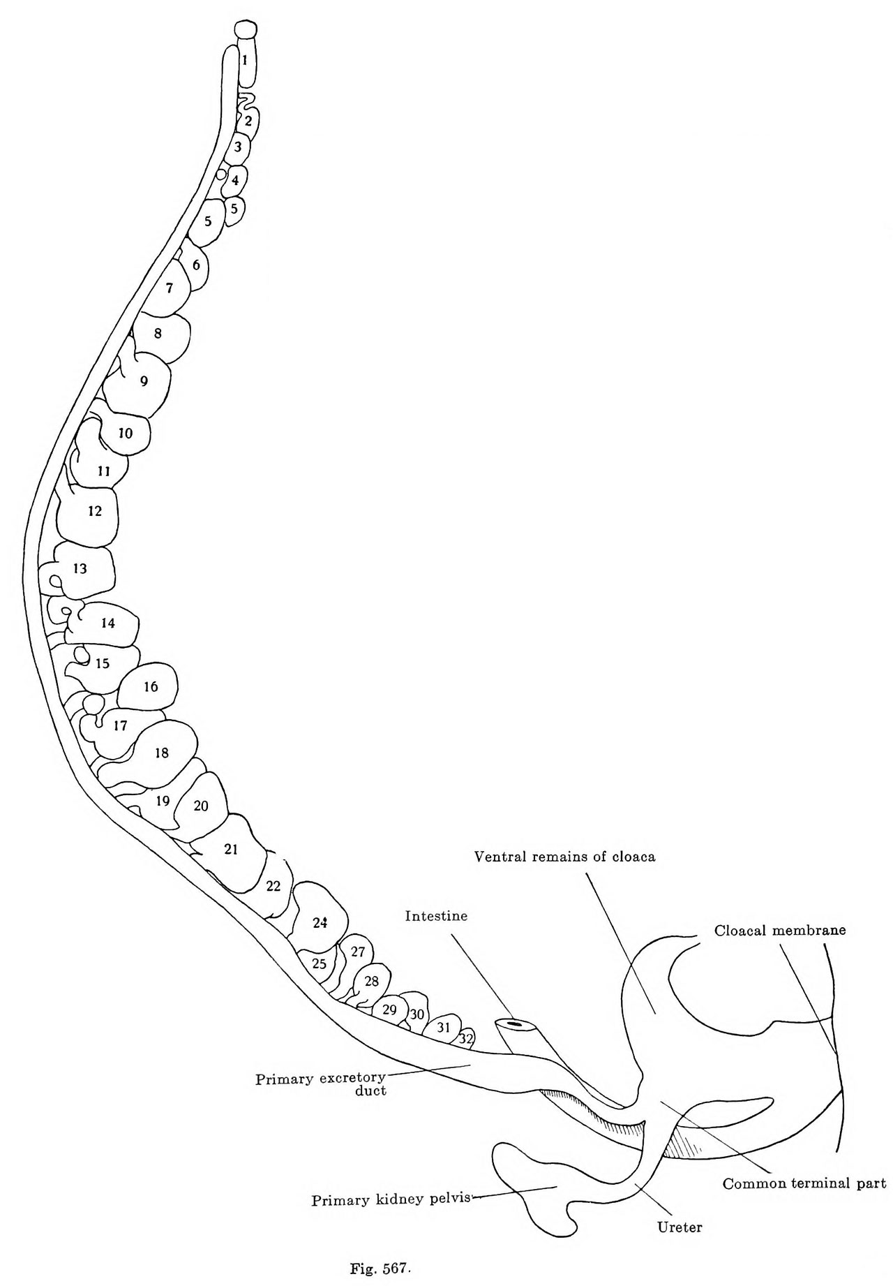
Fig. 567. Mesonephros Human Embryo 9.5 mm
Model of the mesonephros of a human embryo of 9.5 mm greatest length, embryo Ma. 3, from the collection of Professor Hochstetter, Vienna.
The same model that was represented in Fig. 566 seen from the right side.
The figure shows how the Malpighian corpuscles as they enlarge press each other out of the row. The primary excretory duct increases in size in its caudal half, the increase affecting both the dorsoventral and the frontal diameter, the latter to the greater extent It again diminishes before its union with the ureter. This has already developed the primitive pelvis of the kidney, from which the cranial and caudal pole tubules are beginning to grow out. J. The common terminal part of the ureter and the primary excretory duct still persists, but is greatly enlarged. The cloaca is almost completely subdivided.
| Embryology - 26 Apr 2024 |
|---|
| Google Translate - select your language from the list shown below (this will open a new external page) |
|
العربية | català | 中文 | 中國傳統的 | français | Deutsche | עִברִית | हिंदी | bahasa Indonesia | italiano | 日本語 | 한국어 | မြန်မာ | Pilipino | Polskie | português | ਪੰਜਾਬੀ ਦੇ | Română | русский | Español | Swahili | Svensk | ไทย | Türkçe | اردو | ייִדיש | Tiếng Việt These external translations are automated and may not be accurate. (More? About Translations) |
Felix W. The development of the urinogenital organs. In Keibel F. and Mall FP. Manual of Human Embryology II. (1912) J. B. Lippincott Company, Philadelphia. pp 752-979.
| Historic Disclaimer - information about historic embryology pages |
|---|
 |
Cite this page: Hill, M.A. (2024, April 26) Embryology Keibel Mall 2 567.jpg. Retrieved from https://embryology.med.unsw.edu.au/embryology/index.php/File:Keibel_Mall_2_567.jpg
- © Dr Mark Hill 2024, UNSW Embryology ISBN: 978 0 7334 2609 4 - UNSW CRICOS Provider Code No. 00098G
File history
Click on a date/time to view the file as it appeared at that time.
| Date/Time | Thumbnail | Dimensions | User | Comment | |
|---|---|---|---|---|---|
| current | 09:48, 21 February 2017 | | 1,280 × 1,842 (140 KB) | Z8600021 (talk | contribs) | |
| 09:47, 21 February 2017 | 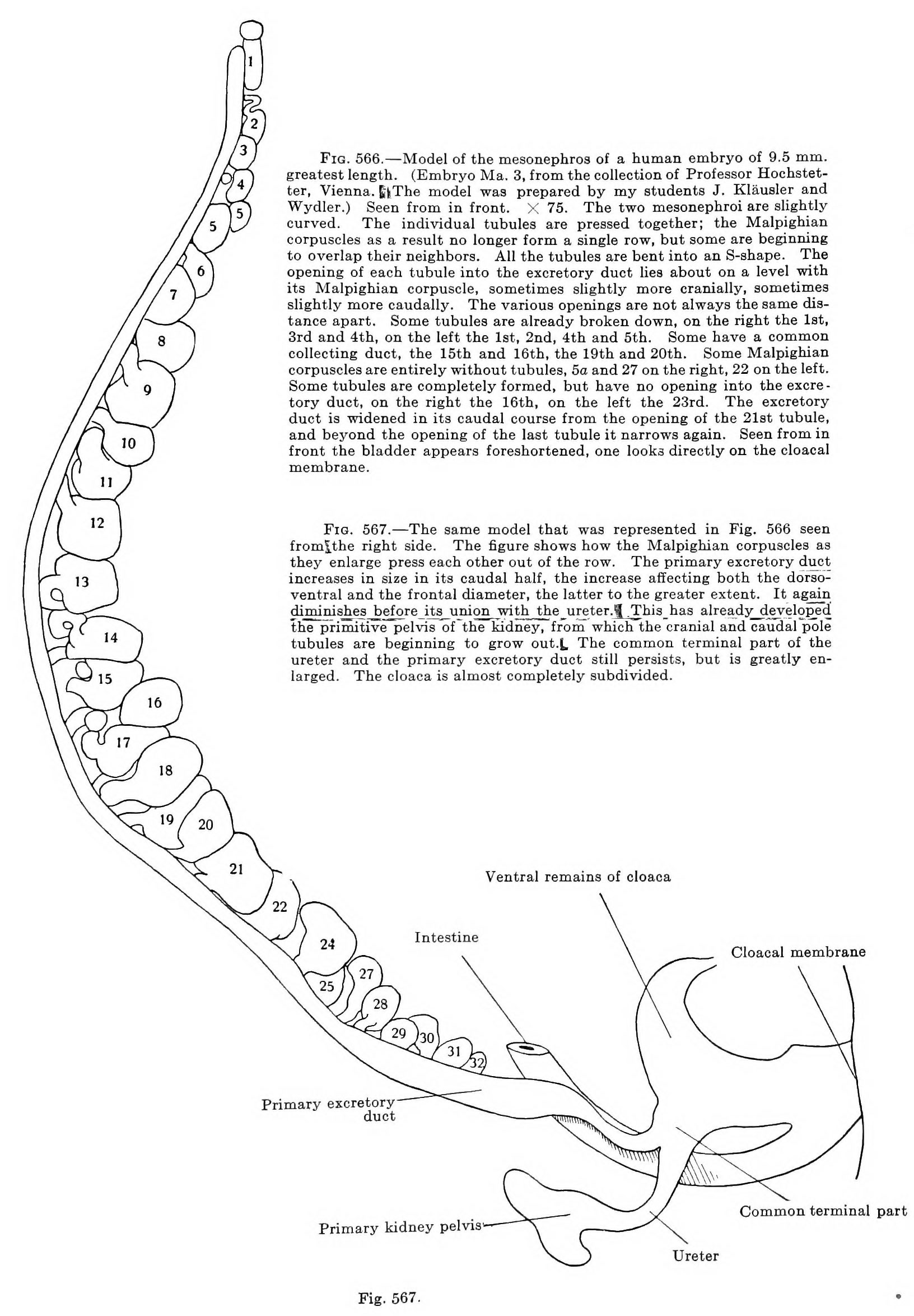 | 1,632 × 2,348 (517 KB) | Z8600021 (talk | contribs) |
You cannot overwrite this file.
File usage
The following 3 pages use this file: