File:Keibel Mall 2 554a.jpg
Original file (1,280 × 933 pixels, file size: 221 KB, MIME type: image/jpeg)
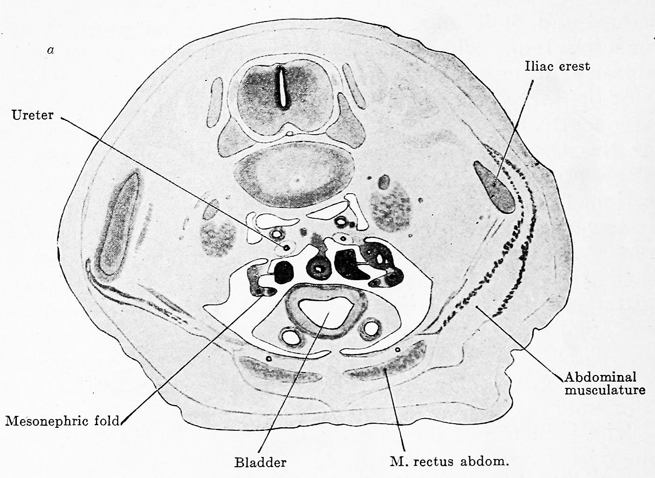
Fig. 554a. Transverse sections through a human embryo of 30 mm trunk length
(Embryo R. Meyer 273, from the collection of Professor Meyer, Berlin; slide 11, row 4, section 2; slide 13, row 5, section 2; slide 14, row 5, section 2.) The three sections show the formation of the genital cord. The legends have been arranged so that different structures are indicated in each section and the reader should first study the legends in all three figures.
a. The mesonephric fold bends between the tubar and gland portions at a right angle and is growing around the reproductive gland, so that the left and right mesonephros come to lie in the same frontal plane. The excretory duct and the tube are shown in the mesonephric fold. The originally lateral tube comes to lie medially, as a result of the bending of the fold. b. An evagination (the inguinal fold) extends from the mesonephric fold between the bladder and the lateral wall of the body to unite with the posterior surface of the anterior abdominal wall (in Fig. c it is cut throughout its entire length). The right and left mesonephric folds have come nearer together.
| Embryology - 26 Apr 2024 |
|---|
| Google Translate - select your language from the list shown below (this will open a new external page) |
|
العربية | català | 中文 | 中國傳統的 | français | Deutsche | עִברִית | हिंदी | bahasa Indonesia | italiano | 日本語 | 한국어 | မြန်မာ | Pilipino | Polskie | português | ਪੰਜਾਬੀ ਦੇ | Română | русский | Español | Swahili | Svensk | ไทย | Türkçe | اردو | ייִדיש | Tiếng Việt These external translations are automated and may not be accurate. (More? About Translations) |
Felix W. The development of the urinogenital organs. In Keibel F. and Mall FP. Manual of Human Embryology II. (1912) J. B. Lippincott Company, Philadelphia. pp 752-979.
| Historic Disclaimer - information about historic embryology pages |
|---|
 |
Cite this page: Hill, M.A. (2024, April 26) Embryology Keibel Mall 2 554a.jpg. Retrieved from https://embryology.med.unsw.edu.au/embryology/index.php/File:Keibel_Mall_2_554a.jpg
- © Dr Mark Hill 2024, UNSW Embryology ISBN: 978 0 7334 2609 4 - UNSW CRICOS Provider Code No. 00098G
File history
Click on a date/time to view the file as it appeared at that time.
| Date/Time | Thumbnail | Dimensions | User | Comment | |
|---|---|---|---|---|---|
| current | 08:02, 15 November 2018 | | 1,280 × 933 (221 KB) | Z8600021 (talk | contribs) | |
| 07:21, 15 November 2018 |  | 1,992 × 1,609 (372 KB) | Z8600021 (talk | contribs) |
You cannot overwrite this file.
File usage
The following 2 pages use this file: