File:Human Stage22 spinal cord02.jpg
Original file (1,044 × 889 pixels, file size: 290 KB, MIME type: image/jpeg)
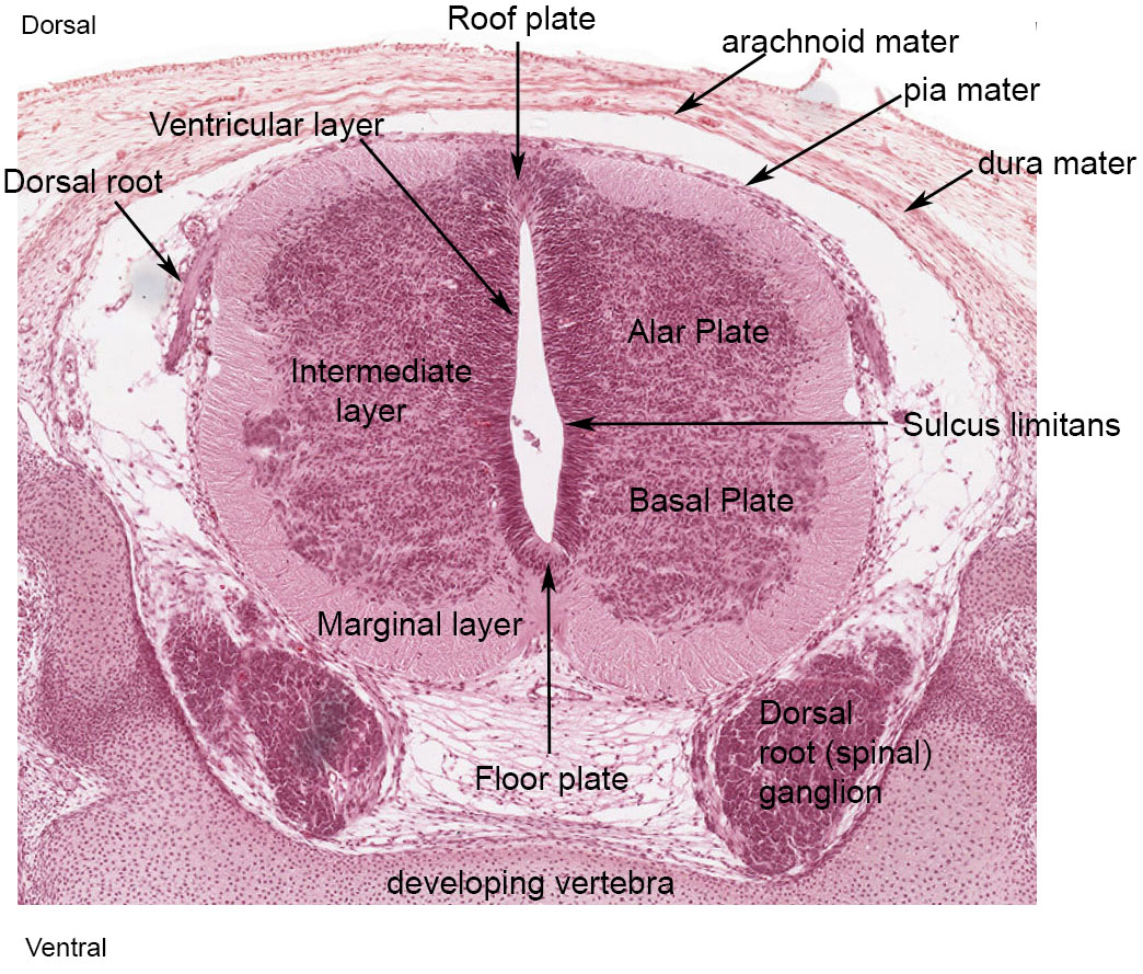
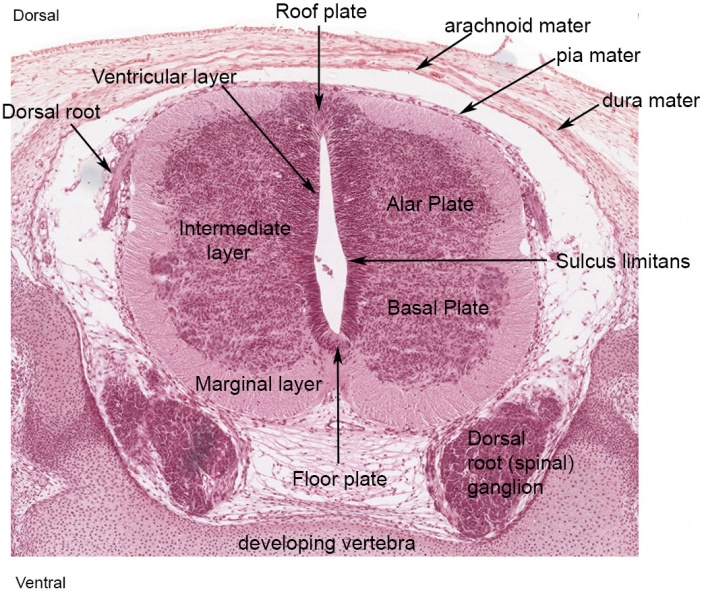
Human Embryo (Carnegie stage 22) Spinal Cord
See also unlabeled image 22. Note - this current image of the week 8 spinal cord has been rotated 180o to give the "neuroscience" view.
- Roof plate - thin wall region that underlies the dorsal ectoderm epithelium. Dorsal patterns the spinal cord, the roof plate produces Bone morphogenetic proteins (BMPs). [1][2]
- Alar plate - thick wall region lying either side of the roof floor plate. The sensory dorsal horn develops there and receives axons from the sensory structures outside the spinal cord. The adult horn is divided into 6 laminae (I to VI). Tracts formed by axons surround these horns and project both up and down the spinal cord.
- Lumen - neuroepithelium lined fluid-filled space continuous with the brain ventricular system.
- Basal plate - thick wall region lying either side of the floor floor plate. The ventral horn motor neurons develop here and extend axons out of the spinal cord to innervate developing skeletal muscle. Tracts formed by axons surround these horns and project both up and down the spinal cord.
- Floor plate - thin wall region that overlies the notochord. Ventral patterns the spinal cord, both floor plate and notochord produce Sonic hedgehog (Shh) (see also Notochord)
Stage 22 Spinal Cord Links: image unlabeled | image labeled | image low resolution | ANAT3411 | spinal cord | meninges
| Virtual Slide Features - Stage 22 Spinal Cord | ||
|---|---|---|
Spinal Cord Features |
The links shown are to specific features shown on the Human embryo (stage 22) Spinal Cord virtual slide. See also notes on Spinal Cord Development
Clicking the text will open the slide at a detailed view with the structure generally located in the centre of the view. The slide then can also be zoomed out from the set magnification using the controls in the upper left or the mouse. Use your browser back button to return to this table. Other Features
| |
| All Virtual Slides | Making your own Link - You can also make your own selected feature view. (See also Permalink help)
|
|
References
- ↑ Chizhikov VV & Millen KJ. (2004). Mechanisms of roof plate formation in the vertebrate CNS. Nat. Rev. Neurosci. , 5, 808-12. PMID: 15378040 DOI.
- ↑ Chizhikov VV & Millen KJ. (2005). Roof plate-dependent patterning of the vertebrate dorsal central nervous system. Dev. Biol. , 277, 287-95. PMID: 15617675 DOI.
Cite this page: Hill, M.A. (2024, May 9) Embryology Human Stage22 spinal cord02.jpg. Retrieved from https://embryology.med.unsw.edu.au/embryology/index.php/File:Human_Stage22_spinal_cord02.jpg
- © Dr Mark Hill 2024, UNSW Embryology ISBN: 978 0 7334 2609 4 - UNSW CRICOS Provider Code No. 00098G
File history
Click on a date/time to view the file as it appeared at that time.
| Date/Time | Thumbnail | Dimensions | User | Comment | |
|---|---|---|---|---|---|
| current | 15:01, 10 February 2016 | | 1,044 × 889 (290 KB) | Z8600021 (talk | contribs) | |
| 12:42, 10 February 2016 | 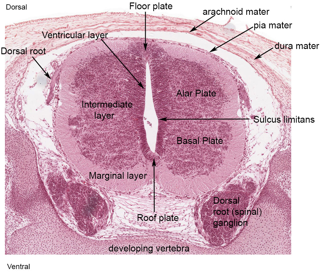 | 1,044 × 889 (290 KB) | Z8600021 (talk | contribs) | ||
| 12:27, 10 February 2016 | 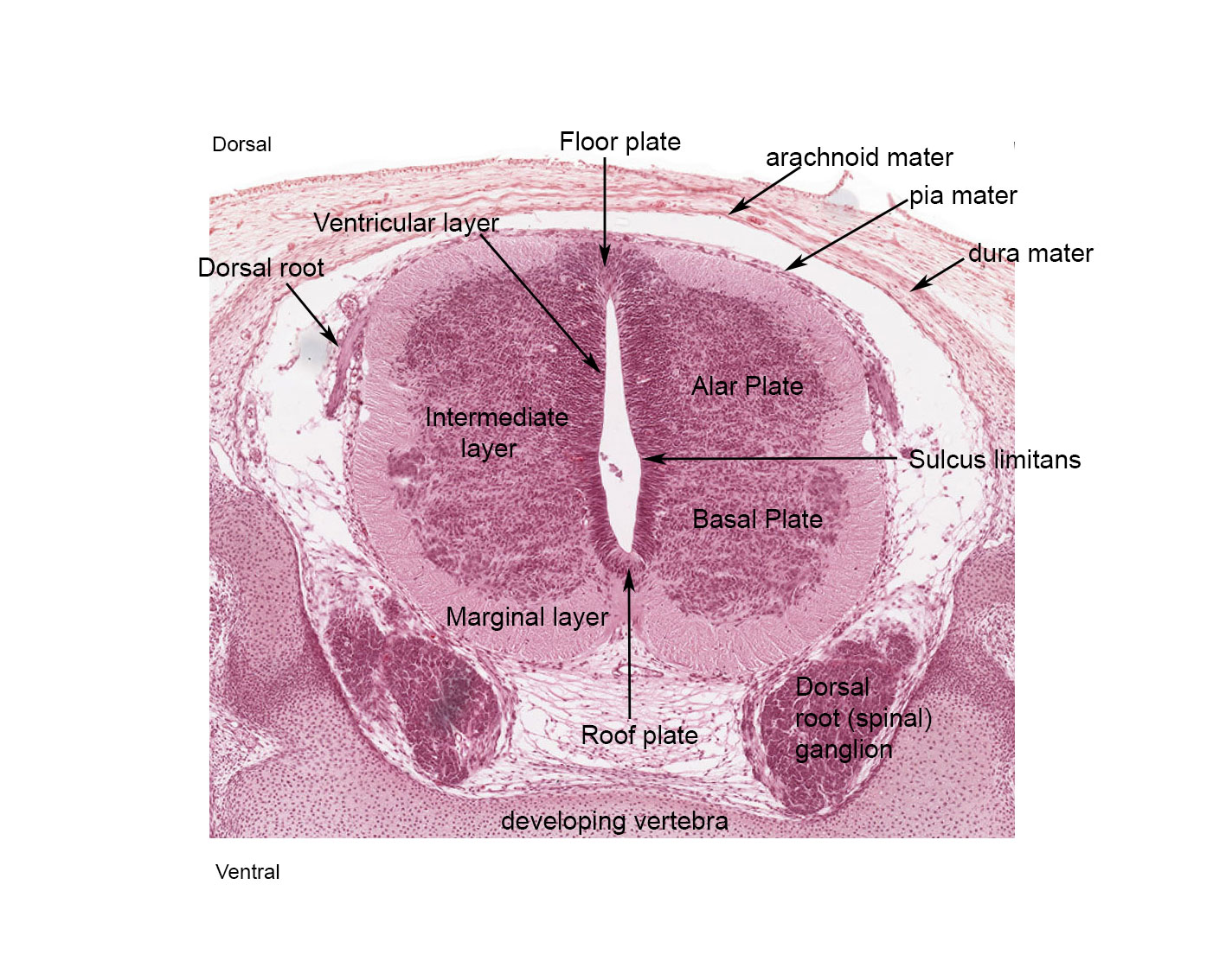 | 1,417 × 1,134 (302 KB) | Z8600021 (talk | contribs) | ==Human Embryo (Carnegie stage 22) Spinal Cord== See also unlabeled image. Note - this image of the week 8 spinal cord has been rotated 108<sup>o</sup> to give the "neuroscience" view. {{Footer}} |
You cannot overwrite this file.
File usage
The following 11 pages use this file: