File:Fetal brain MRI01.jpg

Original file (1,280 × 465 pixels, file size: 45 KB, MIME type: image/jpeg)
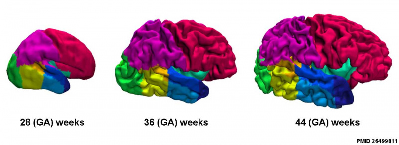
Fetal Brain MRI
Cortical surfaces for neonates at 28, 36 and 44 weeks PMA at scan with the labels overlaid.
- Links: 28-44 week MRI | 28 week MRI | 36 week MRI | 44 week MRI | fetal neural | neural | MRI
Reference
Makropoulos A, Aljabar P, Wright R, Hüning B, Merchant N, Arichi T, Tusor N, Hajnal JV, Edwards AD, Counsell SJ & Rueckert D. (2016). Regional growth and atlasing of the developing human brain. Neuroimage , 125, 456-478. PMID: 26499811 DOI.
Copyright
https://creativecommons.org/licenses/by/4.0/
Fig. 8. resized and relabelled
Cite this page: Hill, M.A. (2024, April 26) Embryology Fetal brain MRI01.jpg. Retrieved from https://embryology.med.unsw.edu.au/embryology/index.php/File:Fetal_brain_MRI01.jpg
- © Dr Mark Hill 2024, UNSW Embryology ISBN: 978 0 7334 2609 4 - UNSW CRICOS Provider Code No. 00098G
File history
Click on a date/time to view the file as it appeared at that time.
| Date/Time | Thumbnail | Dimensions | User | Comment | |
|---|---|---|---|---|---|
| current | 13:02, 7 July 2018 | 1,280 × 465 (45 KB) | Z8600021 (talk | contribs) | ||
| 13:01, 7 July 2018 | 1,280 × 465 (47 KB) | Z8600021 (talk | contribs) | Fig. 8. Example cortical surfaces for neonates at 28, 36 and 44 weeks PMA at scan with the labels overlaid. PMID--26499811 https://creativecommons.org/licenses/by/4.0/ |
You cannot overwrite this file.
File usage
The following 2 pages use this file: