File:Chicken-neural-crest-migration-06.jpg
Chicken-neural-crest-migration-06.jpg (600 × 398 pixels, file size: 40 KB, MIME type: image/jpeg)
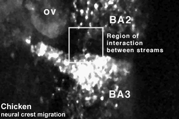
Neural crest migration
Chicken embryo sequence sequence shows the migration of DiI-labeled neural crest cells zoomed in on the region of neural crest cell interactions lateral to the otic vesicle. This sequence of images shows neural crest cells from the second and third branchial arch streams interacting.
r = rhombomere
Duration: 3 hrs Time interval between images: 3 min
Each image in the movie represents 10 confocal sections separated by 10 microns each, projected onto 1 image.
Links: Movies - Chicken Neural Crest | Neural Crest Development | File:Chicken-neural_crest_migration_06.flv | all Development movies
<pubmed>10683170</pubmed>
Original Neural Crest movies kindly provided by Paul Kulesa
Reproduced with Permission Development, © The Company of Biologists Ltd. 2000.
File history
Click on a date/time to view the file as it appeared at that time.
| Date/Time | Thumbnail | Dimensions | User | Comment | |
|---|---|---|---|---|---|
| current | 12:33, 12 August 2010 | | 600 × 398 (40 KB) | S8600021 (talk | contribs) | ==Neural crest migration== Chicken embryo sequence sequence shows the migration of DiI-labeled neural crest cells zoomed in on the region of neural crest cell interactions lateral to the otic vesicle. This sequence of images shows neural crest cells from |
You cannot overwrite this file.
File usage
The following 26 pages use this file:
- 2010 Lab 3
- 2010 Lecture 12
- 2011 Lab 3 - Week 4
- ANAT2341 Lab 3 - Week 4
- Chicken Development
- Chicken Neural Crest Migration Movie 1
- Chicken Neural Crest Migration Movie 2
- Chicken Neural Crest Migration Movie 3
- Chicken Neural Crest Migration Movie 4
- Chicken Neural Crest Migration Movie 5
- Chicken Neural Crest Migration Movie 6
- Chicken Neural Crest Migration Movie 7
- Developmental Mechanisms
- Lecture - Neural Crest Development
- Movie - Chicken Neural Crest Migration 01
- Movies
- Movies - Chicken Neural Crest
- Neural Crest - Cranial Nerve Development
- Neural Crest - Cranial Nerves
- Neural Crest Development
- Talk:2011 Lab 3
- Talk:Flash Movies
- Talk:Quicktime Movies
- Template:Chicken neural crest movies
- Template:Neural Crest movie 6
- Template talk:Chicken neural crest movies