File:Cerebellar oligodendrocyte cartoon.jpg

Original file (1,200 × 682 pixels, file size: 328 KB, MIME type: image/jpeg)
Cerebellar Oligodendrocyte
This drawing of a oligodendrocyte, a type of glial cell, was based upon electron micrograph images of the rat cerebellum.
This drawing shows what the author believes to be the relationship between the oligodendrocyte and myelin. The oligodendrocyte cytoplasm contains numerous RNP particles, an extensive granular endoplasmic reticulum (er), and a prominent Golgi apparatus (G). At the bottom, a thin process (p) is seen arising from the cell body and running along the plasma membrane. Over a large portion of the length of this process, the two membranes are shown as being fused, as apparently occurs in many in- stances. The interperiod line shown between the process and the plasma membrane has been seen on several occasions but is not invariably present. At the lower left, the process spirals around an axon (a) to form a myelin sheath. A larger cell process containing canaliculi (c) is seen at the upper right.
- Links: image - granule cell | image - Bergmann astrocyte | image - oligodendrocyte | Glial Development | Cerebellum Development
Reference
<pubmed>14222815</pubmed>| PMC2106521 | J Cell Biol.
FIGURE 16.
Copyright
Rockefeller University Press - Copyright Policy This article is distributed under the terms of an Attribution–Noncommercial–Share Alike–No Mirror Sites license for the first six months after the publication date (see http://www.jcb.org/misc/terms.shtml). After six months it is available under a Creative Commons License (Attribution–Noncommercial–Share Alike 4.0 Unported license, as described at https://creativecommons.org/licenses/by-nc-sa/4.0/ ). (More? Help:Copyright Tutorial)
Cite this page: Hill, M.A. (2024, April 26) Embryology Cerebellar oligodendrocyte cartoon.jpg. Retrieved from https://embryology.med.unsw.edu.au/embryology/index.php/File:Cerebellar_oligodendrocyte_cartoon.jpg
- © Dr Mark Hill 2024, UNSW Embryology ISBN: 978 0 7334 2609 4 - UNSW CRICOS Provider Code No. 00098G
File history
Click on a date/time to view the file as it appeared at that time.
| Date/Time | Thumbnail | Dimensions | User | Comment | |
|---|---|---|---|---|---|
| current | 10:43, 23 June 2015 | | 1,200 × 682 (328 KB) | Z8600021 (talk | contribs) | |
| 10:43, 23 June 2015 | 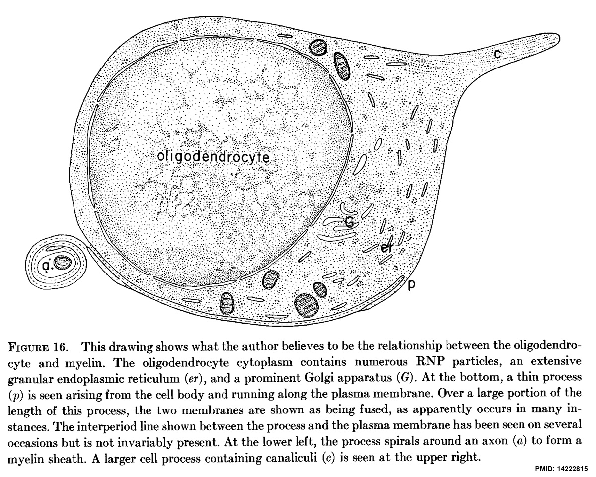 | 1,200 × 961 (475 KB) | Z8600021 (talk | contribs) | ==Cerebellar Oligodendrocyte== This drawing of a granule cell was based upon electron micrograph images of the rat cerebellum. This drawing shows what the author believes to be the relationship between the oligodendrocyte and myelin. The oligodendro... |
You cannot overwrite this file.
File usage
The following page uses this file: