Book - The Elements of Embryology - Chicken 6
| Embryology - 28 Apr 2024 |
|---|
| Google Translate - select your language from the list shown below (this will open a new external page) |
|
العربية | català | 中文 | 中國傳統的 | français | Deutsche | עִברִית | हिंदी | bahasa Indonesia | italiano | 日本語 | 한국어 | မြန်မာ | Pilipino | Polskie | português | ਪੰਜਾਬੀ ਦੇ | Română | русский | Español | Swahili | Svensk | ไทย | Türkçe | اردو | ייִדיש | Tiếng Việt These external translations are automated and may not be accurate. (More? About Translations) |
Foster M. Balfour FM. Sedgwick A. and Heape W. The Elements of Embryology (1883) Vol. 1. (2nd ed.). London: Macmillan and Co.
| The Elements of Embryology 1883
1 Chicken : Hen's egg and the beginning of incubation | Whole history of incubation | day 1 of incubation | first half of day 2 | second half of day 2 | day 3 | day 4 | day 5 | day 6-21 | Appendix | Figures as Gallery
|
| Historic Disclaimer - information about historic embryology pages |
|---|
| Pages where the terms "Historic" (textbooks, papers, people, recommendations) appear on this site, and sections within pages where this disclaimer appears, indicate that the content and scientific understanding are specific to the time of publication. This means that while some scientific descriptions are still accurate, the terminology and interpretation of the developmental mechanisms reflect the understanding at the time of original publication and those of the preceding periods, these terms, interpretations and recommendations may not reflect our current scientific understanding. (More? Embryology History | Historic Embryology Papers) |
The changes which take place during the third day
OF all days in the history of the chick within the egg this perhaps is the most eventful; the rudiments of so many important organs now first make their appearance.
In many instances we shall trace the history of these organs beyond the third day of incubation, in order ta give the reader a complete view of their development.
On opening an egg on the third day the first thing which attracts notice is the diminution of the white of the egg. This seems to be one of the consequences of the functional activity of the newly-established vascular area whose blood-vessel are engaged either in directly absorbing the white or, as is more probable, in absorbing the yolk, which is in turn replenished at the expense of the white. The absorption, once begun, goes on so actively that, by the end of the day, the decrease of the white is very striking.
The blastoderm has now spread over about half the yolk, the extreme margin of the opaque area reaching about half-way towards the pole of the yolk opposite to the embryo.
The vascular area, though still increasing, is much smaller than the total opaque area, being in average-sized eggs about as large as a florin. Still smaller than the vascular area is the pellucid area in the centre of which lies the rapidly growing embryo.
During the third day the vascular area is not only a means for providing the embryo with nourishment from the yolk, but also, inasmuch as by the diminution of the white it is brought close under the shell and therefore fully exposed to the influence of the atmosphere, serves as the chief organ of respiration.
This in fact is the period at which the vascular area may be said to be in the stage of its most complete development; for though it will afterwards become larger, it will at the same time become less definite and relatively less important. We may therefore, before we proceed, add a few words to the description of it given in the last chapter.
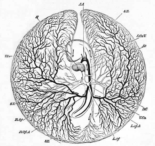
The blood leaving the body of the embryo by the vitelline arteries (Fig. 36, R. Of. A., L. Of. A.} is carried to the small vessels and capillaries of the vascular area, a small portion only being appropriated by the pellucid area.
From the vascular area part of the blood returns directly to the heart by the main lateral trunks of the vitelline veins, R. Of., L. Of. During the second day these venous trunks joined the body of the embryo considerably in front of, that is, nearer the head than, the corresponding arterial ones. Towards the end of the third day, owing to the continued lengthening of the heart, the veins and arteries run not only parallel to each other, but almost in the same line, the points at which they respectively join and leave the body being nearly at the same distance from the head.
Fig. 36. Diagram of the circulation of the yolk-sack at the end of the third day of incubation.
- H. heart. A A. the second, third and fourth aortic arches ; the first has become obliterated in its median portion, but is continued at its proximal end as the external carotid, and at its distal end as the internal carotid. AO. dorsal aorta. L. Of. A. left vitelline artery. R. Of. A. right vitelline artery. S. T. sinus terminalis. L. Of. left vitelline vein. R. Of. right vitelline vein. S. V. sinus venosus. D. C. ductus Cuvieri. S. Ca. V. superior cardinal or jugular vein. V. Ca. inferior cardinal vein. The veins are marked in outline and the arteries are made black. The whole blastoderm has been removed from the egg and is supposed to be viewed from below. Hence the left is seen on the right, and vice verse.
The rest of the blood brought by the vitelline arteries finds its way into the lateral portions of the sinus terminalis, S.T., and there divides on each side into two streams. Of these, the two which, one on each side, flow backward, meet at a point about opposite to the tail of the embryo, and are conveyed along a distinct vein which, running straight forward parallel to the axis of the embryo, empties itself into the left vitelline vein. The two forward streams reaching the gap in the front part of the sinus terminalis fall into either one, or in some cases two veins, which run straight backward parallel to the axis of the embryo, and so reach the roots of the heart. When one such vein only is present, it joins the left vitelline trunk; where there are two they join the left and right vitelline trunks respectively. The left vein is always considerably larger than the right; and the latter when present rapidly gets smaller and speedily disappears.
The chief differences, then, between the peripheral circulation of the second and of the third day are due to the greater prominence of the sinus terminalis and the more complete arrangements for returning the blood from it to the heart. After this day, although the vascular area will go on increasing in size until it finally all but encompasses the yolk, the prominence of the sinus terminalis will become less and less in proportion as the respiratory work of the vascular area is shifted on to the allantois, and its activities confined to absorbing nutritive matter from the yolk.
The folding-in of the embryo makes great progress during this day. Both head and tail have become most distinct, and the side folds which are to constitute the lateral walls have advanced so rapidly that the embryo is now a bond fide tubular sac, connected with the rest of the yolk by a broad stalk. This stalk, as was explained in Chap. II, is double, and consists of an inner splanchnic stalk continuous with the alimentary canal, which is now a tube closed at both ends and open to the stalk along its middle third only, and an outer somatic stalk continuous with the body-walls of the embryo, which have not closed nearly to the same extent as the walls of the alimentary canal. (Compare Fig. 9, A and J5, which may be taken as diagrammatic representations of longitudinal and transverse sections of an embryo of this period.)
The embryo is almost completely covered by the amnion. Early in this day the several amniotic folds will have met and completely coalesced along a line over the back of the embryo in the manner already explained in the last chapter.
During this day a most remarkable change takes place in the position of the embryo. Up to this time it has been lying symmetrically upon the yolk with the part which will be its mouth directed straight downwards. It now turns round so as to lie on its left side.
Fig. 37. chick of the third day (fifty-four hours) viewed from underneath as a transparent object.
- a'. the outer amniotic fold or false amnion. This is very conspicuous around the head, but may also be seen at the tail.
- a. the true amnion, very closely enveloping the head, and here seen only between the projections of the several cerebral vesicles. It may also be traced at the tail.
- In the embryo of which this is a drawing, the head-fold of the amnion reached a little farther backward than the reference u, but its limit could not be distinctly seen through the body of the embryo. The prominence of the false arnnion at the head is apt to puzzle the student ; but if he bears in mind the fact, which could not well be shewn in Fig. 9, that the whole amniotic fold, both the true and the false limb, is tucked in underneath the head, the matter will on reflection become intelligible.
- C. H. cerebral hemisphere. F. B. thalamencephalon or vesicle of the third ventricle. M. B. mid-brain. H. B. hind-brain. Op. optic vesicle. Ot. otic vesicle. Of V. vitelline veins forming the venous roots of the heart. The trunk on the right hand (left trunk when the embryo is viewed in its natural position from above) receives a large branch, shewn by dotted lines, coming from the anterior portion of the sinus terminalis. Ht. the heart, now completely twisted on itself. Ao. the bulbus arteriosus, the three aortic arches being dimly seen stretching from it across the throat, and uniting into the aorta, still more dimly seen as a curved dark line running along the body. The other curved dark line by its side, ending near the reference y, is the notochord ch.
- About opposite the line of reference x the aorta divides into two trunks, which, running in the line of the somewhat opaque mesoblastic somites on either side, are not clearly seen. Their branches however, Ofa, the vitelline arteries, are conspicuous and are seen to curve round the commencing side folds.
- Pv. mesoblastic somites. Below the level of the vitelline arteries the vertebral plates are but imperfectly cut up into mesoblastic somites, and lower down still, not at all.
- x is placed at the "point of divergence" of the splanchnopleure folds. The blind foregut begins here and extends about up to y. x therefore marks the present hind limit of the splanchnopleure folds. The limit of the more transparent somatopleure folds is not shewn.
It will be of course understood that all the body of the embryo above the level of the reference #, is seen through the portion of the yolk-sac (vascular and pellucid area), which has been removed with the embryo from the egg, as well as through the double amniotic fold.
We may repeat that, the view being from below, whatever is described in the natural position as being to the right here appears to be left, and vice versa.
This important change of position at first affects only the head (Fig. 37), but subsequently extends also to the trunk. It is not usually completed till the fourth day. At the same time the left vitelline vein, the one on the side on which the embryo comes to lie, grows very much larger than the right, which henceforward gradually dwindles and finally disappears.
Coincidently with the change of position the whole embryo begins to be curved on itself in a slightly spiral manner.' This curvature of the body becomes still more marked on the fourth day, Fig. 67.
In the head very important changes take place. One of these is the cranial flexure, Figs. 37, 38. This (which must not be confounded with the curvature of the body just referred to) we have already seen was commenced in the course of the second day, by the bending downwards of the head round a point which may be considered as the extreme end either of the notochord or of the alimentary canal.
The flexure progresses rapidly, the front-brain being more and more folded down till, at the end of the third day, it is no longer the first vesicle or fore-brain, but the second cerebral vesicle or mid-brain, which occupies the extreme front of the long axis of the embryo. In fact a straight line through the long axis of the embryo would now pass through the mid-brain instead of, as at the beginning of the second day, through the fore-brain, so completely has the front end of the neural canal been folded over the end of the notochord. The commencement of this cranial flexure gives the body of an embryo of the third day somewhat the appearance of a retort, the head of the embryo corresponding to the bulb. On the fourth day the flexure is still greater than on the third, but on the fifth and succeeding days it becomes less obvious, owing to the filling up of the parts of the skull.
The brain
The vesicle of the cerebral hemispheres, which on the second day began to grow out from the front of the fore -brain, increases rapidly in size during the third day, growing out laterally, so as to form two vesicles, so much so that by the end of the day it (Fig. 37, CH, Fig. 38) is as large or larger than the original vesicle from which it sprang, and forms the most conspicuous part of the brain. In its growth it pushes aside the optic vesicles, and thus contributes largely to the roundness which the head is now acquiring. Each lateral vesicle possesses a cavity, which afterwards becomes one of the lateral ventricles. These cavities are continuous behind with the cavity of the fore-brain.
Owing to the development of the cerebral vesicle the original fore -brain no longer occupies the front position (Fig. 37, FB, Fig. 38, /&), and ceases to be the conspicuous object that it was. Inasmuch as its walls will hereafter be developed into the parts surrounding the so-called third ventricle of the brain, we shall henceforward speak of it as the vesicle of the third ventricle, or thalamencephalon.
On the summit of the thalamencephalon there may now be seen a small conical projection, the rudiment of the pineal gland (Fig. 38, e), while the centre of the floor is produced into a funnel-shaped process, the infundibulum (Fig. 39, In), which, stretching towards the extreme end of the oral invagination or stomodceum, joins a diverticulum of this which becomes the pituitary body.
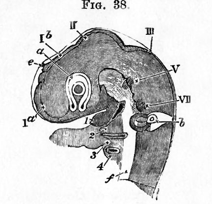
Fig. 38. Head of a chick of the third day viewed sideways as a transparent object. (From Huxley.)
- I a. the vesicle of the cerebral hemisphere. 1 6. the vesicle of the third ventricle (the original fore-brain) ; at its summit is seen the projection of the pineal gland e.
- Below this portion of the brain is seen, in optical section, the optic vesicle a already involuted with its thick inner and thinner outer wall (the letter a is placed on the junction of the two, the primary cavity being almost obliterated). In the centre of the vesicle lies the lens, the shaded portion being the expression of its cavity. Below the lens between the two limbs of the horseshoe is the choroidal fissure.
- II. the mid-brain. III. the hind-brain. V. the rudiments of the fifth cranial nerve, VII. of the seventh. Below the seventh nerve is seen the auditory vesicle b. The head having been subjected to pressure, the vesicle appears somewhat distorted as if squeezed out of place. The orifice is not yet quite closed up.
- I, the inferior maxillary process of the first visceral or mandibular fold. Below, and to the right of this, is seen the first visceral cleft, below that again the second visceral fold (2), and lower down the third (3) and fourth (4) visceral folds. In front of the folds (i.e. to the left) is seen the arterial end of the heart r the aortic arches being buried in their respective visceral folds.
- f. represents the mesoblast of the base of the brain and spinal cord.
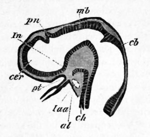
The development of the pituitary body or hypophysis cerebri has been the subject of considerable controversy amongst embryologists, and it is only within the last few years that its origin from the oral epithelium has been satisfactorily established.
In the course of cranial flexure the epiblast on the under side of the head becomes tucked in between the blind end of the throat and the base of the brain. The part so tucked in constitutes a kind of bay, and forms the stomodaeum or primitive buccal cavity already spoken of. The blind end of this bay becomes produced as a papilliform diverticulum which may be called the pituitary diverticulum. It is represented as it appears in a lower vertebrate embryo (Elasmobranch) in Fig. 39, but is in all important respects exactly similar in the chick. Very shortly after the pituitary diverticulum becomes first established the boundary wall between the stomodseum and the throat becomes perforated, and the limits of the stomodaeum obliterated, so that the pituitary diverticulum looks as if it had arisen from the hypoblast. During the third day of incubation the front part of the notochord becomes bent downward, and, ending in a somewhat enlarged extremity, comes in contact with the termination of the pituitary diverticulum. The mesoblast around increases and grows up, in front of the notochord and behind the vesicle of the third ventricle, to form the posterior clinoid process. The base of the vesicle of the third ventricle at the same time grows downwards towards the pituitary diverticulum, and forms what is known as the infundibulum. On the fourth day the mesoblastic tissue around the notochord increases in quantity, and the end of the notochord, though still bent downwards, recedes a little from the termination of the pituitary diverticulum, which is still a triangular space with a wide opening into the alimentary canal.
On the fifth day, the opening of the pituitary diverticulum into the alimentary canal has become narrowed, and around the whole diverticulum an investment of mesoblast-cells has appeared. Behind it the clinoid process has become cartilaginous, while to the sides and in front it is enclosed by the trabeculse. At this stage, in fact, we have a diverticulum from the alimentary canal passing through the base of skull to the infundibulum.
On the seventh day the communication between the cavity of the diverticttlum and that of the throat has become still narrower. The diverticulum is all but converted into a vesicle, and its epiblastic walls have commenced to send out into the mesoblastic investment solid processes. The infundibulum now appears as a narrow process from the base of the vesicle of the third ventricle, which approaches, but does not unite with, the pituitary vesicle.
By the tenth day the opening of the pituitary vesicle into the throat becomes almost obliterated, and the lumen of the vesicle itself very much diminished. The body consists of anastomosing cords of epiblast-cells, the mesoblast between which has already commenced to become vascular. The cords or masses of epiblast cells are surrounded by a delicate membrana propria, and a few of them possess a small lumen. The infundibulum has increased in length. The relative positions of the pituitary body and infundibulum are shewn in the figure of the brain in Chapter vni.
On the twelfth day the communication between the pituitary vesicle and the throat is entirely obliterated, but a solid cord of cells still connects the two. The vessels of the pia mater of the vesicle of the third ventricle have become connected with the pituitary body, and the infundibulum has grown down along its posterior border.
In the later stages all connection is lost between the pituitary body and the throat, and the former becomes attached to the elongated processus infundibuli.
The real nature of the pituitary body is still extremely obscure, but it is not improbably the remnant of a glandular structure which may have opened into the mouth in primitive vertebrate forms, but which has ceased to have a function in existing vertebrates1.
Beyond an increase in size, which it shares with nearly all parts of the embryo, and the change of position to which we have already referred, the midbrain undergoes no great alteration during the third day. Its roof will ultimately become developed into the corpora Ugemina or optic lobes, its floor will form the crura cerebri, and its cavity will be reduced to the narrow canal known as the iter a tertio ad quartum ventriculum.
In the hind-brain, or third cerebral vesicle, that part which lies nearest to the mid-brain, is during the third day marked off from the rest by a slight constriction. This distinction, which becomes much more evident later on by a thickening of the walls and roof of the front portion, separates the hind-brain into the cerebellum in front, and the medulla oblongata behind (Figs. 38 and 39). While the walls of the cerebellar portion of the hind-brain become very much thickened as well at the roof as at the floor and sides, the roof of the posterior or medulla oblongata portion thins out into a mere membrane, forming a delicate covering to the cavity of the vesicle (Fig. 40, iv), which here becoming broad and shallow with greatly thickened floor and sides, is known as the fourth ventricle, subsequently overhung by the largely developed posterior portion of the cerebellum.
(1Wilhelm M tiller Ueber die Entwicklung und Ban der Hypophysis und des Processus Infundibuli Cerebri. Jenaische Zeitschrift, Bd. vi. 1871, and V. von Mihalkovics, Wirbelsaite u. Hirnanhang , Archiv f. mikr. Anat. Vol. xi. 1875.)
The third day, therefore, marks the differentiation of the brain into five distinct parts : the cerebral hemispheres, the central masses round the third ventricle, the corpora bigemina or optic lobes, the cerebellum and the medulla oblongata ; the original cavity of the neural canal at the same time passing from its temporary division of three single cavities into the permanent arrangement of a series of connected ventricles, viz. the lateral ventricles, the third ventricle, the iter (with a prolongation into the optic lobe on each side), and the fourth ventricle.
At the same time that the outward external shape of the brain is thus being moulded, internal changes are taking place in the whole neural canal. These are best seen in sections.
At its first formation, the section of the cavity of the neural canal is round, or nearly so.
About this time, however, the lining of involuted epiblast along the length of the whole spinal cord becomes very much thickened at each side, while increasing but little at the mid-points above and below. The result of this is that the cavity as seen in section (Figs. 64 and 65), instead of being circular, has become a narrow vertical slit, almost completely filled in on each side.
In the region of the brain the thickening of the lining epiblast follows a somewhat different course. While almost everywhere the sides and floor of the canal are greatly thickened, the roof in the region of the various ventricles, especially of the third and fourth, becomes excessively thin, so as to form a membrane reduced to almost a single layer of cells. (Fig. 40, IV.)
Cranial and spinal nerves
A most important event which takes place during the second and third days, is the formation of the cranial and spinal nerves. Till within a comparatively recent period embryologists were nearly unanimous in believing that the peripheral nerves originated from the mesoblast at the sides of the brain and spinal cord. This view has now however been definitely disproved, and it has been established that both the cranial and spinal nerves take their origin as outgrowths of the central nervous system.
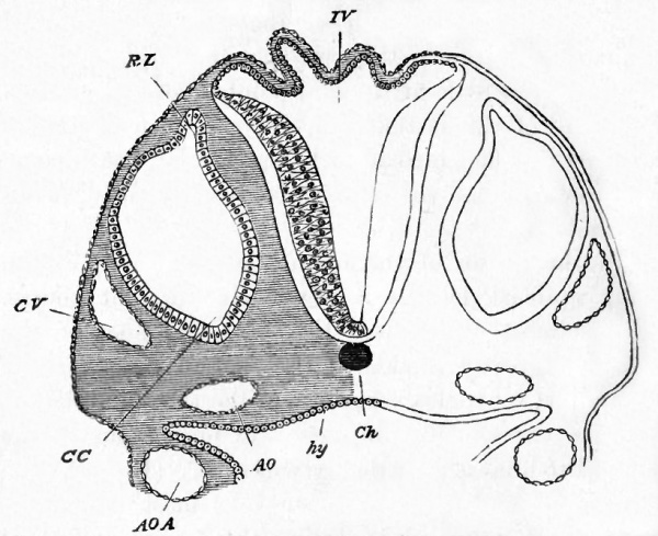
Fig. 40. Section through the hind-brain of a chick at the end of the third day of incubation.
- IV. Fourth ventricle. The section shews the very thin roof and thicker sides of the ventricle.
- Ch. Notochord (diagrammatic shading).
- CV. Anterior cardinal or jugular vein.
- CC. Involuted auditory vesicle. CC points to the end which will form the cochlear canal. RL. Kecessus labyrinthi. hy. hypoblast lining the alimentary canal, hy is itself placed in the cavity of the alimentary canal, in that part of the canal which will become the throat. The ventral (anterior) wall of the canal is not shewn in the section, but on each side are seen portions of a pair of visceral arches. In each arch is seen the section of the aortic arch AOA belonging to the visceral arch. The vessel thus cut through is running upwards towards the head, being about to join the dorsal aorta AO. Had the section been nearer the head, and carried through the plane at which the aortic arch curves round the alimentary canal to reach, the mesoblast above it, AOA and AO would have formed one continuous curved space. In sections lower down in the back the two aortse, AO, one on each side, would be found fused into one median canal.
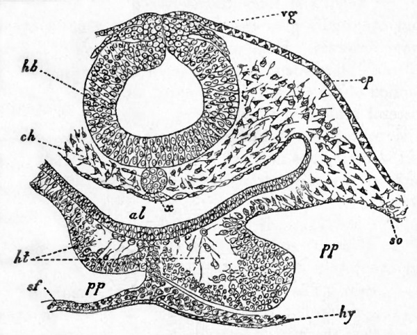
The cranial nerves are the first to be developed and arise before the complete closure of the neural groove. They are formed as paired outgrowths of a continuous band known as the neural band, composed of two laminae, which connects the dorsal edges of the incompletely closed neural canal with the external epiblast. This mode of development will best be understood by an examination of Fig. 41, where the two roots of the vagus nerve (vg) are shewn growing out from the neural band. Shortly after this stage the neural band becomes separated from the external epiblast, and constitutes a crest attached to the roof of the brain, while its two laminae become fused.
Fig. 41. Transverse section through the posterior part of the head of an embryo chick of thirty hours.
- hb. hind-brain ; vg. vagus nerve ; ep. epiblast ; ch. notochord ; x. thickening of hypoblast (possibly a rudiment of the subnotochordal rod) ; al. throat ; ht. heart ; pp. body cavity ; so. somatic mesoblast ; sf. splanchnic mesoblast ; hy. hypoblast.
- Anteriorly, the neural crest extends as far as the roof of the mid-brain. The pairs of nerves which undoubtedly grow out from it are the fifth pair, the seventh and auditory (as a single root), the glossopharyngeal and the various elements of the vagus (as a single root).
- After the roots of these nerves have become established, the crest connecting them becomes partially obliterated. The roots themselves grow centrifugally, and eventually give rise to the whole of each of the cranial nerves. Each complete root develops a ganglionic enlargement near its base, and (with the exception of the third nerve) is distributed to one of the visceral arches, of which we shall say more hereafter. The primitive attachment of the nerves is to the roof of the brain, but in most instances this attachment is replaced by a secondary attachment to the sides or floor.
- The rudiments of four cranial nerves, of which two lie in front of and two behind the auditory vesicle, are easily seen during the third day at the sides of the hind-brain. They form a series of four small opaque masses, somewhat pearshaped, with the stalk directed away from the middle line.
- The most anterior of these is the rudiment of the fifth nerve (Figs. 42 and 67, V). Its narrowed outer portion or stalk divides into two bands or nerves. Of these one passing towards the eye terminates at present in the immediate neighbourhood of that organ. The other branch (the rudiment of the inferior maxillary branch of the fifth nerve) is distributed to the first visceral arch.
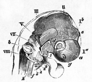
The second mass (Figs. 42 and 67, VII) is the rudiment of the seventh, or facial nerve, and of the auditory nerve. It is the nerve of the second visceral arch.
The two masses behind the auditory vesicle represent the glossopharyngeal and pneumogastric nerves (Fig. 42, VIII, Fig. 67, G. Ph. and Pg). At first united, they subsequently become separate. The glossopharyngeal supplies the third arch, and the pneumogastric the fourth and succeeding arches.
The later development of the cranial nerves has only been partially worked out, and we will confine ourselves here to a very brief statement of some of the main results arrived at. The outgrowth for the vagus nerve supplies in the embryo the fourth and succeeding visceral arches, and from what we know of it in the lower vertebrate types, we may conclude that it is a compound nerve, composed of as many primitively distinct nerves as there are branches to the visceral arches.
The glossopharyngeal nerve is the nerve supplying the third visceral arch, the homologue of the first branchial arch of Fishes. The development of the hypoglossal nerve is not known, but it is perhaps the anterior root of a spinal nerve. The spinal accessory nerve has still smaller claims than the hypoglossal to be regarded as a true cranial nerve. The primitively single root of the seventh auditory nerves divides almost at once into two branches. The anterior of these pursues a straight course to the hyoid arch and forms the rudiment of the facial nerve, Fig. 67, vn ; the second of the two, which is the rudiment of the auditory nerve, develops a ganglionic enlargement, and, turning backwards, closely hugs the ventral wall of the auditory involution. The sixth nerve appears to arise later than the seventh nerve from the ventral part of the hind-brain, and has no ganglion near its root.
Shortly after its development the root of the fifth nerve shifts so as to be attached about half-way down the side of the brain. A large ganglion is developed close to the root, which becomes the Gasserian ganglion. The main branch of the nerve grows into the-mandibular arch (Fig. 67), maintaining towards it similar relations to those of the nerves behind it to their respective arches.
An important branch becomes early developed which is directed straight towards the eye (Fig. 67), near which it meets and unites with the third nerve, where the ciliary ganglion is developed. This branch is usually called the ophthalmic branch of the fifth nerve, and may perhaps represent an independent nerve.
Later than these two branches there is developed a third branch, passing the upper process of the first visceral arch. It forms the superior maxillary branch of the adult.
Nothing is known with reference to the development of the fourth nerve.
The history of the third nerve is still imperfectly known. There is developed early on the second day from the neural crest, on the roof of the mid-brain, an outgrowth on each side, very similar to the rudiment of the posterior nerves. This outgrowth is believed by Marshall to be the third nerve, but it must be borne in mind that there is no direct evidence on the point, the fate of the outgrowth in question not having been satisfactorily followed.
At a very considerably later period a nerve may be found springing from the floor of the mid-brain, which is undoubtedly the third nerve. If identical with the outgrowth just spoken of, it must have shifted its attachment from the roof to the floor of the brain.
The nerve when it springs from the floor of the brain runs directly backwards till it terminates in the ciliary ganglion, from which two branches to the eye-muscles are given off.
[A. Marshall. " The development of the cranial nerves in the Chick." Quart. Journal of Microscop. Science, Vol. xvin.]
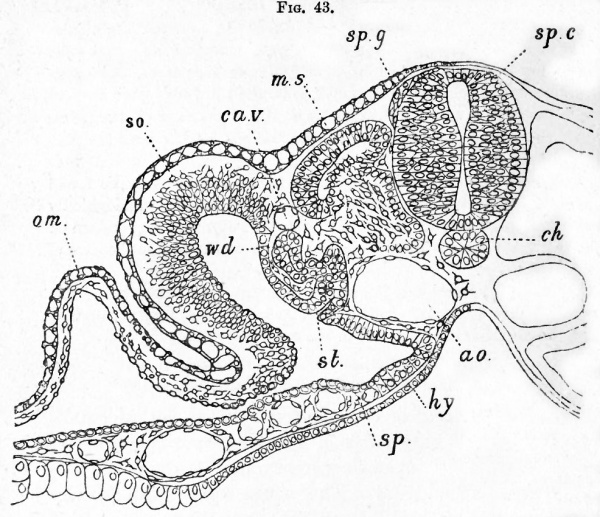
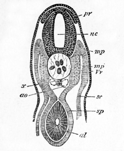
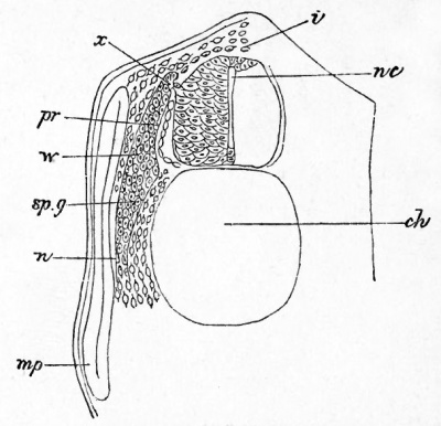
In the case of the spinal nerves the posterior roots originate as outgrowths of a series of median processes of cells, which make their appearance on the dorsal side of the spinal cord. The outgrowths, symmetrically placed on each side, soon take a pyriform aspect, and apply themselves to the walls of the spinal cord. They are represented as they appear in birds in Fig. 43, sp. g. } and as they appear in a lower vertebrate form in Fig. 44.
Fig. 43. Transverse section through the trunk of a duck embryo with about twenty-four mesoblastic somites.
- am. amnion ; so. somatopleure ; sp. splanchnopleure ; wd. Wolffian duct ; st. segment al tube ; ca.v. cardinal vein ; ms. muscle plate ; sp.g. spinal ganglion; sp.c. spinal cord; ch. notochord; ao. aorta ; hy. hypoblast.
The original attachment of the nerve -rudiment to the medullary wall is not permanent. It becomes, in fact, very soon either extremely delicate or absolutely interrupted.
The nerve-rudiment now becomes divided into three parts, (1) a proximal rounded portion; (2) an enlarged middle portion, forming the rudiment of a ganglion ; (3) a distal portion, forming the commencement of the nerve. The proximal portion may very soon be observed to be united with the side of the spinal cord at a very considerable distance from its original point of origin. It is moreover attached, not by its extremity, but by its side. The above points, which are much more easily studied in some of the lower vertebrate forms than in Birds, are illustrated by the subjoined section of an Elasmobranch embryo, Fig. 45.
The origin of the anterior roots of the spinal nerves has not as yet been satisfactorily made out in Birds ; but it appears probable that they grow from the ventral corner of the spinal cord, considerably later than the posterior roots, as a number of strands for each nerve, which subsequently join the posterior roots below the ganglia. The shape of the root of a completely formed spinal nerve, as it appears in an embryo of the fourth day, is represented in Fig. 68.
The Eye
In the preceding chapter we saw how the first cerebral vesicle, by means of lateral outgrowths followed by constrictions, gave rise to the optic vesicles. These and the parts surrounding them undergo on the third day changes which result in the formation of the eyeball.
At their first appearance the optic vesicles stand out at nearly right angles to the long axis of the embryo (Fig. 27), and the stalks which connect them with the fore-brain are short and wide. The constrictions which give rise to the stalks take place chiefly from above downwards, and also somewhat inwards and backwards. Thus from the first the vesicles appear to spring from the under part of the fore-brain.
These stalks soon become comparatively narrow, and constitute the rudiments of the optic nerves (Fig. 46 b). The constriction to which the stalk or optic nerve is due takes place obliquely downwards and backwards, so that the optic nerves open into the base of the front part of the thalamencephalon (Fig. 46 b).
While these changes have been going on in the optic stalks, development has also proceeded in the region of the vesicles themselves, and given rise to the rudiments of the retina, lens, vitreous humour, and other parts of the eye.
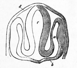
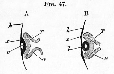
Towards the end of the second day the external or superficial epiblast which covers, and is in all but immediate contact with, the most projecting portion of the optic vesicle, becomes thickened. This thickened portion is then driven inwards in the form of a shallow open pit with thick walls (Fig. 47 A, o), carrying before it the front wall (r) of the optic vesicle. To such an extent does this involution of the superficial epiblast take place, that the front wall of the optic vesicle is pushed close up to the hind wall, and the cavity of the vesicle becomes almost obliterated (Fig. 47, #).
The bulb of the optic vesicle is thus converted into a cup with double walls, containing in its cavity the portion of involuted epiblast. This cup, in order to distinguish its cavity from that of the original optic vesicle, is generally called the secondary optic vesicle. We may, for the sake of brevity, speak of it as the optic cup; in reality it never is a vesicle, since it always remains widely open in front. Of its double walls the inner or anterior (Fig. 47 B, r) is formed from the front portion, the outer or posterior (Fig. 47 5, u) from the hind portion of the wall of the primary optic vesicle. The inner or anterior (r), which very speedily becomes thicker than the other, is converted into the retina; in the outer or posterior (u), which remains thin, pigment is eventually deposited, and it ultimately becomes the tesselated pigment-layer of the choroid.
By the closure of its mouth the pit of involuted epiblast becomes a completely closed sac with thick walls and a small central cavity (Fig. 47 B, I}. At the same time it breaks away from the external epiblast, which forms a continuous layer in front of it, all traces of the original opening being lost. There is thus left lying in the cup of the secondary optic vesicle, an isolated elliptical mass of epiblast. This is the rudiment of the lens. The small cavity within it speedily becomes still less by the thickening of the walls, especially of the hinder one.
Fig. 47. Diagrammatic sections illustrating the formation of the eye. (After Kemak.)
- In A, the thin superficial epiblast h is seen to be thickened at #, in front of the optic vesicle, and involuted so as to form a pit o, the mouth of which has already begun to close in. Owing to this involution, which forms the rudiment of the lens, the optic vesicle is doubled in, its front portion r being pushed against the back portion u, and the original cavity of the vesicle thus reduced in size. The stalk of the vesicle is shewn as still broad.
- In B, the optic vesicle is still further doubled in so as to form a cup with a posterior wall u and an anterior wall r. In the hollow of this cup lies the lens , now completely detached from the superficial epiblast x. Its cavity is still shewn. The cavity of the stalk of the optic vesicle is already much narrowed.
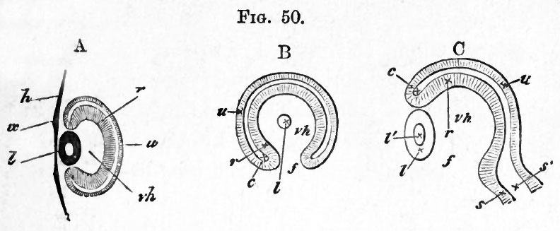
At its first appearance the lens is in immediate contact with the anterior wall of the secondary optic vesicle (Fig. 47 B}. In a short time, however, the lens is seen to lie in the mouth of the cup (Fig. 50 A), a space (vh) (which is occupied by the vitreous humour) making its appearance between the lens and anterior wall of the vesicle.
In order to understand how this space is developed, the position of the optic vesicle and the relations of its stalk must be borne in mind.
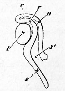
The vesicle lies at the side of the head, and its stalk is directed downwards, inwards and backwards. The stalk in fact slants away from the vesicle. Hence when the involution of the lens takes place, the direction in which the front wall of the vesicle is pushed in is not in a line with the axis of the stalk, as for simplicity's sake has been represented in the diagram Fig. 47, but forms an obtuse angle with that axis, after the manner of Fig. 48, where s represents the cavity of the stalk leading away from the almost obliterated cavity of the primary vesicle.
Fig. 48 represents the early stage at which the lens fills the whole cup of the secondary vesicle. The subsequent state of affairs is brought about through the growth of the walls of the cup taking place more rapidly than that of the lens. But this growth or this dilatation does not take place equally in all parts of the cup. The walls of the cup rise up all round except that part of the circumference of the cup which adjoins the stalk. While elsewhere the walls increase rapidly in height, carrying so to speak the lens with them, at this spot, which in the natural position of the eye is on its under surface, there is no growth: the wall is here imperfect, and a gap is left. Through this gap, which afterwards receives the name of the choroidal fissure, a way is open from the mesoblastic tissue surrounding the optic vesicle and stalk into the interior of the cavity of the cup.
From the manner of its formation the gap or fissure is evidently in a line with the axis of the optic stalk, and in order to be seen must be looked for on the under surface of the optic vesicle. In this position it is readily recognized in the transparent embryo of the third day, Figs. 37 and 48.
Bearing in mind these relations of the gap to the optic stalk, the reader will understand how sections of the optic vesicle at this stage present very different appearances according to the plane in which the sections are taken.
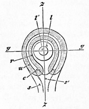
When the head of the chick is viewed from underneath as a transparent object the eye presents very much the appearance represented in the diagram Fig. 49.
A section of such an eye taken along the line y, perpendicular to the plane of the paper, would give a figure corresponding to that of Fig. 50 A. The lens, the cavity and double walls of the secondary vesicle, and the remains of the primary cavity, would all be represented (the superficial epiblast of the head would also be shewn) ; but there would be nothing seen of either the stalk or the fissure. If on the other hand the section were taken in a plane parallel to the plane of the paper, at some distance above the level of the stalk, some such figure would be gained as that shewn in Fig. 50 B. Here the fissure / is obvious, and the communication of the cavity vh of the secondary vesicle with the outside of the eye evident; the section of course would not go through the superficial epiblast,
FIG. 50.
- A. Diagrammatic section taken perpendicular to the plane of the paper, along the line y, y, Fig. 49. The stalk is not seen, the* section falling quite out of its region, vh, hollow of optic cup filled with vitreous humour ; other letters as in Fig. 47 B.
- B. Section taken parallel to the plane of paper through Fig. 49, so far behind the front surface of the eye as to shave off a small portion of the posterior surface of the lens I, but so far in front as not to be carried at all through the stalk. Letters as before ; /, the choroidal fissure.
- C. Section along the line z, z, perpendicular to the plane of the paper, to shew the choroidal fissure /, and the continuity of the cavity of the optic stalk with that of the primary optic vesicle. Had this section been taken a little to either side of the line z, 2, the wall of the optic cup would have extended up to the lens below as well as above. Letters as above.
Lastly, a section, taken perpendicular to the plane of the paper along the line z, i.e. through the fissure itself, would present the appearances of Fig. 50 C, where the wall of the vesicle is entirely wanting in the region of the fissure marked by the position of the letter /. The external epiblast has been omitted in the figure.
The fissure such as we have described it exists for a short time only. Its lips come into contact, and unite (in the neighbourhood of the lens, directly, but in the neighbourhood of the stalk, by the intervention of a structure which we shall describe presently), and thus the cup-like cavity of the secondary optic vesicle is furnished with a complete wall all round. The interior of the cavity is filled by the vitreous humour, a clear fluid in which are a few scattered cells.
With reference to the above description, two points require to be noticed. Firstly it is extremely doubtful whether the invagination of the secondary optic vesicle is to be viewed as an actual mechanical result of the ingrowth of the lens. Secondly it seems probable that the choroid fissure is not simply due to a deficiency in the growth of part of the walls of the secondary optic cup, but is partly due to a more complicated inequality of growth resulting in a doubling up of the primary vesicle from the side along the line of the fissure, at the same time that the lens is being thrust in in front. In Mammalia, the doubling up involves the optic stalk, which becomes flattened (whereby its original cavity is obliterated) and then folded in on itself, so as to embrace a new central cavity continuous with the cavity of the vitreous humour.
During the changes in the optic vesicle just described, the surrounding mesoblast takes on the characters of a distinct investment, whereby the outline of the eyeball is definitely formed. The internal portions of this investment, nearest to the retina, become the choroid (i.e. the chorio-capillaris, and the lamina fusca, the pigment epithelium, as we have seen, being derived from the epiblastic optic cup), and pigment is subsequently deposited in it. The remaining external portion of the investment forms the sclerotic.
The complete differentiation of these two coats of the eye does not however take place till a late period.
In front of the optic cup the mesoblastic investment grows forwards, between the lens and the superficial epiblast, and so gives rise to the substance of the cornea; the epiblast supplying only the anterior epithelium.
We may now proceed to give some further details with reference to the histological differentiation of the parts, whose general development has been dealt with in the preceding pages.
The histological condition of the eye in its earliest stages is very simple. Both the epiblast forming the walls of the optic vesicle, and the superficial layer which is thickened to become the lens, are composed of simple columnar cells. The surrounding mesoblast is made up of cells whose protoplasm is more or less branched and irregular. These simple elements are gradually modified into the complicated tissues of the adult eye, the changes undergone being most marked in the cases of the retina, the optic nerve, and the lens with its appendages.
The optic vesicle
We left the original cavity of the primary optic vesicle as a nearly obliterated space between the two walls of the optic cup. By the end of the third day the obliteration is complete, and the two walls are in immediate contact.
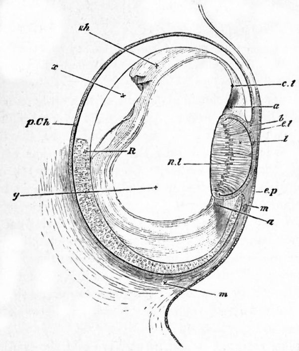
The inner or anterior wall is, from the first, thicker than the outer or posterior ; and over the greater part of the cup this contrast increases with the growth of the eye, the anterior wall becoming markedly thicker and undergoing changes of which we shall have to speak directly (Fig. 51).
In the front portion however, along, so to speak, the lip of the cup, anterior to a line which afterwards becomes the ora serrata, both layers not only cease to take part in the increased thickening, accompanied by peculiar histological changes, which the rest of the cup is undergoing, but also completely coalesce together. Thus a hind portion or true retina is marked off from a front portion.
The front portion, accompanied by the choroid which immediately overlays it, is, behind the lens, thrown into folds, the ciliary ridges ; while further forward it bends in between the lens and the cornea to form the iris. The original wide opening of the optic cup is thus narrowed to a smaller orifice, the pupil ; and the lens, which before lay in the open mouth, is now inclosed in the cavity of the cup. While in the hind portion of the cup, or retina proper, no deposit of black pigment takes place in the layer formed out of the inner or anterior wall of the vesicle, in the front portion we are speaking of, pigment is largely deposited throughout both layers, so that eventually this portion seems to become nothing more than a forward prolongation of the pigment-epithelium of the choroid.
Fig. 51. Section of the eye of chick at the fourth day.
- ep. superficial epiblast of the side of the head.
- R. true retina : anterior wall of the optic cup. p. Gh. pigmentepithelium of the choroid : posterior wall of the optic cup. 6 is placed at the extreme lip of the optic cup at what will become the margin of the iris.
- I. the lens. The hind wall, the nuclei of whose elongated cells are shewn at ril, now forms nearly the whole mass of the lens, the front wall being reduced to a layer of flattened cells el.
- m. the mesoblast surrounding the optic cup and about to form the choroid and sclerotic. It is seen to pass forward between the lip of the optic cup and the superficial epiblast.
- Filling up a large part of the hollow of the optic cup is seen a hyaline mass forming the hyaloid membrane and the coagulum of the vitreous humour. In the neighbourhood of the lens it seems to be continuous as at d with the tissue a, which in turn is continuous with the mesoblast m, and appears to be the rudiment of the capsule of the lens and suspensory ligament.
Thus while the hind moiety of the optic cup becomes the retina proper, including the choroid-pigment in which the rods and cones are imbedded, the front moiety is converted into the ciliary portion of the retina, covering the ciliary processes, and into the uvea of the iris ; the bodies of the ciliary processes and the substance of the iris, their vessels, muscles, connective tissue and ramified pigment, being derived from the mesoblastic choroid. The margin of the pupil marks the extreme lip of the optic vesicle, where the outer or posterior wall turns round to join the inner or anterior.
The ciliary muscle and the ligamentum pectinatum are both derived from the mesoblast between the cornea and the iris.
The retina
At first, as we have said, the two walls of the optic cup do not greatly differ in thickness. On the third day the outer or posterior becomes much thinner than the inner or anterior, and by the middle of the fourth day is reduced to a single layer of flattened cells (Fig. 51, p. Gh.). At about the 80th hour its cells commence to receive a deposit of pigment, and eventually form the so-called pigmentary epithelium of the choroid ; from them no part of the true retina (or no other part of the retina, if the pigment-layer in question be supposed to belong more truly to the retina than to the choroid) is derived.
On the fourth day, the inner (anterior) wall of the optic cup (Fig. 51, R) is perfectly uniform in structure, being composed of elongated somewhat spindle-shaped cells, with distinct nuclei. On its external (posterior) surface a distinct cuticular membrane, the membrana limitans externa, early appears.
As the wall increases in thickness, its cells multiply rapidly, so that it soon appears to be several cells thick : each cell being however probably continued through the whole thickness of the layer. The wall at this stage corresponds closely in its structure with the brain, of which it may properly be looked upon as part. According to the usual view, which is not however fully supported by recent observations, the retina becomes divided in its subsequent growth into (1) an outer part, corresponding morphologically to the epithelial lining of the cerebro-spinal canal, composed of what may be called the visual tells of the eye, i. e. the cells forming the outer granular (nuclear) layer and the rods and cones attached to them ; and (2) an inner portion consisting of the inner granular (nuclear) layer, the inner molecular layer, the ganglionic layer and the layer of nerve-fibres corresponding morphologically to the substance of the brain and spinal cord.
The actual development of the retina is not thoroughly understood. According to the usual statements (Kolliker 1) the layer of ganglion cells and the inner molecular layer are first differentiated, while the remaining cells give rise to the rest of the retina proper, and are bounded externally by the membrana limitans externa. On the inner side of the ganglionic layer the stratum of nerve-fibres is also very early established. The rods and cones are formed as prolongations or cuticularizations of the cells which eventually form the outer granular layer. The layer of cells external to the molecular layer is not divided till comparatively late into the inner and outer granular (nuclear) layers, and the interposed outer molecular layer.
(1Entwick. d. Menschen, etc., 1879. Archiv fur mikr. Anat. Vol. xv.)
Lowe 1 has recently written an elaborate paper on this subject in which he arrives at very different results from Kolliker and other observers.
According to him only the outer limbs of the rods and cones, which he holds to be metamorphosed cells, correspond to the epithelial layer of the brain.
The changes described above are confined to that portion of the retina which, lies behind the ora serrata. In front of this both walls of the cup coalesce as we have said into a cellular layer in which a deposit of pigment takes place.
At a very early period a membrane appears on the side of the retina adjoining the vitreous humour. This membrane is the hyaloid membrane. It is formed at a time when there is no trace of mesoblastic structures in the cavity of the vitreous humour, and must therefore be regarded as a cuticular deposit of the cells of the optic cup.
The optic nerve. The optic nerves are derived, as we have said, from the at first hollow stalks of the optic vesicles. Their cavities gradually become obliterated by a thickening of the walls, the obliteration proceeding from the retinal end inwards towards the brain. While the proximal ends of the optic stalks are still hollow, the rudiments of the optic chiasma are formed at the roots of the stalks, the fibres of the one stalk growing over into the attachment of the other. The decussation of the fibres would appear to be complete. The fibres arise in the remainder of the nerves somewhat later. At first the optic nerve is equally continuous with both walls of the optic cup ; as must of necessity be the case, since the interval which primarily exists between the two walls is continuous with the cavity of the stalk. When the cavity within the optic nerve vanishes, and the fibres of the optic nerve appear, all connection between the outer wall of the optic cup and the optic nerve disappears, and the optic nerve simply perforates the outer wall, remaining continuous with the inner one.
The choroid fissure
During the third day of incubation there passes in through the choroid slit a vascular loop, which no doubt supplies the transuded material for the growth of the vitreous humour. Up to the fifth day this vascular loop is the only structure passing through the choroid slit. On this day however a new structure appears, which remains permanently through life, and is known as the pecten. It consists of a lamellar process of the mesoblast cells round the eye, passing through the choroid slit near the optic nerve, and enveloping part of the afferent branch of the vascular loop above mentioned. The proximal part of the free edge of the pecten is somewhat swollen, and sections through this part have a club-shaped form. On the sixth day the choroid slit becomes rapidly closed, so that at the end of the sixth day it is reduced to a mere seam. There are however two parts of this seam where the edges of the optic cup have not coalesced. The proximal of these adjoins the optic nerve, and permits the passage of the pecten, and at a later period of the optic nerve ; and the second or distal one is placed near the ciliary edge of the slit, and is traversed by the efferent branch of the above-mentioned vascular loop. This vessel soon atrophies, and with it the distal opening in the choroid slit completely vanishes. In some varieties of domestic Fowl (Lieberkiihn) the opening however persists. The seam which marks the original site of the choroid slit is at first conspicuous by the absence of pigment, and at a later period by the deep colour of its pigment. Finally, a little after the ninth day, no trace of it is to be seen.
Up to the eighth day the pecten remains as a simple lamina; by the tenth or twelfth day it begins to be folded or rather puckered, and by the seventeenth or eighteenth day it is richly pigmented, and the puckerings have become nearly as numerous as in the adult, there being in all seventeen or eighteen. The pecten is now almost entirely composed of vascular coils, which are supported by a sparse pigmented connective tissue ; and in the adult the pecten is still extremely vascular. The original artery which became enveloped at the formation of the pecten continues, when the latter becomes vascular, to supply it with blood. The vein is practically a fresh development after the atrophy of the distal portion of the primitive vascular loop of the vitreous humour.
There are no true retinal blood-vessels.
The permanent opening in the choroid fissure for the pecten is intimately related to the entrance of the optic nerve into the eyeball; the fibres of the optic nerve passing in at the inner border of the pecten, coursing along its sides to its outer border, and radiating from it as from a centre to all parts of the retina.
The lens
This when first formed is somewhat elliptical in section with a small central cavity of a similar shape, the front and hind walls being of nearly equal thickness, each consisting of a single layer of elongated columnar cells.
In the subsequent growth of the lens, the development of the hind wall is of a precisely opposite character to that of the front wall. The hind wall becomes much thicker, and tends to obliterate the central cavity by becoming convex on its front surface. At the same time its cells, still remaining as a single layer, become elongated and fibre-like. The front wall on the contrary becomes thinner and thinner and its cells more and more flattened and pavement-like.
These modes of growth continue until at the end of the fourth day, as shewn in Fig. 51, the convex hind wall I comes into absolute contact with the front wall el and the cavity is thus entirely obliterated. The cells of the hind wall have by this time become veritable fibres, which, when seen in section, appear to be arranged nearly parallel to the optic axis, their nuclei nl being seen in a row along their middle. The front wall, somewhat thickened at either side where it becomes continuous with the hind wall, is now a single layer of flattened cells separating the -hind wall of the lens, or as we may now say the lens itself, from the front limb of the lens-capsule ; of this it becomes the epithelium.
The subsequent changes undergone consist chiefly in the continued elongation and multiplication of the lensfibres, with the partial disappearance of their nuclei.
During their multiplication they become arranged in the manner characteristic of the adult lens.
The lens capsule is probably formed as a cuticular membrane deposited by the epithelial cells of the lens. But it should be stated that many embryologists regard it as a product of the mesoblast.
The vitreous humour
The vitreous humour is a mesoblastic product, entering the cavity of the optic cup by the choroid slit just spoken of. It is nourished by the vascular ingrowths through the choroid slit. Its exact nature has been much disputed. It arises as a kind of transudation, but frequently however contains blood-corpuscles and embryonic mesoblastic cells. It is therefore intermediate in its character between ordinary intercellular substance, and the fluids contained in serous cavities.
The integral parts of the eye in front of the lens are the cornea, the aqueous humour, and the iris. The development of the latter has already been sufficiently described in connection with the retina, and there remain to be dealt with the cornea, and the cavity containing the aqueous humour.
The cornea
The cornea is formed by the coalescence of two structures, viz. the epithelium of the cornea and the cornea proper. The former is directly derived from the external epiblast, which covers the eye after the invagination of the lens. The latter is formed in a somewhat remarkable manner, first clearly made out by Kessler.
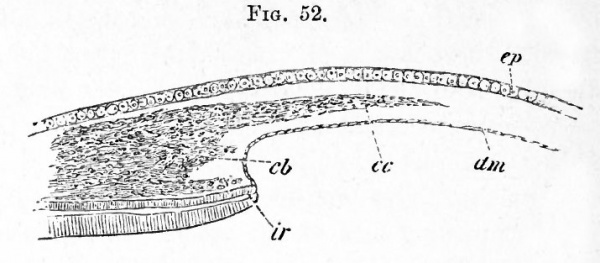
When the lens is completely separated from the epidermis the central part of its outer wall remains directly in contact with the epidermis (future corneal epithelium). At its edge there is a small ring-shaped space bounded by the outer skin, the lens and the edge of the optic cup. There appears, at about the time when the cavity of the lens is completely obliterated, a structureless layer external to the above ring-like space and immediately adjoining the inner face of the epidermis. This layer, which forms the commencement of the cornea proper, at first only forms a ring at the border of the lens, thickest at its outer edge, and gradually thinning away towards the centre. It soon however becomes broader, and finally forms a continuous stratum of considerable thickness, interposed between the external skin and the lens. As soon as this stratum has reached a certain thickness, a layer of flattened cells grows in along its inner side from the mesoblast surrounding the optic cup (Fig. 52, dm). This layer is the epithelioid layer of the membrane of Descemet 1 . After it has become completely established, the mesoblast around the edge of the cornea becomes divided into two strata ; an inner one (Fig. 52 cb) destined to form the mesoblastic tissue of the iris already described, and an outer one (Fig. 52 cc) adjoining the epidermis.
1 It appears possible that Lieberkiihn may be right in stating that the epithelium of Descemet 's membrane grows in between the lens and the epiblast before the formation of the cornea proper, and that Kessler's account, given above, may on this point require correction. From the structure of the eye in some of the lower forms it seems probable that Descemet's membrane is continuous with the choroid.)
Fig. 52. Section through the eye of a fowl on the eighth dat of development, to shew the iris and cornea in the process of formation. (After Kessler.)
- ep. epiblastic epithelium of cornea ; cc. corneal corpuscles growing into the structureless matrix of the cornea ; dm. Descemet's membrane ; ir. iris ; cb. mesoblast of the iris (this reference letter points a little too high). The space between the layers dm. and ep. is filled with the structureless matrix of the cornea.
The outer stratum gives rise to the corneal corpuscles, which are the only constituents of the cornea not yet developed. The corneal corpuscles make their way through the structureless corneal layer, and divide it into two strata, one adjoining the epiblast, and the other adjoining the inner epithelium. The two strata become gradually thinner as the corpuscles invade a larger and larger portion of their substance, and finally the outermost portion of each alone remains to form above and below the membrana elastica anterior and posterior (Descemet's membrane) of the cornea. The corneal corpuscles, which have grown in from the sides, thus form a layer which becomes continually thicker, and gives rise to the main substance of the cornea.
Whether the increase in the thickness of the layer is due to the immigration of fresh corpuscles, or to the division of those already there, is not clear. After the cellular elements have made their way into the cornea, the latter becomes continuous at its edge with the mesoblast which forms the sclerotic.
The derivation of the original structureless layer of the cornea is still uncertain. Kessler derives it from the epiblast, but it appears more probable that Kolliker 1 is right in regarding it as derived from the mesoblast. The grounds for this view are, (1) the fact of its growth inwards from the border of the mesoblast round the edge of the eye, (2) the peculiar relations between it and the corneal corpuscles at a later period. This view would receive still further support if a layer of mesoblast between the lens and the epiblast were really present as believed by Lieberkiihn. It must however be admitted that the objections to Kessler's view of its epiblastic nature are rather a priori than founded on definite observation.
The observations of Kessler, which have been mainly followed in the above account, are strongly opposed by Lieberkiihn and other observers, and are not entirely accepted by Kolliker. It is however especially on the development of these parts in Mammalia (to be spoken of in the sequel) that the above authors found their objections.
The aqueous humour. The cavity for the aqueous humour has its origin in the ring-shaped space round the front of the lens, which, as already mentioned, is bounded by the external skin, the edge of the optic cup, and the lens. By the formation of the cornea this space is shut off from the external skin, and on the appearance of the epithelioid layer of Descemet's membrane a continuous cavity is developed between the cornea and the lens. This cavity enlarges and receives its final form upon the full development of the iris.
(1 L. Kessler, Zur Entwick. d. Auges d. Wirbelthiere. Leipzig, 1874. N. Lieberkiihn, " Beitrage z. Anat. d. embryonalen Auges," Archiv f. Anat. u. Phys., 1879. Kolliker, Entwick. d. Henschen, etc. Leipzig, 1879.)
Summary. We may briefly recapitulate the main facts in the development of the eye as follows.
The eye commences as a lateral outgrowth of the fore-brain, in the form of a stalked vesicle.
The stalk, becoming narrowed and subsequently solid, is converted into the optic nerve.
An involution of the superficial epiblast over the front of the optic vesicle, in the form first of a pit, then of a closed sac with thick walls, and lastly, of a solid rounded mass (the small central cavity being entirely obliterated by the thickening of the hind wall), gives rise to the lens. Coincidently with this involution of the lens, the optic vesicle is doubled up on itself, and its cavity obliterated ; thus a secondary optic vesicle or optic cup with a thick anterior and a thin posterior wall is produced. As a result of the manner in which the doubling up takes place, or of the mode of growth afterwards, the cup of the secondary optic vesicle is at first imperfect along its under surface, where a gap, the choroidal fissure, exists for some little time, but subsequently closes up.
The mesoblast in which the eye is imbedded gathers itself together around the optic cup into a distinct investment, of which the internal layers become the choroid, the external the sclerotic. An ingrowth of this investment between the front surface of the lens and the superficial epiblast furnishes the body of the cornea, the epiblast itself remaining as the anterior corneal epithelium.
The mesoblast entering on the under side through the choroidal fissure gives rise to the vitreous humour, while at a later stage a definite process of this mesoblast becomes the pecten.
Of the walls of the optic cup, the thinner outer (posterior) wall becomes, behind the line of the ora serrata, the pigment-epithelium of the choroid, while the thicker inner (anterior) wall supplies all the elements of the retina, including the rods and cones which grow out from it into the pigment-epithelium.
In front of the line of the ora serrata, both walls of the optic cup, quite thin and wholly fused together, give rise to the pigment- epithelium of the ciliary processes and iris, the bodies of both these organs being formed from the mesoblastic investment.
Accessory Organs connected with the Eye
Eyelids
The most important accessory structures connected with the eye are the eyelids. They are developed as simple folds of the integument with a mesoblastic prolongation between their two laminae. They are three in number, viz. an upper and lower, and a lateral one the nictitating membrane springing from the inner or anterior border of the eye. Their inner face is lined by a prolongation of conjunctiva, which is the modified epiblast covering the cornea and part of the sclerotic.
The Lacrymal glands and Lacrymal duct
The lacrymal glands are formed as solid ingrowths of the conjunctival epithelium. They appear on the eighth day of incubation.
The lacrymal duct begins as a solid ridge of the epidermis, projecting inwards along the line of the so-called lacrymal groove, from the eye to the nasal pit.
At the end of the sixth day this ridge begins to be separated from the epidermis, remaining however united with it on the inner side of the lower eyelid.
After it has become free, it forms a solid cord, the lower end of which unites with the wall of the nasal cavity. The cord so formed gives rise directly to the whole of the duct proper and to the lower branch of the collecting tube. The upper branch of the collecting tube is formed as an outgrowth from it. A lumen begins to be formed in it on the twelfth day of incubation, and first appears at the nasal end. It arises as a space amongst the cells of the cord, but is not due to an absorption of the central cells 1 .
Organ of hearing
During the second day the ear first made its appearance on either side of the hindbrain as an involution of the external epiblast, thrust down into the mass of mesoblast rapidly developing between the epiblast of the skin and that of the neuralcanal (Fig. 27, au. >.). It then had the form of a shallow pit with a widely open mouth, similar in form to that shewn for an embryo dog-fish in Fig. 53, au. p. Before the end of the third day, its mouth closes up and all signs of the opening are obliterated. The pit thus becomes converted into a closed vesicle, lined with epiblast, and surrounded by mesoblast. This vesicle is the otic vesicle, whose cavity rapidly enlarges while its walls become thickened (Fig. 54, CC).
+++++++++++++++++++++++++++++++
FIG. 53. SECTION THROUGH THE HEAD OF AN ELASMOBRANCH EMBRYO, AT THE LEVEL OF THE AUDITORY INVOLUTION.
aup. auditory pit ; aun. ganglion of auditory nerve ; iv.v. roof of fourth ventricle ; a.c.v. anterior cardinal vein ; aa. aorta ;
1 G. Born: "Die Nasenhohlen u. Thranennasengang d. amnioten Wirbelthiere, i. Lacertilia n. Aves." Morphologisches Jahrbuch, Vol. v., 1879.
Laa. aortic trunk of mandibular arch ; pp. head cavity of mandibular arch ; Ivc. alimentary pouch which will form the first visceral cleft ; Th. rudiment of thyroid body.
++++++++++++++++++++++++++++++++++++++
+++++++++++++++++++++++++++++
FIG. 54. SECTION THROUGH THE HlND-BRAIN OF A CHICK AT THE END OF THE THIRD DAY OF INCUBATION.
IV. Fourth ventricle. The section shews the very thin roof and thicker sides of the ventricle.
Ch. Notochord (diagrammatic shading).
CV. Anterior cardinal or jugular vein.
CO. Involuted auditory vesicle. CO points to the end which will form the cochlear canal. RL. Recessus labyrinthi. hy. hypoblast lining the alimentary canal, hy is itself placed in the cavity of the alimentary canal, in that part of the canal which will become the throat. The lower (anterior) wall of the canal is not shewn in the section, but on each side are seen portions of a pair of visceral arches. In each arch is seen the section of the aortic arch AOA belonging to the visceral arch. The vessel thus cut through is running upwards towards the head, being about to join the dorsal aorta AO. Had the section been nearer the head, and carried through the plane at which the aortic arch curves round the alimentary canal to reach the mesoblast above it, AOA and AO would have formed one continuous curved space. In sections lower down in the back the two aorta, AO, one on either side, would be found fused into one median canal.
++++++++++++++++++++++++++++++++++
The changes by which this simple otic vesicle is converted into the complicated system of parts known as the internal ear, have been much more completely worked out for Mammals than for Birds. We shall therefore reserve a full account of them for a later portion of this work. Meanwhile a brief statement of the essential nature of the changes may be useful ; and will be most conveniently introduced here.
The internal ear consists essentially of an inner membranous labyrinth lying loosely in and only partially attached to an outer osseous labyrinth.
The membranous labyrinth (Fig. 55) consists of two parts : (1) the vestibule, with which are connected three pairs of semicircular canals, pag', fr, hor ', and a long narrow hollow process, the aqueductus or recessus vestibuli, and (2) the ductus cochlearis, which in birds is a flask-shaped cavity slightly bent on itself, the dilated end of which is called the lagena. The several parts of each of these cavities freely communicate, and the two are joined together by a narrow canal, the canalis reuniens, cr.
++++++++++++++++++++++++
FIG. 55. TWO VIEWS OF THE MEMBRANOUS LABYRINTH OF COLUMBA
DOMESTICA (copied from Hasse). A, from the exterior, B, from the interior.
hor'. horizontal semicircular canal, hor. ampulla of ditto, pag'. posterior vertical semicircular canal, pag. ampulla of ditto, //. anterior vertical semicircular canal, fr. ampulla of ditto, u. utriculus, ru. recessus utriculi, v. the connecting tube between the ampulla of the anterior vertical semicircular canal and the utriculus, de. ductus endolymphaticus (recessus vestibuli), s. sacculus hemisphericus, cr. canalis reuniens, lag. lagena, mr. membrane of Reissner, pb. Basilar membrane.
++++++++++++++++++++++++
The osseous labyrinth has a corresponding form, and may be similarly divided into parts : into a bony vestibule, with its bony semicircular canals and recessus vestibuli, and into a bony cochlea; but the junction between the cochlea and the bony vestibule is much wider than the membranous canalis reuniens.
The cavity of the osseous cochlea is partially divided lengthways by the ductus cochlearis into a scala tympani and a scala vestibuli, which do not however extend to the lagena.
The auditory nerve, piercing the osseous labyrinth in various points, is distributed in the walls of the membranous labyrinth.
All these complicated structures are derived from the simple primary otic vesicle and the surrounding mesoblast by changes in its form and differentiation of its walls. . All the epiblast of the vesicle goes to form the epithelium of the membranous labyrinth, whose cavity, filled with endolymph, represents the original cavity which was first open to the surface but subsequently covered in. It gradually attains its curiously twisted form by a series of peculiar processes of unequal growth in the, at first, simple walls of the vesicle. The corium of the membranous labyrinth, and all the tissues of the osseous labyrinth, are developed out of the mesoblastic investment of the vesicle. The space between the osseous and membranous labyrinths, including the scala vestibuli and scala tympani, may be regarded as essentially a series of lymphatic cavities hollowed out in the mesoblast.
It will be seen then that the ear, while resembling the eye in so far as the peculiar structures in which the sensory nerve in each case terminates are formed of involuted epiblast, differs from it inasmuch as it arises by an independent involution of the superficial epiblast, whereas the eye is a constricted portion of the general involution which gives rise to the central nervous system.
The origin of the auditory nerve has already been described. It is shewn in close contact with the walls of the auditory pit in Fig. 53.
Organ of Smell
The organ of smell makes its appearance during the third day, as two depressions or pits, on the under surface of the head, a little in front of the eye (Fig. 56, N).
Like the lens and the labyrinth of the ear, they are formed from the external epiblast; unlike them they are never closed up.
The olfactory nerves arise as outgrowths of the front end of the cerebral hemispheres, before any trace of a special division of the brain, forming an olfactory lobe, has become established. Their peripheral extremities unite with the walls of the olfactory pits during the third day. The olfactory lobes arise as outgrowths of the cerebral hemispheres on the seventh day of incubation. ++++++++++++++++++++++++++++++++
FIG. 56. HEAD OP AN EMBRYO CHICK OF THE THIRD DAY VIEWED
SIDEWAYS AS AN OPAQUE OBJECT.
(Chromic acid preparation.)
C.H. Cerebral hemispheres. F.B. Vesicle of third ventricle. M.B. Mid-brain. Cb. Cerebellum. H.B. Medulla oblongata.
N. Nasal pit. ot. otic vesicle in the stage of a pit with the opening not yet closed up. op. Optic vesicle, with I. lens and ch.f. choroidal fissure. The superficial epiblast moulds itself to the form of the optic vesicle and the lens ; hence the choroidal fissure, though formed entirely underneath the superficial epiblast, is distinctly visible from the outside.
1 F. The first visceral fold; above it is seen a slight indication of the superior maxillary process.
2, 3, 4 F. Second, third and fourth visceral folds, with the visceral clefts between them.
+++++++++++++++++++++++++++++++++++++++
Visceral Arches and Visceral Clefts
It must be borne in mind that, especially in the early stages of development, owing to the very unequal growth of different parts, the relative position of the various structures is continually shifting. This is very well seen in the instance of the heart. At its first appearance, the heart is lodged immediately beneath the extreme front of the alimentary canal, so far forwards as to underlie that portion of the medullary canal which will form the brain. It is, in fact, at that epoch a part of the head. From that early position it gradually recedes farther and farther backward, until, at the end of the third day, a considerable interval is observed between it and the actual head. In other words, a distinct neck has been formed, in which most important changes take place.
The neck is distinguished from the trunk in which the heart now lies by the important feature that in it there is no cleavage of the mesoblast into soinatopleure and splanchnopleure, and consequently no pleuroperitoneal cavity. In passing from the exterior into the alimentary canal, the three layers of the blastoderm are successively traversed, without any breach of continuity, save such as is caused by the cavities of the blood vessels. In this neck, so constituted, there appear on the third day certain fissures or clefts, the visceral or branchial clefts. These are real clefts or slits passing right through the walls of the throat, and are placed in series on either side across the axis of the alimentary canal, lying not quite at right angles to that axis and parallel to each other, but converging somewhat to the middle of the throat in front (Fig. 56). Viewed from the outside in either fresh or preserved embryos they are not very distinctly seen to be clefts ; but when they are seen from within, after laying open the throat, their characters as elongated oval slits can easily be recognised.
Four in number on either side, the most anterior is the first to be formed, the other three following in succession. Their formation takes place from within outwards. The hypoblast is pushed outwards as a pouch, which grows till it meets the epiblast, which is then broken through, while the hypoblast forms a junction with the epiblast at the outside of the throat.
No sooner has a cleft been formed than its anterior border (i.e. the border nearer the head) becomes raised into a thick lip or fold, the visceral or branchial fold. Each cleft has its own fold on its anterior border, and in addition the posterior border of the fourth or last visceral cleft is raised into a similar fold. There are thus five visceral folds to four visceral clefts (Fig. 56). The last two folds however, and especially the last, are not nearly so thick and prominent as the other three, the second being the broadest and most conspicuous of all. The first fold meets, or nearly meets, its fellow in the middle line in front, but the second falls short of reaching the middle line, and the third, fourth and fifth do so in an increasing degree. Thus in front views of the neck a triangular space with its apex directed towards the head is observed between the ends of the several folds.
Into this space the pleuroperitoneal cavity extends, the somatopleure separating from the splanchnopleure along the ends of the folds ; and it is here that the aorta plunges into the mesobkst of the body.
The visceral clefts and arches to a large extent disappear in the adult, and constitute examples of an interesting class of embryonic organs, whose presence is only to be explained by the fact that, in the ancestors of the types in which they are now developed in the embryo, they performed an important function in the adult. The visceral arches and clefts are in fact the homologues of the branchial arches and branchial clefts of Fishes, which continue to be formed in the embryos of the higher vertebrate types, although they have ceased to serve as organs of respiration. The skeletal structures developed in the visceral arches persist as the jaw-bones and hyoid bone, but the clefts, with the exception of the first, become obliterated.
Of the history of the skeletal elements we shall speak in detail hereafter; meanwhile we may briefly deal with the general history of these parts.
The first fold on either side, increasing rapidly in size and prominence, does not, like the others, remain single, but sends off in the course of the third day a branch or bud-like process from its anterior edge. This branch, starting from near the dorsal beginning of the fold, runs ventralwards and forwards, tending to meet the corresponding branch from the fold on the other side, at a point in the middle line nearer the front of the head than the junction of the main folds. The two branches do not quite meet, being separated by a median process, which at the same time grows down from the extreme front of the head, and against which they abut. Between the main folds, which are directed somewhat backwards and the branches which slant forwards, a somewhat lozenge-shaped space is developed which, as the folds become more and more prominent, grows deeper and deeper. In the main folds are developed the mandibles, and in the branches the superior maxillce : the lozenge-shaped cavity between them is the cavity of the mouth, and the descending process which helps to complete the upper margin of this cavity is called, from the parts which will be formed out of it, the frontonasal process.
Part of the mesoblast of the two succeeding pairs of visceral folds is transformed into the hyoid bone, which will be best considered in connection with the development of the skull. The two last arches disappear without giving rise to any permanent structures.
With the exception of the first the visceral clefts become obliterated at an early stage of embryonic life ; but the first persists, although it loses all trace of its original branchial function and becomes intimately connected with the organ of hearing, of which in fact it forms a most essential part, becoming converted into the Eustachian tube and tympanic cavity. The outer opening and the outer part also of the cleft become obliterated at an early date, but from the inner part of the cleft a diverticulum is given off towards the exterior, which becomes the tympanic cavity. The inner part of the cleft itself forms the Eustachian tube, while its mouth forms the oral aperture of this tube.
The meatus auditorius externus first appears as a shallow depression at the region where the closure of the first visceral cleft takes place. It is in part formed by the tissue surrounding this depression growing up in the form of a wall, but the blind end of the meatus also becomes actually pushed in towards the tympanic cavity.
The tympanic membrane is derived from the tissue which separates the meatus auditorius externus from the tympanic cavity. This tissue is obviously constituted of an hypoblastic epithelium on its inner aspect, an epiblastic epithelium on its outer aspect, and a layer of mesoblast between them, and these three layers give rise to the three layers of which this membrane is formed in the adult. During the greater part of foetal life it is relatively very thick, and presents a structure bearing but little resemblance to that in the adult.
The tympanic cavity is bounded on its inner aspect by the osseous investment of the internal ear, but at two points, known as the fenestra ovalis and fenestra rotunda, the bone is deficient and its place is taken by a membrane.
These two fenestrse appear early, and are probably formed by the nonchrondrification of a -small area of the embryonic cartilage. The upper of the two, or fenestra ovalis, contains the base of a bone, known as the columella. The main part of the columella is formed of a stalk which is held by Parker to be derived from part of the skeleton of the visceral arches, while the base, forming the stapes, appears to be an independent formation.
The stalk of the columella extends to the tympanic membrane; its outer end becoming imbedded in this membrane, and serving to transmit the vibrations of the membrane to the fluid in the internal ear.
Vascular system
By the end of the second day three pairs of aortic arches had been established in connection with the heart. When the visceral folds and clefts are formed, a definite arrangement between them and the aortic arches is always observed. The first visceral cleft runs between the first and second aortic arches. Consequently the first aortic arch runs in the first visceral fold, and the second in the second. In the same way, the second visceral cleft lies between the second and third aortic arches, the third aortic arch running in the third visceral fold. Each aortic arch runs in the thickened mesoblast of the corresponding fold.
Arrived at the dorsal surface of the alimentary canal, these arches unite at acute angles to form a common trunk, the dorsal aorta (Fig. 57, A.0\ which runs along the back immediately under the notochord. The length of this common single trunk is not great, as it soon divides into two main branches, each of which, after giving off the large vitelline artery, Of. A., pursues its course with diminished calibre to the tail, where it is finally lost in the capillaries of that part.
The heart is now completely doubled up on itself. Its mode of curvature is apparently somewhat complicated. Starting from the point of junction of the vitelline veins (Fig. 37, Ht), there is first a slight curvature towards the left; this is followed by a turn to the right, and then the heart is completely bent on itself, so that afterwards it pursues a course directed from behind quite straight forwards (except perhaps for a little inclination to the left) to the point where the aortic arches branch off. In this way, as shewn in section in Fig. 59, A, the end of the bulbus arteriosus (v) comes to lie just underneath (or in front of according to the position of the embryo) that part which has already been marked off by the lateral bulgings as the auricular portion (au).
+++++++++++++++++++++++++++++
Fia 57. DIAGRAM OF THE ARTERIAL CIRCULATION ON THE THIRD DAY.
1, 2, 3. The first three pairs of aortic arches. A. The vessel formed by the junction of the three pairs of arches. A.O. Dorsal aorta formed by the junction of the two branches A and A ; it quickly divides again into two branches which pass down one on each side of the notochord. From each of these is given off a large branch Of. A., the vitelline artery. E.CA, LCA, external and internal carotid arteries.
+++++++++++++++++++++++++++++++++++++++
That part of the heart which is turned to the right, including the point of doubling up, is the ventricular portion, and is even at this stage separated from the auricular portion by a slight neck. This external constriction corresponds to an internal narrowing of the lumen of the heart, and marks the position of the future canalis auric u laris.
The ventricular portion is, on the other hand, likewise separated by a fainter constriction from the anterior continuation of the heart which forms the bulbus arteriosus. The projecting part where the doubling takes place is at this stage still quite round ; we shall see that later on it becomes pointed and forms the apex of the heart.
The whole venous portion of the heart (if we may so speak of it, though of course at this stage blood of the same quality passes right along the whole cardiac canal) lies in a plane which is more dorsal than the arterial portion. The point at which the venous roots of the heart, i.e. the two vitelline trunks, unite into a single canal, is on this day carried farther and farther away from the heart itself. By the end of the day there is a considerable distance between the auricular portion of the actual heart and the point where the venous roots separate, each to pursue its course along the splanchnopleure-fold of its own side. This distance is traversed by a single venous trunk, of which the portion close to the auricles is called the sinus venosus, and the more distant the ductus venosus. We shall give to the whole trunk the name used by the older observers, the meatus venosus.
Small arteries to various parts of the body are now being given off by the aorta and its branches. The capillaries in which these end are gathered into veins which unite to form two main trunks on either side, the cardinal veins, anterior and posterior (Fig. 36, Fig. 58J and (7), which run parallel to the long axis of the body in the upper part of the mesoblast, a little external to the mesoblastic somites.
+++++++++++++++++++++++
FIG. 58. DIAGRAM OF THE VENOUS CIRCULATION ON THE THIRD DAY.
H. Heart. J. Jugular or anterior cardinal vein. C. Inferior or posterior cardinal vein. Of. Vitelline vein. dc. Ductus Cuvieri.
+++++++++++++++++++++++++++
These veins, which do not attain to any great importance till well on in the third day, unite opposite to the heart, on each side, into a short common trunk at right angles to themselves. The two short trunks thus formed, which bear the name of ductus Cuvieri (Fig. 36, Fig. 58, dc), running ventralwards and then transversely straight inwards towards the middle line fall into the sinus venosus.
The two ductus Cuvieri pass from the heart to the body walls in a special horizontal mesentery, whose formation and function we shall return to in speaking of the formation of the pericardial cavity. The position of one of them is shewn in section in Fig. 59 B, dc.
++++++++++++++++++++++
FIG. 59. TRANSVERSE SECTIONS THROUGH A CHICK EMBRYO WITH TWENTY-ONE MESOBLASTIC SOMITES TO SHEW THE FORMATION OF THE PERICARDIAL CAVITY, A. BEING THE ANTERIOR SECTION.
pp. body cavity ; pc. pericardial cavity ; al. alimentary cavity ; au. auricle ; v. ventricle ; sv. sinus venosus ; dc. ductu& Cuvieri ; ao. aorta ; mp. muscle-plate ; me. medullary cord.
+++++++++++++++++++++++
The alimentary canal
As we stated above, the folding in of the splanchnopleure to form the alimentary canal is proceeding with great rapidity, the tail-fold as well as the head-fold contributing largely to this result.
The formation of the tail-fold is very similar to that of the head-fold. The tail is a solid, somewhat curved,, blunt cone of mesoblast, immediately coated with the superficial epiblast except at the upper surface (corresponding to the back of the embryo), where lies the pointed termination of the neural tube.
So rapid is the closure of the splanchnopleure both in front and behind, that two of the three parts into which the digestive tract may be divided, are brought, on this day, to the condition of complete tubes.
The first division, including the region from the mouth to the duodenum, is completely folded in by the end of the day; so likewise is the third division comprising the large intestine and the cloaca. The middle division, corresponding to the future small intestine, still remains quite open to the yolk-sac below.
The attachment of the newly formed alimentary canal to the body above is at first very broad, and only a thin stratum of mesoblast separates the hypoblast of the canal from the notochord and mesoblastic somites; even that maybe absent under the notochord. During the third day, however, along such portions of the canal as have become regularly enclosed, i.e. the hinder division and the posterior moiety of the anterior division, the mesoblastic attachment becomes narrower and (in a vertical direction) longer, the canal appearing to be drawn more ventralwards (or according to the position of the embryo forwards), away from the vertebral column.
In what may be regarded as the pleural division of the general pleuroperitoneal space, along that part of the alimentary canal which will form the oesophagus, this withdrawal is very slight (Fig. 59), but it is very marked in the peritoneal space. Here such parts of the digestive canal as are formed come to be suspended from the body above by a narrow flattened band of mesoblastic tissue which reaches from the neighbourhood of the notochord, and becomes continuous with the mesoblastic coating which wraps round the hypoblast of the canal. This flattened band is the mesentery, shewn commencing in Fig. 65, and much more advanced in Fig. 68, M. It is covered on either side by a layer of flat cells forming the epithelioid lining of the peritoneal membrane, while its interior is composed of indifferent tissue.
The front division of the digestive tract consists of three parts. The most anterior part, the oesophagus, still ending blindly in front reaches back as far as the level of the hind end of the heart ; and is divided into two regions, viz. an anterior region, characterized by the presence of the visceral clefts, whose development has already been dealt with, and a posterior region without such clefts.
Its transverse section, which up to the end of the second day was somewhat crescent-shaped, with the convexity downwards, becomes on this day more nearly circular. Close to its hinder limit, the lungs (Fig. 60, Ig), of whose formation we shall speak directly, take their origin.
The portion of the digestive canal which succeeds the oesophagus, becomes towards the close of the third day somewhat dilated (Fig. 60, St) ; the region of the stomach is thus indicated.
The hinder or pyloric end of the stomach is separated by a very small interval from the point where the complete closing in of the alimentary canal ceases, and where the splanchnopleure-folds spread out over the yolk, This short tract is nevertheless clearly marked out as the duodenum by the fact that from it, as we shall presently point out, the rudiments of the ducts of the liver and pancreas are beginning to be formed.
++++++++++++++++++++++++++
FIG. 60. DIAGRAM OF A PORTION OF THE DIGESTIVE TRACT OF A CHICK UPON THE FOURTH DAY. (Copied from Gotte.)
The black inner line represents the hypoblast, the outer shading the mesoblast. Ig. lung-diverticulum with expanded termination, forming the primary lung-vesicle. St. stomach. I. two hepatic diverticula with their terminations united by cords of hypoblast cells, p. diverticulum of the pancreas with the vesicular diverticula coming from it.
+++++++++++++++++++++++++++
The posterior division of the digestive tract, corresponding to the great intestine and cloaca, is from its very first formation nearly circular in section and of a larger bore than the oesophagus.
During part of the third day the hinder end of this section of the gut is in communication with the neural tube by the neur enteric canal already spoken of (Fig. 61, ne). The communication between the two tubes does not last long, but even after its rupture there remains a portion of the canal continuous with the gut ; this, however, constitutes a purely embryonic and transient section of the alimentary canal, and is known as the postanal gut. Immediately in front of it is a deep infolding of the epiblast, which nearly meets the hypoblast (Fig. 61, an) and forms the rudiment of the anus and of the outer section of the cloaca into which the bursa Fabricii opens in the adult. It is known to embryologists as the proctodceum, but does not open into the alimentary tract till considerably later. The section of the alimentary tract immediately in front of the postanal gut is somewhat enlarged, and becomes the inner section of the adult cloaca receiving the urinary and genital ducts. The allantois, to whose development we shall return directly, opens into it ventrally.
+++++++++++++++++++++++++++++++++
FIG. 61. DIAGRAMMATIC LONGITUDINAL SECTION THROUGH THE POSTERIOR END OF AN EMBRYO BIRD, AT THE TIME OF THE FORMATION ON THE ALLANTOIS.
ep. epiblast; Sp.c. spinal canal ; ch. notochord ; n.e. neurenteric canal ; hy. hypoblast ; p.a.g. postanal gut ; pr. remains of primitive streak folded in on the ventral side ; al. allantois ; me. mesoblast ; an. point where anus will be formed ; p.c. perivisceral cavity ; am. amnion ; so. somatopleure ; sp. splanchnopleure.
+++++++++++++++++++++++++++++++
It is to be noted that the two sections of the cloaca of adult birds have a different origin. The inner section being part of the primitive alimentary tract and lined by hypoblast ; the outer being part of an involution of the outer skin and lined by epiblast.
The lungs are in their origin essentially buds or processes from the primitive oesophagus.
At a point immediately behind the region of the visceral clefts the cavity of the alimentary canal becomes compressed laterally, and at the same time constricted in the middle so that its transverse section (Fig. 62, 1) is somewhat hourglass-shaped, and shews an upper or dorsal chamber d, joining on to a lower or ventral chamber I by a short narrow neck.
The hinder end of the lower tube enlarges (Fig. 62, 2), and then becomes partially divided into two lobes (Fig. 62, 3). All these parts at first freely communicate, but the two lobes behind, partly by their own growth, and partly by a process of constriction, soon become isolated posteriorly (Fig. 60, lg)\ while in front they open into the lower chamber of the oesophagus.
By a continuation forwards of the process of constriction the lower chamber of the oesophagus, carrying with it the two lobes above mentioned, becomes gradually transformed into an independent tube, opening in front by a narrow slit-like aperture into the oesophagus. The single tube in front is the rudiment of the trachea and larynx, while the two diverticula behind (Fig. 60, Ig) become the bronchial tubes and lungs.
While the above changes are taking place in the hypoblastic walls of the alimentary tract, the splanchnic mesoblast surrounding these structures becomes very much thickened; but otherwise bears no marks of the internal changes which are going on, so that the above formation of the lungs and trachea cannot be seen from the surface. As the paired diverticula of the lungs grow backwards, the mesoblast around them takes however the form of two lobes, into which they gradually bore their way.
++++++++++++++++++++
FIG. 62. FOUR DIAGRAMS ILLUSTRATING THE FORMATION OF THE LUNGS. (After Gotte.)
a. mesoblast ; b. hypoblast ; d. cavity of digestive canal ; I. cavity of the pulmonary diverticulum.
In (1) the digestive canal has commenced to be constricted into a dorsal and ventral canal ; the former the true alimentary canal, the latter the pulmonary tube ; the two tubes communicate with each other in the centre.
In (2) the ventral (pulmonary) tube has become expanded.
In (3) the expanded portion of the tube has become constricted into two tubes, still communicating with each other and with the digestive canal.
In (4) these are completely separated from each other and from the digestive canal, and the mesoblast has also begun to exhibit externally changes corresponding to the internal changes which have been going on.
++++++++++++++++++++++++++++++++
The further development of the lungs is, at first, essentially similar to that of a racemose gland. From each primitive diverticulum numerous branches are given off. These branches, which are mainly confined to the dorsal and lateral parts, penetrate into the surrounding mesoblast and continue to give rise to secondary and tertiary branches. At right angles to the finest of these the arborescent branches so characteristic of the avian lung are given off. In the mesoblast around them numerous capillaries make their appearance.
The air sacs, which form such important adjuncts of the avian lungs, are the dilated extremities of the primary pulmonary diverticula and of their main branches.
The whole pulmonary structure is therefore the result of the growth by budding of a system of branched hypoblastic tubes in the midst of a mass of mesoblastic tissue, the hypoblastic elements giving rise to the epithelium of the tubes and the mesoblast providing the elastic, muscular, cartilaginous, connective and other tissues of the tracheal and bronchial walls.
The liver is the first formed chylopoietic appendage of the digestive canal, and arises between the 55th and 60th hour as a couple of diverticula one from either side of the duodenum immediately behind the stomach (Fig. 60, I). These diverticula are of course lined by hypoblast. The right one is, in all cases, from the first longer, but of smaller diameter than the left. Situated a little behind the heart, they embrace between them the two vitelline veins forming the roots of the meatus venosus.
The diverticula soon give rise to numerous hollow branches or processes, which extend into the surrounding mesoblast.
Towards the end of the third day there may further be observed in the greatly thickened mesoblastic investment of either diverticulum a number of cylindrical solid cords of hypoblast which are apparently outgrowths from the hypoblast of the branches of the diverticula. These cylinders rapidly increase in number, apparently by a process of sprouting, and their somewhat swollen peripheral extremities come into contact and unite. And thus, about the ninetieth hour, a sort of network of solid thick strings of hypoblastic cells is formed, the mesoblast in the meshes of the network becoming at the same time largely converted into blood-vessels. Each diverticulum becomes in this way surrounded by a thick mass composed partly of solid cylinders, and to a less extent of hollow processes, continuous with the cylinders on the one hand, and the main diverticulum on the other, all knit together with commencing blood-vessels and unchanged mesoblastic tissue. Between the two masses runs the now fused roots of the meatus venosus with which the bloodvessels in each mass are connected.
Early on the fourth day each mass sends out ventral to the meatus venosus a solid projection of hypoblastic cylinders towards its fellow, that from the left side being much the longest. The two projections unite and form a long solid wedge, which passes obliquely down from the right (or from the embryo lying on its left side, the upper) mass to the left (or lower) one. In this new wedge may be seen the same arrangement of a network of hypoblastic cylinders filled in with vascular mesoblast as in the rest of the liver. The two original diverticula with their investing masses represent respectively the right and left lobes of the liver, and the wedgelike bridge connecting them is the middle lobe.
During the fourth and fifth days the growth of the solid, lobed liver thus formed is very considerable; the hypoblastic cylinders multiply rapidly, and the network formed by them becomes very close, the meshes containing little more than blood-vessels. The hollow processes of the diverticula also ramify widely, each branch being composed of a lining of hypoblast enveloped in a coating of spindle-shaped mesoblastic cells. The blood-vessels are in direct connection with the meatus venosus have become, in fact, branches of it. It may soon be observed, that in those vessels which are connected with the posterior part of the liver (Fig. 74), the stream of blood is directed from the meatus venosus into the network of the liver. In those connected with the anterior part the reverse is the case ; here the blood flows from the liver into the meatus venosus. The thick network of solid cylinders represents the hepatic parenchyma of the adult liver, while the hollow processes of the diverticula are the rudiments of the biliary ducts; and we may suppose each solid cylinder to represent a duct with its lumen almost, but perhaps not quite, completely obliterated.
During the fifth day, a special sac or pouch is developed from the right primary diverticulum. This pouch, consisting of an inner coat of hypoblast, and an outer of mesoblast, is the rudiment of the gall-bladder.
The Pancreas
The pancreas arises nearly at the same time as the liver in the form of an almost solid outgrowth from the dorsal side of the intestine nearly opposite but slightly behind the hepatic outgrowths (Fig. 60, p). Its blind end becomes somewhat enlarged and from it numerous diverticula grow out into the passive splanchnic mesoblast.
As the ductules grow longer and become branched, vascular processes grow in between them, and the whole forms a compact glandular body in the mesentery on the dorsal side of the alimentary tract. The primitive outgrowth elongates and assumes the character of a duct.
On the sixth day a new similar outgrowth from the duodenum takes place between the primary diverticulum and the stomach. This, which ultimately coalesces with its predecessor, gives rise to the second duct, and forms a considerable part of the adult pancreas. A third duct is formed at a much later period.
The Thyroid body
The thyroid body arises at the end of the second or beginning of the third day as an outgrowth from the hypoblast of the ventral wall of the throat opposite the point of origin of the anterior aortic arch. It has at first the form of a groove extending forwards up to the future mouth, and in its front part extending ventrally to the epiblast. It has not been made out whether the whole groove becomes converted into the permanent thyroid. By the fourth day it becomes a solid mass of cells, and by the fifth ceases to be connected with the epithelium of the throat, becoming at the same time bilobed. By the seventh day it has travelled somewhat backwards, and the two lobes have completely separated from each other. By the ninth day the whole is invested by a capsule of connective tissue, which sends in septa dividing it into a number of lobes or solid masses of cells, and by the sixteenth day its two lobes are composed of a number of follicles, each with a 'meinbrana propria,' and separated from each other by septa of connective tissue, much as in the adult x .
The spleen
Although the spleen cannot be reckoned amongst the glands of the alimentary tract its development may conveniently be dealt with here. It is formed shortly after the first appearance of the pancreas, as a thickening of the mesentery of the stomach (mesogastrium) and is therefore entirely a mesoblastic structure. The mass of mesoblast which forms the spleen becomes early separated by a groove on the one side from the pancreas and on the other from the mesentery. Some of its cells become elongated, and send out processes which, uniting with like processes from other cells, form the trabecular system. From the remainder of the tissue are derived the cells of the spleen pulp, which frequently contain more than one nucleus. Especial accumulations of these take place at a later period to form the so-called Malpighian corpuscles of the spleen.
The Allantois
We have already had occasion to point out that the allantois is essentially a diverticulum of the alimentary tract into which it opens immediately in front of the anus. Its walls are formed of vascular splanchnic mesoblast, within which is a lining of hypoblast. It becomes a conspicuous object on the third day of incubation, but its first development takes place at an earlier period, and is intimately connected with the formation of the posterior section of the gut.
At the time of the folding in of the hinder end of the gut the splitting of the mesoblast into somatopleure and splanchnopleure has extended up to the border of the hinder division of the primitive streak. The ventral wall of what we have already termed the postanal section of the alimentary tract is formed by the primitive streak. Immediately in front of this is the involution which forms the proctodseum; while the wall of the hindgut in front of the proctodseum owes its origin to a folding in of the splanchnopleure.
(1 Miiller Ueber die Entwiclcelung der Schilddruse. Jenaischa Zeitschrift, 1871.)
The allantois first appears as a narrow diverticulum formed by a special fold of the splanchnopleure just in front of the proctodaeum. This protuberance arises, however, before the splanchnopleure has begun to be tucked in so as to form the ventral wall of the hindgut ; and it then forms a diverticulum (Fig. 63 A, All) the open end of which is directed forward, while its blind end points somewhat dorsalwards and towards the peritoneal space behind the embryo.
As the hindgut becomes folded in the allantois shifts its position, and forms (Figs. 63 B and 61) a rather wide vesicle lying immediately ventral to the hind end of the digestive canal, with which it communicates freely by a still considerable opening; its blind end projects into the pleuroperitoneal cavity below.
Still later the allantois grows forward, and becomes a large spherical vesicle, still however remaining connected with the cloaca by a narrow canal which forms its neck or stalk (Fig. 9 G, al). From the first the allantois lies in the pleuroperitoneal cavity. In this cavity it grows forwards till it reaches the front limit of the hindgut, where the splanchnopleure turns back to enclose the yolk-sac. It does not during the third
+++++++++++++++++++++
FIG. 63. TWO LONGITUDINAL SECTIONS OF THE TAIL-END OF AN EMBRYO CHICK TO SHEW THE ORIGIN OF THE ALLANTOIS. A AT THE BEGINNING OF THE THIRD DAY; B AT THE MIDDLE OF THE THIRD DAY. (After Dobrynin.)
t. the tail ; m. the mesoblast ; cc f . the epiblast ; x". the neural , canal ; Dd. the dorsal wall of the hindgut ; SO. somatopleure ; Spl. splanchnopleure ; u. the mesoblast of the splanchnopleure carrying the vessels of the yolk-sac ; pp. pleuroperitoneal cavity ; Df. the epithelium lining the pleuroperitoneal cavity; All. the commencing allantois ; w. projection formed by anterior and posterior divisions of the primitive streak; y. hypoblast which will form the ventral wall of the hindgut ; v. anal invagination (proctodseum) ; 6f. cloaca.
++++++++++++++++++++++++++++++++
day project beyond this point ; but on the fourth day begins to pass out beyond the body of the chick, along the as yet wide space between the splanchnic and somatic stalks of the embryo, on its way to the space between the external and internal folds of the amnion, which it will be remembered, is directly continuous with the pleuroperitoneal cavity (Fig. 9 K). In this space it eventually spreads out over the whole body of the chick. On the first half of the fourth day the vesicle is still very small, and its growth is not very rapid. Its mesoblast wall still remains very thick. In the latter half of the day its growth becomes very rapid, and it forms a very conspicuous object in a chick of that date (Fig. 67, Al}. At the same time its blood-vessels become important. It receives its supply of blood from two branches of the aorta known as the allantoic arteries, and the blood is brought back from it by two allantoic veins which run along in the body walls, and after uniting into a single trunk fall into the vitelline vein close behind the liver.
Mesoblast of the trunk
Coincidently with the appearance of these several rudiments of important organs in the more or less modified splanchnopleurefolds, the solid trunk of the embryo is undergoing marked changes.
When we compare a transverse section taken through say the middle of the trunk at the end of the third day (Fig. 65), with a similar one of the second day (Fig. 34), or even the commencement of the third day (Fig. 64), we are struck with the great increase of depth (from dorsal to ventral surface) in proportion to breadth. This is partly due to the slope of the side walls of the body having become much steeper> as a direct result of the rapidly progressing folding off of the embryo from the yolk-sac. But it is also brought about by the great changes both of shape and structure which are taking place in the mesoblastic somites, as well as by the development of a mass of tissue between the notochord and the hypoblast of the alimentary canal.
It will be remembered that the horizontal splitting of the mesoblast into somatic and splanchnic layers extends at first to the dorsal summit of the vertebral plates, but after the formation of the somites the split between the somatic and splanchnic layers becomes to a large extent obliterated, though in the anterior somites it appears in part to persist. The somites on the second day, as seen in a transverse section (Fig. 34, P.v), are somewhat quadrilateral in form but broader than they are deep.
+++++++++++++++++++++++++++++
FIG. 64. TRANSVERSE SECTION THROUGH THE TRUNK OF A DUCK EMBRYO WITH ABOUT TWENTY-FOUR MESOBLASTIC SOMITES.
am. amniou ; so. somatopleure ; sp. splanchnopleure ; wd. Wolfnan duct ; st. segmental tube ; ca.v. cardinal vein ; ms. muscle -plate ; sp.g. spinal ganglion ; sp.c. spinal cord ; ch, notochord ; ao. aorta ; Jiy. hypoblast.
+++++++++++++++++++++++++++++++
Each at that time consists of a somewhat thick cortex of radiating rather granular columnar cells, enclosing a small kernel of spherical cells. They are not, as may be seen in the above figure, completely separated from the ventral (or rather at this period lateral) parts of the mesoblastic plate, and the dorsal and outer layer of the cortex of the somites is continuous with the somatic layer of mesoblast, the remainder of the cortex, with the central kernel, being continuous with the splanchnic layer. Towards the end of the second and beginning of the third day the dorsal and outer layer of the cortex, together probably with some of the central cells of the kernel, becomes separated off as a special plate. From this plate, which is shewn in the act of being formed in Fig. 64, ms, the greater part of the voluntary muscular system of the trunk is developed. When once formed the muscleplates have in surface views a somewhat oblong form, and consist of two layers, an inner and an outer, which enclose between them an almost obliterated central cavity (Fig. 65, mp). No sooner is the muscle-plate formed than the middle portion of the inner layer becomes converted into longitudinal muscles. The central space in the muscle -plates is clearly a remnant of the vertebral portion of the body cavity, which, though it wholly or partially disappears in a previous stage, reappears again on the formation of the muscle-plate.
It is especially interesting to note that the first formed muscles in embryo birds have an arrangement like that which is permanent in fishes; being longitudinal in direction, and divided into segments.
+++++++++++++++++++++++++
FIG. 65. SECTION THROUGH THE DORSAL REGION OF AN EMBRYO CHICK AT THE END OF THE THIRD DAY.
Am. amnion. m. p. muscle-plate. C. V. cardinal vein. Ao. dorsal aorta. The section passes through the point where the dorsal aorta is just commencing to divide into two branches. Ch. notochord. W. d. Wolman duct. W. b. commencing differentiation of the mesoblast cells to form the Wolman body. ep. epiblast. SO. somatopleure. Sp. splaiichnopleure. hy. hypoblast. The section passes through the point where the digestive canal communicates with the yolksac, and is consequently still open below.
This section should be compared with the section through the dorsal region of an embryo at the commencement of the third day (Fig. 64). The chief differences between them arise from the great increase in the space (now filled with mesoblast-cells) between the notochord and the hypoblast. In addition to this we have in the later section the completely formed amnion, the separation of the muscle-plate from the mesoblastic somites, the formation of the Wolman body, etc.
The mesoblast including the Wolman body and the muscleplate (m.p.) is represented in a purely diagrammatic manner. The amnion, of which only the inner limb or true amnion is represented in the figure, is seen to be composed of epiblast and a layer of mesoblast ; though in contact with the body above the top of the medullary canal, it does not in any way coalesce with it, as might be concluded from the figure.
++++++++++++++++++++++++++++++++++
The remainder of the somites, after the formation of the muscle-plates, is of very considerable bulk ; the cells of the cortex belonging to them lose their distinctive characters, and their major part becomes converted, in a manner which will be more particularly described in a future chapter, into the bodies of the permanent vertebrae.
We may merely add here that each of these bodies sends a process inwards ventral to the medullary cord, and that the processes from each pair of these bodies envelope between them the notochord.
The intermediate cell-mass and the Wolffian body.
In a transverse section of a 45 hours' embryo a considerable mass of cells may be seen collected between the mesoblastic somites and the point where the divergence into somatopleure and splanchnopleure begins (Fig. 34, just below W.d). This mass of cells, which we have already spoken of as the intermediate cell-mass, is at first indistinguishable from the cells lining the inner end of the body cavity ; but on the third day, a special peritoneal lining of epithelioid cells is developed which is more or less sharply marked off from the adjoining part of the intermediate cell-mass. This latter now also passes without any very sharp line of demarcation into the mesoblastic somite itself; and as the folding in of the side wall progresses, the mass of cells in this position increases in size and grows in between the notochord and the hypoblast, but does not accumulate to a sufficient extent to separate them widely until the end of the third or beginning of the fourth day.
The fusion between the intermediate cell-mass and the inner portions of the somites becomes so complete on the third day that it is almost impossible to say which of the cells in the neighbourhood of the notochord are derived from the somites and which form the intermediate cell-mass. It seems almost certain however that the cells which form the immediate investment of the notochord really belong to the somites.
The intermediate cell-mass is of special importance to the embryologist, in that the excretory and generative systems are developed from it.
We have already described (p. 106) the development of the Wolffian duct, and we have now to deal with the Wolffian body which is, as the reader has no doubt gathered, the embryonic excretory organ.
The structure of the fully developed Wolffian body is fundamentally similar to that of the permanent kidneys, and consists essentially of convoluted tubules, commencing in Malpighian bodies with vascular glomeruli, and opening into the duct.
The tubules of the Wolffian body are developed independently of the Wolffian duct, and are derived from the intermediate cell-mass, shewn in Fig. 34, between the upper end of the body-cavity and the mesoblastic somite. In the chick the mode of development of this mass into the segmental tubules is different in the regions in front of and behind about the sixteenth segment. In front of about the sixteenth segment special parts of the intermediate cell-mass remain attached to the peritoneal epithelium, on this layer becoming differentiated ; there being several such parts to each segment The parts of the intermediate cellmass attached to the peritoneal epithelium become converted into S-shaped cords (Fig. 64 sf) which soon unite with the Wolffian duct (wd), and constitute the primitive Wolffian tubules. Into the commencement of each of these cords the lumen of the body-cavity is for a short distance prolonged, so that this part constitutes a rudimentary peritoneal funnel leading from the body-cavity into the lumen of the Wolffian tubule.
In the foremost Wolffian tubules, which never reach very complete development, the peritoneal funnels riden considerably. The section of the tube adjoining tie wide peritoneal funnel becomes partially invaginated the formation of a vascular ingrowth known as a glomerulus, and this glomerulus soon grows to such an extent as to project through the peritoneal funnel, the neck of which it completely fills, into the body-cavity (Fig. 66, gl). There is thus formed a series of glomeruli belonging to the anterior Wolffian tubuli projecting freely into the body-cavity. These glomeruli with their tubuli become however early aborted.
+++++++++
Fig. 66. SECTION THROUGH THE EXTERNAL GLOMERULUS OF ONE OF THE ANTERIOR SEGMENTAL TUBES OF AN EMBRYO CHICK OF ABOUT 100 HOURS. gl. glomerulus ; ge. peritoneal epithelium ; Wd. Wolffian duct ;
ao. aorta ; me. mesentery.
The Wolffian tubule, and the connection between the external and internal parts of the glomerulus are not shewn in this figure.
++++++++++++++++
In the case of the remaining tubules developed from the S-shaped cords, the attachment to the peritoneal epithelium is very soon lost. The cords acquire a lumen, and open into the Wolffian duct. Their blind extremities constitute the commencements of Malpighian bodies.
In the posterior part of the Wolffian body of the chick the intermediate cell-mass becomes very early detached from the peritoneal epithelium, and at a considerably later period breaks up into oval vesicles, which elongate into Wolffian tubules. In addition to the primary tubules, whose development has just been described, secondary and tertiary tubules are formed on the dorsal side of the primary tubules. They are differentiated out of the mesoblast of the intermediate cell-mass and open independently into the Wolffian duct.
A tubule of the Wolffian body typically consists of the follow ing parts, (1) a section carrying the peritoneal opening, and known as the peritoneal funnel, (2) a dilated vesicle into which this opens, (3) a coiled tubulus proceeding from (2), and terminating in (4) a wider portion opening into the Wolffian duct.
In the chick, the peritoneal funnel is only found in the most anterior tubules and soon atrophies; it is not developed in the tubules of the posterior part of the Wolffian body. Region No. 4 also is not clearly marked off from region No. 3. One part of the wall of the dilated vesicle (2) is invaginated by a bunch of capillaries and gives rise to the Malpighian body.
In consequence of the continual folding in of the somatopleure and especially of the splanchnopleure, as well as owing to the changes taking place in the mesoblastic somites, the Wolffian duct undergoes on the third day a remarkable change of position. Instead of lying, as on the second day, immediately under the epiblast (Fig. 34, W.d), it is soon found to have apparently descended into the middle of the intermediate cell-mass (Fig. 64, w.d) and at the end of the third day occupies a still lower position and even projects somewhat towards the pleuroperitoneal cavity. (Fig. 65, W.d.)
Summary Day 3
The chief events then which take place on the third day are as follows :
- The turning over of the embryo so that it now lies on its left side.
- The cranial flexure round the anterior extremity of the notochord.
- The completion of the circulation of the yolksac; the increased curvature of the heart, and the demarcation of its several parts; the appearance of new aortic arches, and of the cardinal veins.
- The formation of four visceral clefts and five visceral arches.
- The involution to form the lens, and the formation of the secondary optic vesicle.
- The closing in of the otic vesicle.
- The formation of the nasal pits.
- The appearance of the vesicles of the cerebral hemispheres ; the separation of the hind-brain into cerebellum and medulla oblongata.
- The definite establishment of the cranial and spinal nerves as outgrowths of the central nervous system.
- The completion of the fore-gut and of the hind-gut; the division of the former into oasophagus, stomach and duodenum, of the latter into large intestine and cloaca.
- The formation of the lungs from a diverticulum of the alimentary canal immediately in front of the stomach.
- The formation of the liver and pancreas: the former as two diverticula from the duodenum, which subsequently become united by nearly solid outgrowths ; the latter as a single diverticulum also from the duodenum.
- The changes in the mesoblastic somites and the appearance of the muscle-plates.
- The definite formation of the Wolffian bodies and the change in position of the Wolffian duct.
The Elements of Embryology - Volume 1 (1883)
The History of the Chick: Egg structure and incubation beginning | Summary whole incubation | First day | Second day - first half | Second day - second half | Third day | Fourth day | Fifth day | Sixth day to incubation end | Appendix
| Historic Disclaimer - information about historic embryology pages |
|---|
| Pages where the terms "Historic" (textbooks, papers, people, recommendations) appear on this site, and sections within pages where this disclaimer appears, indicate that the content and scientific understanding are specific to the time of publication. This means that while some scientific descriptions are still accurate, the terminology and interpretation of the developmental mechanisms reflect the understanding at the time of original publication and those of the preceding periods, these terms, interpretations and recommendations may not reflect our current scientific understanding. (More? Embryology History | Historic Embryology Papers) |
Glossary Links
- Glossary: A | B | C | D | E | F | G | H | I | J | K | L | M | N | O | P | Q | R | S | T | U | V | W | X | Y | Z | Numbers | Symbols | Term Link
Cite this page: Hill, M.A. (2024, April 28) Embryology Book - The Elements of Embryology - Chicken 6. Retrieved from https://embryology.med.unsw.edu.au/embryology/index.php/Book_-_The_Elements_of_Embryology_-_Chicken_6
- © Dr Mark Hill 2024, UNSW Embryology ISBN: 978 0 7334 2609 4 - UNSW CRICOS Provider Code No. 00098G

