Book - The Elements of Embryology - Chicken 5
| Embryology - 28 Apr 2024 |
|---|
| Google Translate - select your language from the list shown below (this will open a new external page) |
|
العربية | català | 中文 | 中國傳統的 | français | Deutsche | עִברִית | हिंदी | bahasa Indonesia | italiano | 日本語 | 한국어 | မြန်မာ | Pilipino | Polskie | português | ਪੰਜਾਬੀ ਦੇ | Română | русский | Español | Swahili | Svensk | ไทย | Türkçe | اردو | ייִדיש | Tiếng Việt These external translations are automated and may not be accurate. (More? About Translations) |
Foster M. Balfour FM. Sedgwick A. and Heape W. The Elements of Embryology (1883) Vol. 1. (2nd ed.). London: Macmillan and Co.
| The Elements of Embryology 1883
1 Chicken : Hen's egg and the beginning of incubation | Whole history of incubation | day 1 of incubation | first half of day 2 | second half of day 2 | day 3 | day 4 | day 5 | day 6-21 | Appendix | Figures as Gallery
|
| Historic Disclaimer - information about historic embryology pages |
|---|
| Pages where the terms "Historic" (textbooks, papers, people, recommendations) appear on this site, and sections within pages where this disclaimer appears, indicate that the content and scientific understanding are specific to the time of publication. This means that while some scientific descriptions are still accurate, the terminology and interpretation of the developmental mechanisms reflect the understanding at the time of original publication and those of the preceding periods, these terms, interpretations and recommendations may not reflect our current scientific understanding. (More? Embryology History | Historic Embryology Papers) |
The changes which take place during the second half of the second day
ONE important feature of this stage is the rapid increase in the process of the folding-off of the embryo from the plane of the germ, and its consequent conversion into a distinct tubular cavity. At the beginning of the second day, the head alone projected from the rest of the germ, the remainder of the embryo being simply a part of a flat blastoderm, nearly completely level from the front mesoblastic somite to the hind edge of the pellucid area. At this epoch, however, a tail-fold makes its appearance, elevating the tail above the level of the blastoderm in the same way that the head was elevated. Lateral folds also, one on either side, soon begin to be very obvious. By the progress of these, together with the rapid backward extension of the head-fold and the slower forward extension of the tail- fold, the body of the embryo becomes more and more distinctly raised up and marked off from the rest of the blastoderm.
The medullary canal closes up rapidly. The wide sinus rhomboidalis becomes a narrow fusiform space, and at the end of this period is entirely roofed over. The conversion of the original medullary groove into a closed tube is thus completed.
The brain. In the region of the head most important changes now take place. We saw that at the beginning of this day the front end of the medullary canal was dilated into a bulb, the first cerebral vesicle, which by budding off two lateral vesicles became converted into three vesicles : a median one connected by short hollow stalks with a lateral one on either side. The lateral vesicles known as the optic vesicles (Fig. 27, op. v, Fig. 35, a), become converted into parts of the eyes ; the median one still retains the name of the first cerebral vesicle.
The original vesicle being primarily an involution of the epiblast, the walls of all three vesicles are formed of epiblast ; all three vesicles are in addition covered over with the common epiblastic investment which will eventually become the epidermis of the skin of the head. Between this superficial epiblast and the involuted epiblast of the vesicles, there exists a certain quantity of mesoblast to serve as the material out of which will be formed the dermis of the scalp, the skull, and other parts of the head. At this epoch, however, the mesoblast is found chiefly underneath the several vesicles (Fig. 30). A small quantity may in section be seen at the sides ; but at the top the epidermic epiblast is either in close contact with the involuted epiblast of the cerebral and optic vesicles or separated from it by fluid alone, there being as yet in this region between the two no cellular elements representing the mesoblast.
The constrictions marking off the optic vesicles also take place of course beneath the common epiblastic investment, which is not involved in them. As a consequence, though easily seen in the transparent fresh embryo (Fig. 28), they are but slightly indicated in hardened specimens (Fig. 27).
When an embryo of the early part of the second day is examined as a transparent object, that portion of the medullary canal which lies immediately behind the first cerebral vesicle is seen to be conical in shape, with its walls thrown into a number of wrinkles. These wrinkles may vary a good deal in appearance, and shift from time to time, but eventually, before the close of the second day, after the formation of the optical vesicles, settle down into two constrictions, one separating the first cerebral vesicle from that part of the medullary canal which is immediately behind it, and the other separating this second portion from a third. So that instead of there being one cerebral vesicle only r as at the commencement of the second day, there is now, in addition to the optic vesicles, a series of three, one behind the other : a second and third cerebral vesicle have been added to the first (Fig. 27, mb, hb). They may be also called the " fore brain," the " mid brain,"" and the "hind brain," for into these parts will they eventually be developed.
The optic vesicles, lying underneath the epiblast,, towards the end of the day are turned back and pressed somewhat backwards and downwards against the sides of the first cerebral vesicle or fore brain, an elongation of their stalks permitting this movement to take place. The whole head becomes in consequence somewhat thicker and rounder.
Before the end of the day the fore brain elongates anteriorly. The part so established is not at first separate from that behind, but it is . nevertheless the first unpaired commencement of two vesicles which develop into the cerebral hemispheres ; but up to the end of the day it is still very small and inconspicuous.
Early on the second day the commencements of several of the cranial nerves make their appearance as outgrowths of the (Fig. 30, vg) roof of the mid and hind brains, but their development, together with that of the spinal nerves, will be dealt with in the next chapter.
The notochord
The notochord, whose origin was described in the account of the first day, is during the whole of the second day a very conspicuous object. It is seen as a transparent rod, somewhat elliptical in section (Fig. 34, ch), lying immediately underneath the medullary canal for the greater part of its length, and reaching forward in front as far as below the hind border of the first cerebral vesicle.
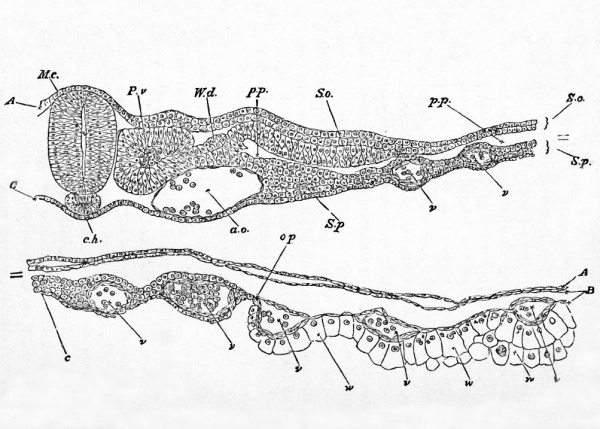
Fig. 34. Transverse section through the dorsal region of an embryo of 45 hours.
- A. epiblast. B. mesoblast. C. hypoblast consisting of a single row of flattened cells. M. c. medullary canal. P. v. mesoblastic somite. W. d. Wolffian duct. S. o. Somatopleure. S.p. Splanchnopleure. p.p. pleuroperitoneal cavity, c. h. notochord. a. o. dorsal aorta, v. blood-vessels of the yolksac, o.p. line of junction between opaque and pellucid areas ; w. palisade-like yolk spheres which constitute the germinal wall. Only one-half of the section is represented in the figure if completed it would be bilaterally symmetrical about the line of the medullary canal.
Cranial flexure
Round the anterior termination of the notochord, the medullary canal, which up to the present time has remained perfectly straight, towards the end of the day begins to curve. The front portion of the canal, i.e. the fore-brain with its optic and cerebral vesicles, becomes slightly bent downwards, so as to form a rounded obtuse angle with the rest of the embryo. This is the commencement of the so-called cranial flexure and is, mechanically speaking, a consequence of the more rapid growth of the dorsal wall of the anterior part of the brain as compared with that of the ventral.
Auditory vesicle

Lastly, as far as the head is concerned, the epiblastic plates forming the rudiments of the auditory vesicles become converted into deep pits opening one on each side of the hind-brain (Fig. 27, au. p).
Heart
We left the heart as a fusiform body slightly bent to the right, attached to the under wall of the foregut by the mesocardium. The curvature now increases so much that the heart becomes almost GQ -shaped, the venous portion being drawn up towards the head so as to lie somewhat above (dorsal to) and behind the arterial portion. (It would perhaps be more correct to say that the free intermediate portion is by its own growth bent downwards, backwards, and somewhat to the right, while the venous root of the heart is at the same time continually being lengthened by the carrying back of that "point of divergence" of the splanchnopleure folds which marks the union of the vitelline veins into a single venous trunk.) The heart then has at this time two bends, the one, the venous bend, the right-hand curve of the uz ; the other, the arterial bend, the left-hand curve of the cc. The venous bend which, as we have said, is placed above and somewhat behind the arterial bend, becomes marked by two bulgings, one on either side. These are the rudiments of the auricles, or rather of the auricular appendages. The ascending limb of the arterial bend soon becomes conspicuous as the bulbus arteriosus > while the rounded point of the bend itself will hereafter grow into the ventricles.
Vascular system
The blood-vessels, whose origin during the first half of this day has been already described, become during the latter part of the day so connected as to form a complete system, through which a definite circulation of the blood is now for the first time (consequently some little while after the commencement of the heart's pulsation) carried on.
The two primitive aortce have already been described as encircling the foregut, and then passing along the body of the embryo immediately beneath the mesoblastic somites on each side of the notochord. They are shewn in Figs. 32 A.o. and 34 a.o in section as two large rounded spaces lined with flattened cells. At first they run as two distinct canals along the whole length of the embryo ; but, after a short time, unite at some little distance behind the head into a single trunk, which lies in the middle line of the body immediately below the notochord (Fig. 57). Lower down, nearer the tail, this single primitive trunk again divides into two aorte, which, getting smaller and smaller, are finally lost in the small blood-vessels of the tail. At this epoch, therefore, there are two aortic arches springing from the bulbus arteriosus, and uniting above the alimentary canal in the back of the embryo to form the single dorsal aorta, which travelling backwards in the median line divides near the tail into two main branches. From each of the two primitive aortae, or from each of the two branches into which the single aorta divides, there is given off on either side a large branch. These have been already spoken of as the vitelline arteries. At this stage they are so large that by far the greater part of the blood passing down the aorta finds its way into them, and a small remnant only pursues a straight course into the continuations of the aorta towards the tail.
Each vitelline artery leaving the aorta at nearly right angles (at a point some little way behind the backward limit of the splanchnopleure fold which is forming the alimentary canal), runs outwards beneath the mesoblastic somites in the lower range of the mesoblast, close to the hypoblast. Consequently, when in its course outwards it reaches the point where the mesoblast is cleft to form the somatopleure and splanchnopleure, it attaches itself to the latter. Travelling along this, and dividing rapidly into branches, it reaches the vascular area in whose network of small vessels (and also to a certain extent in the similar small vessels of the pellucid area) it finally loses itself.
The terminations of the vitelline arteries in the vascular and pellucid areas are further connected with the heart in two different ways. From the network of capillaries, as we may call them, a number of veins take their origin, and finally unite into two main trunks, the vitelline veins. These have already been described as running along the folds of the splanchnopleure to form the venous roots of the heart. Their course is consequently more or less parallel to that of the vitelline arteries, but at some little distance nearer the head, inasmuch as the arteries run in that part of the splanchnopleure which has not yet been folded in to form the alimentary canal. Besides forming the direct roots of the vitelline veins, the terminations of the vitelline arteries in the vascular area are also connected with the sinus terminates spoken of above as running almost completely round, and forming the outer margin of the vascular area. This (Fig. 36, ST.), may be best described as composed of two semicircular canals, which nearly meet at points opposite the head and opposite the tail, thus all but encircling the vascular area between them. At the point opposite the head the end of each semicircle is connected with vessels (Fig. 36), which run straight in towards the heart along the fold of the splanchnopleure, and join the right and left vitelline veins. At the point opposite the tail there is at this stage no such definite connection. At the two sides, midway between their head and tail ends, the two semicircles are especially connected with the vitelline arteries.
The circulation of the blood then during the latter half of the second day may be described as follows. The blood brought by the vitelline veins falls into the twisted cavity of the heart, and is driven thence through the bulbus arteriosus and aortic arches into the aorta. From the aorta, by far the greater part of the blood flows into the vitelline arteries, only a small remnant passing on into the caudal terminations. From the capillary net-work of the vascular and pellucid areas into which the vitelline arteries discharge their contents, part of the blood is gathered up at once into the lateral or direct trunks of the vitelline veins. Part however goes into the middle region of each lateral half of the sinus terminalis, and there divides on each side into two streams. One stream, and that the larger one, flows in a forward direction until it reaches the point opposite the head, thence it returns by the veins spoken of above, straight to the vitelline trunks. The other stream flows backward, and becomes lost at the point opposite to the tail. This is the condition of things during the second day; it becomes considerably changed on the succeeding day.
At the time that the heart first begins to beat the capillary system of the vascular and pellucid areas is not yet completed ; and the fluid which is at first driven by the heart contains, according to most observers, very few corpuscles.
At the close of the second day the single pair of aortic arches into which the bulbus arteriosus divides is found to be accompanied by a second pair, formed in the same way as the first, and occupying a position a little behind it. Sometimes even a third pair is added. Of these aortic arches we shall have to speak more fully later on.
Wolffian duct. During the latter half of the second day the Wolfrlan duct to which we have already alluded becomes fully established, while the first traces of the embryonic excretory organs or kidneys, known as the Wolman bodies, make their appearance. The development of the latter will be dealt with in the history of the third day, but the history of the duct itself may conveniently be completed here.
The first trace of it is visible in an embryo Chick with eight somites, as a ridge projecting from the intermediate cell mass towards the epiblast in the region of the seventh somite. In the course of further development it continues to constitute such a ridge as far as the eleventh somite (Fig. 34 TFd), but from this point it grows backwards by the division of its cells, as a free column in the space between the epiblast and rnesoblast. In an embryo with fourteen somites of about the stage represented in fig. 28 a small lumen has appeared in its middle part, and in front it is connected with rudimentary Wolffian tubules, which develop in continuity with it. In the succeeding stages the lumen of the duct gradually extends backwards and forwards.
and the duct itself also passes inwards relatively to the epiblast (fig. 43 wd). Its hind end elongates till it comes into connection with, and opens on the fourth day into the cloacal section of the hind-gut.
The amnion and allantois. The amnion, especially the anterior or head fold, advances in growth very rapidly during the second day, and at the close of the day completely covers the head and neck of the embryo ; so much so that it is necessary to tear or remove it when the head has to be examined in hardened opaque specimens. The tail and lateral folds of the amnion, though still progressing, lag considerably behind the head-fold.
The side -folds eventually meet in the median dorsal line, and their coalescence proceeds backwards from the head-fold in a linear direction, till there is only a small opening left over the tail of the embryo. This finally becomes closed early on the third day.
In Figs. 32 and 43 am. the folds of the amnion are shewn before they have coalesced. After the coalescence of the folds of the amnion above the embryo the two limbs of which each is formed become, as already explained in chapter IL, separate from each other: the inner, forming a special investment of the embryo, and constituting the amnion proper (Fig. 65), the outer attaching itself to the vitelline membrane and becoming the serous envelope.
The development of the allantois commences during the second day, but since it is mainly completed during the third day we need not dwell upon it further in this place.
Summary Day 2 second half
The chief events, then, which occur during the second half of the second day are as follow:
- The second and third cerebral vesicles make their appearance behind the first.
- The optic vesicles spring as hollow buds from the lateral, and the unpaired commencement of the cerebral hemispheres from the front, portions of the first cerebral vesicle.
- The auditory plate becomes converted into a pit, opening at the side of the hind-brain or third cerebral vesicle.
- The first indications of the cranial flexure become visible.
- The head-fold, and especially the splanchnopleure moiety, advances rapidly backwards ; the head of the embryo is in consequence more definitely formed. The tail-fold also becomes distinct.
- The curvature of the heart increases; the first rudiments of the auricles appear.
- The circulation of the yolk-sac is established.
- The amnion grows rapidly, and the allantois commences to be formed.
The Elements of Embryology - Volume 1 (1883)
The History of the Chick: Egg structure and incubation beginning | Summary whole incubation | First day | Second day - first half | Second day - second half | Third day | Fourth day | Fifth day | Sixth day to incubation end | Appendix
| Historic Disclaimer - information about historic embryology pages |
|---|
| Pages where the terms "Historic" (textbooks, papers, people, recommendations) appear on this site, and sections within pages where this disclaimer appears, indicate that the content and scientific understanding are specific to the time of publication. This means that while some scientific descriptions are still accurate, the terminology and interpretation of the developmental mechanisms reflect the understanding at the time of original publication and those of the preceding periods, these terms, interpretations and recommendations may not reflect our current scientific understanding. (More? Embryology History | Historic Embryology Papers) |
Glossary Links
- Glossary: A | B | C | D | E | F | G | H | I | J | K | L | M | N | O | P | Q | R | S | T | U | V | W | X | Y | Z | Numbers | Symbols | Term Link
Cite this page: Hill, M.A. (2024, April 28) Embryology Book - The Elements of Embryology - Chicken 5. Retrieved from https://embryology.med.unsw.edu.au/embryology/index.php/Book_-_The_Elements_of_Embryology_-_Chicken_5
- © Dr Mark Hill 2024, UNSW Embryology ISBN: 978 0 7334 2609 4 - UNSW CRICOS Provider Code No. 00098G

