Book - The Elements of Embryology - Chicken 4
| Embryology - 28 Apr 2024 |
|---|
| Google Translate - select your language from the list shown below (this will open a new external page) |
|
العربية | català | 中文 | 中國傳統的 | français | Deutsche | עִברִית | हिंदी | bahasa Indonesia | italiano | 日本語 | 한국어 | မြန်မာ | Pilipino | Polskie | português | ਪੰਜਾਬੀ ਦੇ | Română | русский | Español | Swahili | Svensk | ไทย | Türkçe | اردو | ייִדיש | Tiếng Việt These external translations are automated and may not be accurate. (More? About Translations) |
Foster M. Balfour FM. Sedgwick A. and Heape W. The Elements of Embryology (1883) Vol. 1. (2nd ed.). London: Macmillan and Co.
| The Elements of Embryology 1883
1 Chicken : Hen's egg and the beginning of incubation | Whole history of incubation | day 1 of incubation | first half of day 2 | second half of day 2 | day 3 | day 4 | day 5 | day 6-21 | Appendix | Figures as Gallery
|
| Historic Disclaimer - information about historic embryology pages |
|---|
| Pages where the terms "Historic" (textbooks, papers, people, recommendations) appear on this site, and sections within pages where this disclaimer appears, indicate that the content and scientific understanding are specific to the time of publication. This means that while some scientific descriptions are still accurate, the terminology and interpretation of the developmental mechanisms reflect the understanding at the time of original publication and those of the preceding periods, these terms, interpretations and recommendations may not reflect our current scientific understanding. (More? Embryology History | Historic Embryology Papers) |
The changes which take place during the first half of the second day
General development
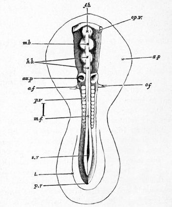
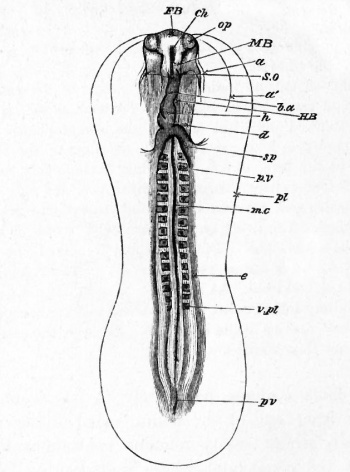
In attempting to remove the blastoderm from an egg which has undergone from 30 to 36 hours' incubation, the observer cannot fail to notice a marked change in the consistency of the blastodermic structures. The excessive delicacy and softness of texture which rendered the extraction of an 18 or 20 hours' blastoderm so difficult, has given place to a considerable amount of firmness; the outlines of the embryo and its appendages are much bolder and more distinct; and the whole blastoderm can be removed from the egg with much greater ease.
In the embryo itself viewed from above one of the features which first attracts attention is the progress in the head-fold (Fig. 27). The upper limb or- head has become much more prominent, while the lower groove is not only proportionately deeper, but is also being carried back beneath the body of the embryo.
The medullary folds are closing rapidly. In the region of the head they have quite coalesced, a slight notch in the middle line at the extreme front marking for some little time their line of junction (Fig. 23). The open medullary groove of the first day has thus become converted into a tube, the neural canal, closed in front, but as yet open behind. Even before the medullary folds coalesce completely in the cephalic region, the front end of the neural canal dilates into a small bulb, whose cavity remains continuous with the rest of the canal, and whose walls are similarly formed of epiblast. This bulb is known as the first cerebral vesicle, Fig. 27, /A, and makes its appearance in the early hours of the second day. From its sides two lateral processes almost at once grow out : they are known as the optic vesicles (Fig. 27, op. v.), and their history will be dealt with at length somewhat later. Behind the first cerebral vesicle a second and a third soon make their appearance ; they are successively formed very shortly after the first vesicle ; but the consideration of them may be conveniently reserved to a later period. At the level of the hind end of the head two shallow pits are visible. They constitute the first rudiments of the organ of hearing, and are known as the auditory pits (Fig. 27, au.p.).
The number of mesoblastic somites increases rapidly by a continued segmentation of the vertebral plates of mesoblast. The four or five pairs formed during the first day have by the middle of the second increased to as many as fifteen. The addition takes place from before backwards ; and the hindermost one is for some time placed nearly on a level with the boundary between the hind end of the trunk of the embryo, and the front end of the primitive streak. For some time the already formed somites do not increase in size, so that at first the embryo clearly elongates by additions to its hinder end.
Immediately behind the level of the last mesoblastic somites there is placed an enlargement of the unclosed portion of the medullary canal. This enlargement is the sinus rhomboidalis already spoken off It is shewn in Fig. 23. On its floor is placed the' front end of the primitive streak. It is a purely embryonic structure which disappears during the second day.
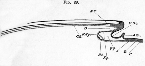
In a former chapter it was pointed out (p. 27) that the embryo is virtually formed by a folding or tucking in of a limited portion of the blastoderm, first at the anterior extremity, and afterwards at the posterior extremity and at the sides. One of the results of this doubling up of the blastoderm to form the head is the appearance, below the anterior extremity of the medullary tube, of a short canal, ending blindly in front, but open widely behind (Fig. 29, Z>), a cul de sac, in fact, lined with hypoblast and reaching from the extreme front of the embryo to the point where the splanchnopleuric leaf of the head-fold (Fig. 29, F. Sp) turns back on itself. This cul de sac, which of course becomes longer and longer the farther back the head-fold is carried, is the rudiment of the front end of the alimentary canal, the fore-gut, as it might be called. In transverse section it appears to be flattened horizontally, and also bent, so as to have its convex surface looking downwards (Fig. 30, al). At first the anterior end is quite blind, there being no mouth as yet; the formation of this at a subsequent date will be described later on.
At the end of the first half of the second day the head-fold has not proceeded very far backwards, and its limits can easily be seen in the fresh embryo both from above and from below (Fig. 28).
The heart
It is in the head-fold that the formation of the heart takes place, its mode of origin being connected with that cleavage of the mesoblast and consequent formation of splanchnopleure and somatopleure of which we have already spoken.
At the extreme end of the embryo (Fig. 29), where the blastoderm begins to be folded back, the mesobtast is never cleft, and here consequently there is neither somatopleure nor splanchnopleure ; but at a point a very little further back, close under the blind end of the foregut, the cleavage (at the stage of which we are speaking) begins, and the somatopleure, F. So, and Splanchnopleure, F. Sp. diverge from each other. They thus enclose between them a cavity, pp, which rapidly increases behind by reason of the fact that the fold of the splanchnopleure is carried on towards the hinder extremity of the embryo considerably in advance of that of the somatopleure. Both folds, after running a certain distance towards the hind end of the embryo, are turned round again, and then course once more forwards over the yolk-sac. As they thus return (the somatopleure having meanwhile given off the fold of the amnion, Am.), they are united again to form the uncleft blastodermic investment of the yolk-sac. In this way the cavity arising from their separation is closed below.
Fig. 29. Diagrammatic longitudinal section through the axis of an embryo.
- The section is supposed to be made at a time when the headfold has commenced but the tail-fold has not yet appeared. N.C. neural canal, closed in front but as yet open behind. Ch. notochord. The section being taken in the middle line, the protovertebrae are of course not shewn. In front of the notochord is seen a mass of uncleft mesoblast, which will eventually form part of the skull. D. the commencing foregut or front part of the alimentary canal. F. So. Somatopleure, raised up in its peripheral portion into the amniotic fold Am. Sp. Splanchnopleure. At Sp. it forms the under wall of the foregut ; at F. Sp. it is turning round and about to run forward. Just at its turning point the cavity of the heart Ht. is being developed in its mesoblast. pp. pleuroperitoneal cavity. A epiblast, B mesoblast, C hypoblast, indicated in the rest of the figure by differences in the shading. At the part where these three lines of reference end the mesoblast is as yet uncleft.
It is in this cavity, which from its mode of formation the reader will recognise as a part (and indeed at this epoch it constitutes the greater part) of the general pleuroperitoneal cavity, that the heart is formed.
This makes its appearance at the under surface and hind end of the foregut, just where the splanchnopleure folds turn round to pursue a forward course (Fig. 29, Ht.) ; and by the end of the first half of the second day (Fig. 28, h) has acquired somewhat the form of a flask with a slight bend to the right. At its anterior end a slight swelling marks the future bulbus arteriosus ; and a bulging behind indicates the position of the auricles. It is hollow, and its cavity opens behind into two vessels called the vitelline veins (Figs. 27, o.f. and 28 sp.), which pass outwards in the folds of the splanchnopleure at nearly right angles to the axis of the embryo. The anterior extremity of the heart is connected with the two aortae.
The heart, including both its muscular wall and its epitheloid lining, is developed out of the splanchnic mesoblast on the ventral side of the throat. But since the first commencements of the heart make their appearance prior to the formation of the throat, the development of this organ is somewhat complicated; and in order to gain a clear conception of the manner in which it takes place the topography of the region where it is formed needs to be very distinctly understood.
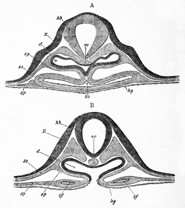
In the region where the heart is about to appear, the splanchnopleure is continually being folded in on either side, and these lateral folds are progressively meeting and uniting in the middle line to form the under or ventral wall of the foregut. At any given moment these folds will be found to have completely united in the middle line along a certain distance measured from the point in front where the cleavage of the mesoblast (i.e. the separation into somatopleure and splanchnopleure) begins, to a particular point farther back. They will here be found to be diverging from the point where they were united, and not only diverging laterally each from the middle line, but also both turning so as to run in a forward direction to regain the surface of the yolk and rejoin the somatopleure, Fig. 29. In a transverse section taken behind this extreme point of union, or point of divergence, as we may call it, the splanchnopleure on either side when traced downwards from the axis of the embryo may be seen to bend in towards the middle so as to approach its fellow, and then to run rapidly outwards, Fig. 31, B. A longitudinal section shews that it runs forwards also at the same time, Fig. 29. A section through the very point of divergence shews the two folds meeting in the middle line and then separating again, so as to form something like the letter x, with the upper limbs converging, and the lower limbs diverging. In a section taken in front of the point of divergence, the lower diverging limbs of the x have disappeared altogether; nothing is left but the upper limbs, which, completely united in the middle line, form the under-wall of the foregut.
As development proceeds, what we have called the point of divergence is continually being carried farther and farther back, so that the distance between it and the point where the somatopleure and splanchnopleure separate from each other in front, i. e. the length of the foregut, is continually increasing.
In the chick, as we have already stated, the heart commences to be formed in a region where the folds of the splanchnopleure have not yet united to form the ventral wall of the throat, and appears in the form of two thickenings of the mesoblast of the splanchnopleure, along the diverging folds, i.e. along the lower limbs of the x, just behind the point of divergence. These thickenings are continued into each other by a similar thickening of the mesoblast extending through the point of divergence itself.
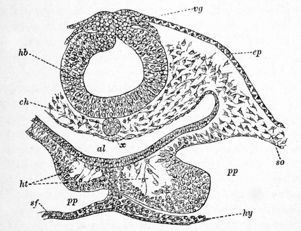
The heart has thus at first the form of an inverted V, and consists of two independent cords of splanchnic mesoblast which meet in front, without however uniting. As the foldirig-in of the splanchnopleure is continued backwards the two diverging halves of the heart are gradually brought together. Thus very soon the developing heart has the form of an inverted Y, consisting of an unpaired portion in front and two diverging limbs behind. The unpaired portion is the true heart, while the diverging limbs are the vitelline veins already spoken of (Fig. 28, sp). While the changes just spoken of have been taking place in the external form of the heart, its internal parts have also become differentiated. A cavity is formed in each of the halves of the heart before even they have coalesced. Each of these cavities has at first the form of an irregular space between the splanchnic mesoblast and the wall of the throat (Fig. 30, fit.). During their formation (Fig. 30), a thin layer of mesoblast remains in contact with the hypoblast, but connected with the main mass of the mesoblast of the heart by protoplasmic processes. A second layer next becomes split from the main mass of mesoblast, being still connected with the first layer by the above-mentioned protoplasmic processes. These two layers unite to form a tube which constitutes the epithelioid lining of the heart ; the lumen of this tube is the cavity of the heart, and soon loses the protoplasmic trabeculse which at first traverse it. The cavity of the heart may thus be described as being formed by a hollowing out of the splanchnic mesoblast. Some of the central cells of the original thickenings probably become blood-corpuscles.
Fig. 30. Transverse section through the posterior part of the head of an embryo chick of thirty hours.
- hb. hind-brain ; vg. vagus nerve ; ep. epiblast ; ch. notochord ; x. thickening of hypoblast (possibly a rudiment of the subnotochordal rod) ; al. throat ; lit. heart ; pp. body cavity ; so. somatic mesoblast ; sf. splanchnic mesoblast ; hy. hypoblast.
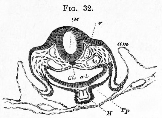
The thick outer part of the cords of splanchnic mesoblast which form the heart become the muscular walls and peritoneal covering of this organ. The muscular wall of each division of the heart has at first the form of a half tube widely open on its dorsal aspect, that is towards the hypoblast of the gut (Fig. 30 and 32). After the two halves of the heart have coalesced in the manner already explained, the muscular walls grow in towards the middle line on the dorsal side until they meet each other and coalesce, thus forming a complete tube as shewn diagrammatically in Fig. 31, A. They remain, however, at first continuous with the splanchnic mesoblast surrounding the throat, thus forming a provisional mesentery the mesocardium attaching the heart to the ventral wall of the throat. The epithelioid tubes formed in the two halves of the heart remain for some time separate, and cause the cavity of the heart to be divided into two tubes even after its two halves have to all appearance completely coalesced1.
Soon after its formation the heart begins to beat ; its at first slow and rare pulsations beginning at the venous and passing on to the arterial end. It is of some interest to note that its functional activity commences long before the cells of which it is composed shew any distinct differentiation into muscular or nervous elements.
Vascular system
To provide channels for the fluid thus pressed by the contractions of the heart, a system of tubes has made its appearance in the mesoblast both of the embryo itself and of the vascular and pellucid areas. In front the single tube of the bulbus arteriosus bifurcates into two primitive aortce, each of which bending round the front end of the foregut, passes from its under to its upper side, the two forming together a sort of incomplete arterial collar imbedded in the mesoblast of the gut. Arrived at the upper side of the gut, they turn sharply round, and run separate but parallel to each other backwards towards the tail, in the mesoblast on each side of the notochord immediately under the mesoblastic somites (Figs. 32, Ao, 34, ao). About half way to the hinder extremity each gives off at right angles to the axis of the embryo a large branch, the vitelline artery (Fig. 36, Of, A.), which, passing outwards, is distributed over the pellucid and vascular areas, the main trunk of each aorta passing on with greatly diminished calibre towards the tail, in which it becomes lost.
(1This is not shewn in the diagram, Fig. 31, A.)
Fig. 31. Two diagrammatic sections of a thirty-six hours' embryo illustrating the structure of the heart shortly after its formation. a is the anterior section.
- hb. hind brain ; nc. notochord ; E. epiblast ; so. somatopleure ; sp. splanchnopleure ; d. alimentary canal ; hy. hypoblast ; hz. (in A) heart ; of. vitelline vein.
- In A the two halves of the heart have coalesced to form an unpaired tube suspended from the ventral wall of the throat.
- In B are seen in the diverging folds of the splanchnopleure the two vitelline veins (of) which will shortly unite to form the ductus venosus.
Fig. 32. Transverse section of an embryo at the end of the second day passing through the region of the bulbus arteriosus. (Copied from His.)
- M. medullary canal in the region of the hind brain ; V. anterior cardinal vein ; Ao. Aorta ; Ch. Notochord ; al. alimentary canal ; H. Heart (bulbus arteriosus) ; Pp. Pleuroperitoneal cavity; am. amnion.
In the vascular and pellucid areas, the formation of vascular channels with a subsequent differentiation into arteries, capillaries and veins, is proceeding rapidly. Blood-corpuscles too are being formed in considerable numbers. The mottled yellow vascular area becomes covered with red patches consisting of aggregations of blood-corpuscles, often spoken of as blood-islands.
Round the extreme margin of the vascular area and nearly completely encircling it, is seen a thin red line, the sinus or vena terminalis (Fig. 36, Sv.). This will soon increase in size and importance.
From the vascular and pellucid area several large channels are seen to unite and form two large trunks, one on either side, which running along the splanchnopleure folds at nearly right angles to the axis of the embryo, unite at the " point of divergence " to join the venous end of the heart. These are the vitelline veins spoken of above.
Both vessels and corpuscles are formed entirely from the cells of the mesoblast; arid in the regions where the mesoblast is cleft, are at first observed exclusively in the splanchnopleure. Ultimately of course they are found in the mesoblast everywhere.

In the pellucid area, where the formation of the blood-vessels may be most easily observed, a number of mesoblastic cells are seen to send out processes (Fig. 33). These processes unite, and by their union a protoplasmic network is formed containing nuclei at the points from which the processes started. The nuclei, which as a rule are much elongated and contain large oval nucleoli, increase very rapidly by division, and thus form groups of nuclei at the, so to speak, nodal points of the network. Several nuclei may also be seen here and there in the processes themselves. The network being completed, these groups, by continued division of the nuclei, increase rapidly in size ; the protoplasm around them acquires a red colour, and the whole mass breaks up into blood-corpuscles (Fig. 33, b.c.) The protoplasm on the outside of each group, as well as that of the uniting processes, remains granular, and together with the nuclei in it forms the walls of the blood-vessels. A plasma is secreted by the walls, and in this the blood-corpuscles float freely.
Each nodal point is thus transformed into a more or less rounded mass of blood-corpuscles floating in plasma but enveloped by a layer of nucleated protoplasm, the several groups being united by strands of nucleated protoplasm. These uniting strands rapidly increase in thickness; new processes are also continually being formed ; and thus the network is kept close and thickset while the area is increasing in size.
By changes similar to those which took place in the nodal points, blood-corpuscles make their appearance in the processes also, the central portions of which become at the same time liquefied.
By the continued widening of the connecting processes and solution of their central portions, accompanied by a corresponding increase in the enveloping nucleated cells, the original protoplasmic network is converted into a system of communicating tubes, the canals of which contain blood-corpuscles and plasma, and the walls of which are formed of flattened nucleated cells.
The blood-corpuscles pass freely from the nodal points into the hollow processes, and thus the network of protoplasm becomes a network of blood-vessels, the nuclei of the corpuscles and of the walls of which have been, by separate paths of development, derived from the nuclei of the original protoplasm.
The formation of the corpuscles does not proceed equally rapidly or to the same extent in all parts of the blastoderm. By far the greater part are formed in the vascular area, but some arise in the pellucid area, especially in the hinder part. In the front of the pellucid area the processes are longer and the network accordingly more open ; the corpuscles also are both later in appearing and less numerous when formed.
Assuming the truth of the above account, it is evident that the blood-vessels of the yolk-sack of the chick do not arise as spaces or channels between adjacent cells of the mesoblast, but are hollowed out in the communicating protoplasmic substance of the cells themselves. The larger vessels of the trunk are however probably formed as spaces between the cells, much as is the case with the heart.
Wolffian duct
About this period there may be seen in transverse sections, taken through the embryo in the region of the seventh to the eleventh somite a small group of cells (Fig. 34, W. d) projecting on either side from the mass of uncleft mesoblast on the outside of the mesoblastic somites, into the somewhat triangular space bounded by the epiblast above, the upper and outer angle of the mesoblastic somite on the inside, and the somatic mesoblast on the outside.
This group of cells is the section of a longitudinal ridge, the rudiment of the Wolffian duct or primitive duct of the excretory system ; while the mass of cells from which it springs is known as the intermediate cell mass. We shall return to them immediately.
Summary Day 2 first half
The most important changes then which take place during the first half of the second day are:
- the closure of the medullary folds, especially in the anterior part, and the dilatation of the canal so formed into the first cerebral vesicle
- the establishment of a certain number of mesoblastic somites
- the elevation of the head from the plane of the blastoderm
- the formation of the tubular heart and of the great blood-vessels
- the appearance of the rudiment of the Wolffian duct.
It is important to remember that the embryo of which we are now speaking is simply a part of the whole germinal membrane, which is gradually spreading over the surface of the yolk. It is important also to bear in mind that all that part of the embryo which is in front of the foremost somite corresponds to the future head, and the rest to the neck, body and tail. During this period the head occupies about a third of the whole length of the embryo.
The Elements of Embryology - Volume 1 (1883)
The History of the Chick: Egg structure and incubation beginning | Summary whole incubation | First day | Second day - first half | Second day - second half | Third day | Fourth day | Fifth day | Sixth day to incubation end | Appendix
| Historic Disclaimer - information about historic embryology pages |
|---|
| Pages where the terms "Historic" (textbooks, papers, people, recommendations) appear on this site, and sections within pages where this disclaimer appears, indicate that the content and scientific understanding are specific to the time of publication. This means that while some scientific descriptions are still accurate, the terminology and interpretation of the developmental mechanisms reflect the understanding at the time of original publication and those of the preceding periods, these terms, interpretations and recommendations may not reflect our current scientific understanding. (More? Embryology History | Historic Embryology Papers) |
Glossary Links
- Glossary: A | B | C | D | E | F | G | H | I | J | K | L | M | N | O | P | Q | R | S | T | U | V | W | X | Y | Z | Numbers | Symbols | Term Link
Cite this page: Hill, M.A. (2024, April 28) Embryology Book - The Elements of Embryology - Chicken 4. Retrieved from https://embryology.med.unsw.edu.au/embryology/index.php/Book_-_The_Elements_of_Embryology_-_Chicken_4
- © Dr Mark Hill 2024, UNSW Embryology ISBN: 978 0 7334 2609 4 - UNSW CRICOS Provider Code No. 00098G

