Book - The Elements of Embryology - Chicken 3
| Embryology - 27 Apr 2024 |
|---|
| Google Translate - select your language from the list shown below (this will open a new external page) |
|
العربية | català | 中文 | 中國傳統的 | français | Deutsche | עִברִית | हिंदी | bahasa Indonesia | italiano | 日本語 | 한국어 | မြန်မာ | Pilipino | Polskie | português | ਪੰਜਾਬੀ ਦੇ | Română | русский | Español | Swahili | Svensk | ไทย | Türkçe | اردو | ייִדיש | Tiếng Việt These external translations are automated and may not be accurate. (More? About Translations) |
Foster M. Balfour FM. Sedgwick A. and Heape W. The Elements of Embryology (1883) Vol. 1. (2nd ed.). London: Macmillan and Co.
| The Elements of Embryology 1883
1 Chicken : Hen's egg and the beginning of incubation | Whole history of incubation | day 1 of incubation | first half of day 2 | second half of day 2 | day 3 | day 4 | day 5 | day 6-21 | Appendix | Figures as Gallery
|
| Historic Disclaimer - information about historic embryology pages |
|---|
| Pages where the terms "Historic" (textbooks, papers, people, recommendations) appear on this site, and sections within pages where this disclaimer appears, indicate that the content and scientific understanding are specific to the time of publication. This means that while some scientific descriptions are still accurate, the terminology and interpretation of the developmental mechanisms reflect the understanding at the time of original publication and those of the preceding periods, these terms, interpretations and recommendations may not reflect our current scientific understanding. (More? Embryology History | Historic Embryology Papers) |
The changes which take place during the first day of incubation
DURING the descent of the egg along the oviduct, where it is exposed to a temperature of about 40 C., the germinal disc, as we have seen, undergoes important changes. When the egg is laid and becomes cold these changes all but entirely cease, and the blastoderm remains inactive until, under the influence of the higher temperature of natural or artificial incubation, the vital activities of the germ are brought back into play, the arrested changes go on again, and usher in the series of events which we have now to describe in detail.
The condition of the blastoderm at the time when the egg is laid is not exactly the same in all eggs ; in some the changes being farther advanced than in others, though the differences of course are slight. In some eggs, especially in warm weather, changes of the same kind as those caused by actual incubation may take place, to a certain extent, in the interval between laying and incubation ; lastly, in all eggs, both under natural and especially under artificial incubation, the dates of the several changes are, within the limits of some hours, very uncertain, particularly in the first few days ; one egg being found, for example, at 36 hours in the same stage as another at 24 or 30 hours, or a third at 40 or 48 hours. When we speak therefore of any event as taking place at any given hour or part of any given day, we are to be understood as meaning that such an event will generally be found to have taken place at about that time. We introduce exact dates for the convenience of description.
The changes which take place during the first day will be most easily considered under several periods.
From the 1st to about the 8th hour. During this period the blastoderm, when viewed from above, is found to have increased in size. The pellucid area, which at the best is but obscurely marked in the unincubated egg, becomes very distinct (the central opacity having disappeared), and contrasts strongly with the opaque area, which has even still more increased both in distinctness and size.
For the first few hours both the pellucid and opaque areas remain approximately circular, and the most important change, besides increase in size and greater distinctness which can be observed in them, is a slight ill-defined opacity or loss of transparency, which makes its appearance in the hinder half of the pellucid area. This is known as the embryonic shield.
Slight as are the changes which can at this stage be 1 seen from surface views, sections taken from hardened specimens bring to light many most important changes in the nature and arrangement of the constituent cells.
Fig. 12. Section of a blastoderm of a fowl's egg at the commencement of incubation.
- The thin but complete upper layer ep composed of columnar cells rests on the incomplete lower layer I, composed of larger and more granular cells. The lower layer is thicker in some places than in others, and is especially thick at the periphery. The line below the under layer marks the upper surface of the white yolk. The larger so-called formative cells are seen at b, lying on the white yolk. The figure does not take in quite the whole breadth of the blastoderm ; but the reader must understand that both to the right hand and the left ep is continued farther than I, so that at the extreme edge it rests directly on the white yolk.
It will be remembered that the blastoderm in the unincubated egg is composed of two layers, an upper (Fig. 12, ep) and an under layer; that the upper is a coherent membrane of columnar nucleated cells, but that the lower one (Fig. 12, 1) is formed of an irregular network of larger cells in which the nuclei are with difficulty visible; and that in addition to this there are certain still larger cells, called 'formative cells' (Fig. 12,6), lying at the bottom of the segmentation-cavity.
Under the influence of incubation changes take place very rapidly, which result in the formation of the three layers of the blastoderm.
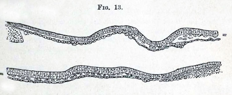
The upper layer, which is the epiblast already spoken of (Fig. 13), takes at first but little share in these changes.
In the lower layer, however, certain of the cells begin to get flattened horizontally, their granules become less numerous, and the nucleus becomes distinct; the cells so altered cohere together and form a membrane. The membrane thus formed, which is first completed in the centre of the pellucid area, constitutes the hypoblast. Between the hypoblastic membrane and the epiblast there remain a number of scattered cells (Fig. 13) which cannot however be said to form a definite layer altogether distinct from the hypoblast. They are almost entirely confined to the posterior part of the area pellucida, and give rise to the opacity of that part, which we have spoken of as the embryonic shield.
Fig. 13. Transverse section through the blastoderm of a chick before the appearance of the primitive streak.
- The epiblast is represented somewhat diagrammatically. The hyphens shew the points of junction of the two halves of the section. The hypoblast is already constituted as a membrane of flattened cells, and a number of scattered cells are seen between it and the epiblast.
At the edge of the area pellucida the hypoblast becomes continuous with a thickened rim of material, underlying the epiblast, and derived from the original thickened edge of the blastoderm and the subjacent yolk. It is mainly formed of yolk granules, with a varying number of cells and nuclei imbedded in it. It is known as the germinal wall, and is spoken of more in detail on pp. 65 and 66.
The epiblast is the Hornblatt (corneal layer), and the hypoblast the Darmdrusenblatt (epithelial glandular layer) of the Germans, while those parts of the mesoblast which take part in the formation of the somatopleure and splanchnopleure correspond respectively to the Haut-musJcel-platte and Darm-faserplatte.
All blood-vessels arise in the mesoblast. Hence the vascular layer of the older writers falls entirely within the mesoblast.
The serous layer of the old authors includes the whole of the epiblast, but also comprises a certain portion of mesoblast ; for they speak of all the organs of animal life (skin, bones, muscle, &c.) as being formed out of the serous layer, whereas the epiblast proper gives rise only to the epidermis and to certain parts of the nervous system. In the same way their mucous layer corresponds to the hypoblast with so much of the mesoblast as takes part in the formation of the organs of organic life. Their vascular layer therefore answers to a part only of the mesoblast viz. that part in which blood-vessels are especially developed.
From the 8th to the 12th hour
The changes which next take place result in the complete differentiation of the embryonic layers, a process which is intimately connected with the formation of a structure known as the primitive streak. The full meaning of the latter structure, and its relation to the embryo, can however only be understood by comparison with the development of the lower forms of vertebrate life.
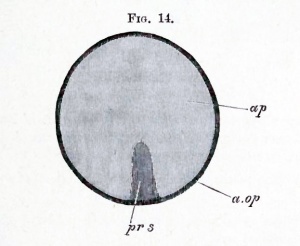
It will be remembered that in surface views of the unincubated blastoderm a small arc, at what we stated to be the posterior end, close to the junction between the area opaca and the area pellucida is distinguished by its more opaque appearance. In the surface view the primitive streak appears as a linear opacity, which gradually grows forwards from the middle of this arc till it reaches about one-third of the diameter of the area pellucida. During the formation of the primitive streak the embryonic shield grows fainter and finally vanishes. When definitely established the primitive streak has the appearance diagrammatically represented in Fig. 14.
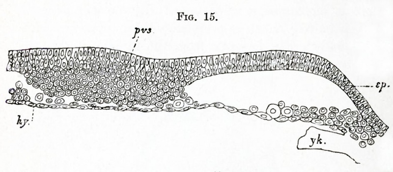
Sections at this stage throw a very important light on the nature and mode of origin of the primitive streak. In the region in front of it the blastoderm is still formed of two layers only, but in the region of the streak itself the structure of the blastoderm is greatly altered. The most important features in it are represented in Fig. 15. This figure shews that the median portion of the blastoderm has become very much thickened (thus producing the opacity of the primitive streak), and that this thickening is caused by a proliferation of rounded cells from the epiblast. In the very young primitive streak, of which Fig. 15 is a section, the rounded cells are still continuous throughout with the epiblast, but they form nevertheless the rudiment of the greater part of a sheet of mesoblast, which will soon arise in this region.
Fig. 15. Transverse section through a blastoderm of about the age represented in flg. 14, shewing the flrst differentiation of the primitive streak.
- The section passes through about the middle of the primitive streak. pvs. primitive streak ; ep. epiblast ; hy. hypoblast ; yk. yolk of the germinal wall.
In addition to the cells clearly derived from the epiblast, there are certain other cells (Fig. 15), closely adjoining the hypoblast ; these are derivatives of the cells, interposed between the epiblast and hypoblast, which gave rise to the appearance of the embryonic shield during the previous stage. In our, opinion these cells also have a share in forming the future mesoblast.
It thus appears that the primitive streak is essentially a linear proliferation of epiblast cells; the cells produced being destined to give rise to the mesoblast. This proliferation first commences at the hinder end of the area pellucida, and thence proceeds forwards.
While the primitive streak is being established, the epiblast becomes two or more rows of cells deep in the region of the area pellucida.
Soon after this, the hitherto circular pellucid area becomes oval (the opaque area remaining circular). The oval is, with remarkable regularity, so placed that its long axis forms a right angle, or very nearly a right angle, with the long axis of the egg itself. Its narrow end corresponds with the future hind end of the embryo. If an egg be placed with its broad end to the right hand of the observer, the head of the embryo will in nearly all cases be found pointing away from him.
The 12th to the 16th hour
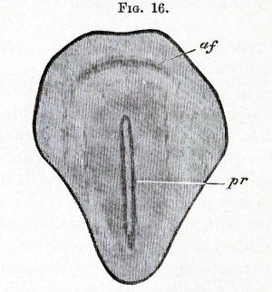
The primitive streak at its first appearance is shadowy and ill-defined; gradually however it becomes more distinct; and during the same period the pellucid area rapidly increases in size, and from being oval becomes pear-shaped (Fig. 16). The primitive streak grows even more rapidly than the pellucid area; so that by the 16th hour it is not only absolutely, but also relatively to the pellucid area, longer than it was at the 12th hour.
It finally occupies about two-thirds of the length of the area pellucida; but its hinder end in many instances appears to stop short of the posterior border of the area pellucida (Fig. 16). The median line of the primitive streak becomes marked by a shallow furrow running along its axis. In fresh specimens, viewed with transmitted light, this furrow appears as a linear transparency, but in hardened specimens seen under reflected light may be distinctly recognized as a narrow groove, the bottom of which, being thinner than the sides, appears more transparent when viewed with transmitted light. It is known as the primitive groove. Its depth and the extent of its development are subject to great variations.
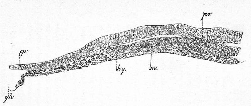
During these changes in external appearance there grow from the edges of the cord of cells constituting the primitive streak two lateral wings of mesoblast cells, whiclj gradually extend till they reach the sides of the area pellucida (Fig. 17). The two wings of mesoblast meet along the line of the primitive streak, where they still remain attached to the epiblast. During this period many sections through the primitive streak give an impression of the mesoblast being involuted along the lips of a groove. The hypoblast below the primitive streak is always quite independent of the mesoblast above, though much more closely attached to it in the median line than at the sides. The part of the mesoblast, which we believe to be derived from the primitive lower layer cells, can generally be distinctly traced. In many cases, especially at the front end of the primitive streak, it forms, as in Fig. 17, a distinct layer of stellate cells, quite unlike the rounded cells of the mesoblastic involution of the primitive streak.
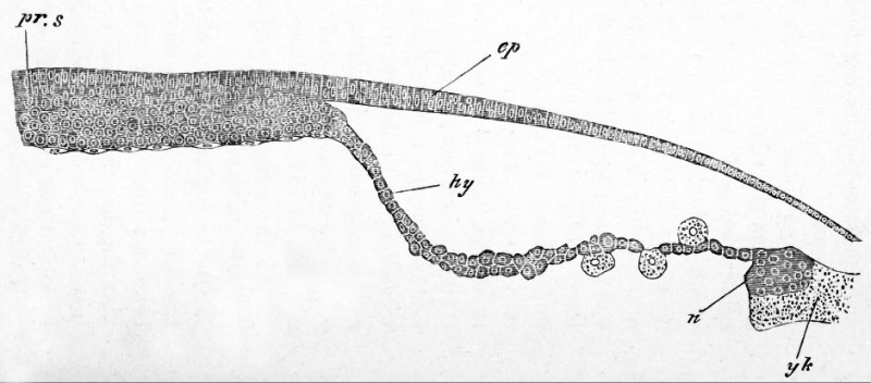
In the region in front of the primitive streak, where the first trace of the embryo will shortly appear, the layers at first undergo no important changes, except that the hypoblast becomes somewhat thicker. Soon, however, as shewn in longitudinal section in Fig. 18, the hypoblast along the axial line becomes continuous behind with the front end of the primitive streak. Thus at this point, which is the future hind end of the embryo, the mesoblast, the epiblast, and the hypoblast all unite together.
|
Fig. 17. Transverse section through the front end op the primitive streak of a blastoderm of the same age as Fig. 16.
|
|
Fig. 18. Longitudinal section through the axial line of the primitive streak, and the part of the blastoderm in front of it, of the blastoderm of a chick somewhat younger than Fig. 19.
|
From the 16th to the 20th hours
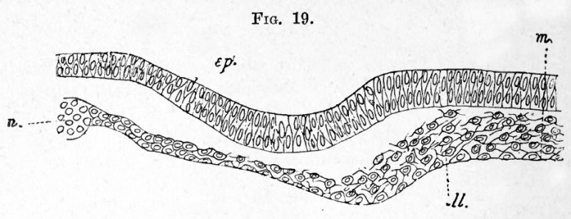
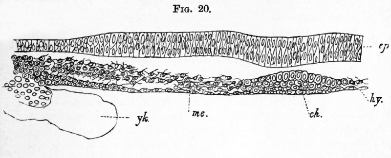
At about the 16th hour, in blastoderms of the stage represented in Fig. 16, an important change takes place in the constitution of the primitive hypoblast in front of the primitive streak. The rounded cells, of which it is at first composed (Fig. 18), break up into (1) a layer formed of a single row of more or less flattened elements below the hypoblast proper and (2) into a layer formed of several rows of stellate elements, between the hypoblast and the epiblast the mesoblast (Fig. 19 m). A separation between these two layers is at first hardly apparent, and before it has become at all well marked, especially in the median line, an axial opaque line makes its appearance in surface views, continued forwards from the front end of the primitive streak, but stopping short at a semicircular fold the future head-fold near the front end of the area pellucida. In section (Fig. 20) this opaque line is seen to be due to a special concentration of cells in the form of a cord. This cord is the commencement of an extremely important structure found in all vertebrate embryos, which is known as the notochord (ch). In most instances the commencing notochord remains attached to the hypoblast, after the mesoblast has at the sides become quite detached (vide Fig. 20), but in other cases the notochord appears to become differentiated in the already separated layer of mesoblast. In all cases the notochord and the hypoblast below it unite with the front end of the primitive streak; with which also the two lateral plates of mesoblast become continuous.
Fig. 19. Transverse section through the embryonic kegion of the blastoderm of a chick shortly prior to the formation of the medullary groove and notochord.
- m. median line of the section ; ep. epiblast ; 11. lower layer cells (primitive hypoblast) not yet completely differentiated into mesoblast and hypoblast ; n. nuclei.
From what has just been said it is clear that in the region of the embryo the mesoblast originates as two lateral plates split off from the primitive hypoblast, and that the notochord originates simultaneously with the mesoblast,with which it is at first continuous, as a median plate similarly of hypoblastic origin.
Fig. 20. Transverse section through the embryonic region of the blastoderm of a chick at the time of the formation of the notochord, but before the appearance of the medullary groove.
- ep. epiblast ; hy. hypoblast ; ch. notochord ; me. mesoblast ; ylc. yolk of germinal wall.
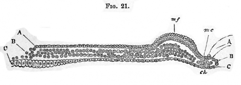
Fig. 21. Transverse section of a blastoderm incubated for 18 hours.
- The section passes through the medullary groove we., at some distance behind its front end. A. Epiblast. B. Mesoblast. C. Hypoblast. m.c. medullary groove ; m.f. medullary fold ; ch. notochord.
Kolliker 1 holds that the mesoblast of the region of the embryo is derived from a forward growth from the primitive streak. There is no theoretical objection to this view, and we think it would be impossible to shew for certain by sections whether or no there is a growth such as he describes ; but such sections as that represented in Fig. 19 (and we have series of such sections from several embryos) appear to us to be conclusive in favour of the view that the mesoblast of the region of the embryo is to a large extent derived from a differentiation of the primitive hypoblast. The mesoblast of the primitive streak forms in part the vascular structures found in the area pellucida, and probably also in part the mesoblast of the allantois.
The differentiation of the embryo may be said to commence with the formation of the notochord and the lateral plates of mesoblast. Very shortly after the formation of these parts, the axial part of the epiblast above the notochord and in front of the primitive streak, being here somewhat thicker than in the lateral parts, becomes differentiated into a distinct medullary plate, the sides of which form two folds known as the medullary folds, enclosing between them a groove known as the medullary groove. The medullary plate itself constitutes that portion of the epiblast which gives rise to the central nervous system.
Between the 18th to the 20th hour the medullary groove, with its medullary folds or laminae dorsales, is fully established. It then presents the appearance, towards the hinder extremity of the embryo, of a shallow groove with sloping diverging walls, which embrace between them the front end of the primitive streak. Passing forwards towards what will become the head of the embryo the groove becomes narrower and deeper with steeper walls. On reaching the head-fold (Fig. 22), which continually becomes more and more prominent, the medullary folds curve round and meet each other in the middle line, so as to form a somewhat rounded end to the groove. In front therefore the canal does not become lost by the gradual flattening and divergence of its walls, as is the case behind, but has a definite termination, the limit being marked by the head-fold.
(1Entwick. d. Menschen u. hoheren Thiere. Leipzig, 1879.)
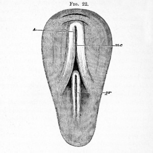
In front of the head-fold, quite out of the region of the medullary folds, there is usually another small fold formed earlier than the head-fold, which is the beginning of the amnion (Fig. 22).
The appearance of the embryo and its relation to the surrounding parts are somewhat diagrammatically represented in Fig. 22. The primitive streak now ends with an anterior swelling (not represented in the figure), and is usually somewhat unsymmetrical. In most cases its axis is more nearly continuous with the left, or rarely the right, medullary fold than with the medullary groove. In sections its front end appears as a ridge on one side or rarely in the middle of the floor of the wide medullary groove.
The general structure of the developing embryo at the present stage is best understood from such a section as that represented in Fig. 21. The medullary groove (m. c.) lined by thickened epiblast is seen in the median line of the section. Below it is placed the notochord (cA), which at this stage is a mere rod of cells, and on each side are situated the mesoblastic plates (B). The hypoblast forms a continuous and nearly flat layer below.
While the changes just described have been occurring in the area pellucida, the growth of the area opaca has also progressed actively. The epiblast has greatly extended itself, and important changes have taken place in the constitution of the germinal wall already spoken of.
The mesoblast and hypoblast of the area opaca do not arise by simple extension of the corresponding layers of the area pellucida ; but the whole of the hypoblast of the area opaca, and a large portion of the mesoblast, and possibly even some of the epiblast, take their origin from the peculiar material which forms the germinal wall and which is continuous with the hypoblast at the edge of the area opaca (vide figs. 15, 17, 18, 19, 20).
The exact nature of this material has been the subject of many controversies. Into these controversies it is not our purpose to enter, but subjoined are the results of our own examination. The germinal wall first consists, as already mentioned, of the lower cells of the thickened edge of the blastoderm, and of the subjacent yolk material with nuclei. During the period before the formation of the primitive streak the epiblast appears to extend itself over the yolk, partly at the expense of the cells of the germinal wall, and possibly even of cells formed around the nuclei in this part. The cells of the germinal wall, which are at first well separated from the yolk below, become gradually absorbed in the growth of the hypoblast, and the remaining cells and yolk then become mingled together, and constitute a compound structure, continuous at its inner border with the hypoblast. This structure is the germinal wall usually so described. It is mainly formed of yolk granules with numerous nuclei, and a somewhat variable number of rather large cells imbedded amongst them. The nuclei, some of which are probably enclosed by a definite cell body, typically form a special layer immediately below the epiblast. A special mass of nuclei (vide Figs. 18 and 20, n) is usually present at the junction of the hypoblast with the germinal wall.
The germinal wall retains the characters just enumerated till near the close of the first day of incubation. One function of its cells appears to be the absorption of yolk material for the growth of the embryo.
The chief events then of the second period of the first day are the appearance of the medullary folds and groove, the formation of the notochord and lateral plates of mesoblast, the beginning of the head-fold and amnion, and the histological changes taking place in the several layers.
From the 20th to the 24th hour
A view of the embryo during this period is given in Fig. 23. The head-fold enlarges rapidly, the crescentic groove becoming deeper, while at the same time the overhanging margin of the groove (the upper limb of the g), rises up above the level of the blastoderm ; in fact, the formation of the head of the embryo may now be said to have definitely begun.
The medullary folds, increasing in size in every dimension, but especially in height, lean over from either side towards the middle line, and thus tend more and more to roof in the medullary canal, especially near the head. About the end of the first day they come into direct contact in the region which will afterwards become the brain, though they do not as yet coalesce. In this way a tubular canal is formed. This is the medullary or neural canal (Fig. 23, Fig. 24, Mc.).
It is not completely closed in till a period considerably later than the one we are considering.
Meanwhile important changes are taking place in the axial portions of the mesoblast, which lie on each side of the notochord beneath the medullary folds.
In an embryo of the middle period of this day, examined with transmitted light, the notochord is seen at the bottom of the medullary groove between the medullary folds, as a transparent line shining through the floor of the groove when the embryo is viewed from above. On either side of the notochord the body of the embryo appears somewhat opaque, owing to the thickness of the medullary folds; as these folds slope away outwards on either side, so the opacity gradually fades away in the pellucid area. There is present at the sides no sharp line of demarcation between the body of the embryo and the rest of the area; nor will there be any till the lateral folds make their appearance ; and transverse vertical sections shew (Fig. 21) that there is no break in the mesoblast, from the notochord to the margin of the pellucid area, but only a gradual thinning.
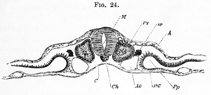
During the latter period of the day, however, the plates of mesoblast on either side of the notochord begin to be split horizontally into two layers, the one of which attaching itself to the epiblast, forms with it the somatopleure (shewn for a somewhat later stage in Fig. 24), while the other, attaching itself to the hypoblast, forms with it the splanchnopleure. By the separation of these two layers from each other, a cavity (Pp), containing fluid only, and more conspicuous in certain parts of the embryo than in others, is developed. This cavity is the beginning of that great serous cavity of the body which afterwards becomes divided into separate cavities. We shall speak of it as the pleuro-peritoneal cavity.
This cleavage into somatopleure and splanchnopleure extends close up to the walls of the medullary canal, but close to the medullary canal a central or axial portion of each plate becomes marked off by a slight constriction from the peripheral (Fig. 24), and receives the name of vertebral plate, the more external mesoblast being called the lateral plate. The cavity between the two layers of the lateral plate rapidly enlarges, while that in the vertebral plate remains in the condition of a mere split.
Fig. 24. Transverse section through the dorsal region op an embryo of the second day.
- (copied from His), introduced here to illustrate the formation of the mesoblastic somite, and the cleavage of the mesoblast. M. medullary canal ; Pv. mesoblastic somite ; w. rudiment of Wolffian duct; A. epiblast; C. hypoblast ; Ch. notochord ; Ao. aorta ; BO. splanchnopleure.
At first each vertebral plate is not only unbroken along its length, but also continuous at its outer edge with the upper and lower layers of the lateral plate of the same side. Very soon, however, clear transverse lines are seen, in surface views (Fig. 23), stretching inwards across each vertebral plate from the edge of the lateral plate towards the notochord ; while a transparent longitudinal line makes its appearance on either side of the notochord along the line of junction of the lateral with the vertebral plate.
The transverse lines are caused by the formation of vertical clefts, that is to say, narrow spaces containing nothing but clear fluid ; and sections shew that they are due to breaches of continuity in the mesoblast only, the epiblast and hypoblast having no share in the matter.
Thus each vertebral plate appears in surface views to be cut up into a series of square plots, bounded by transparent lines (Fig. 23). Each square plot is the surface of a corresponding cubical mass (Fig. 24, Pv.). The two such cubical masses first formed, lying one on each side of the notochord, beneath and a little to the outside of the medullary folds, are the first pair of mesoblastic somites1.
The mesoblastic somites form the basis out of which the voluntary muscles of the trunk and the bodies of the vertebrae are formed.
The first somite rises close to the anterior extremity of the primitive streak, but the next is stated to arise in front of this, so that the first-formed somite corresponds to the second permanent vertebra. The region of the embryo in front of the second formed somite at first the largest part of the whole embryo is the cephalic region (Fig. 23). The somites following the second are formed in regular succession from before backwards, out of the unsegmented mesoblast of the posterior end of the embryo, which rapidly grows in length to supply the necessary material. With the growth of the embryo the primitive streak is continually carried back, the lengthening of the embryo always taking place between the front end of the primitive streak and the last somite ; and during this (1These bodies are frequently called protovertebra, but we shall employ for them the term mesoblastic somites.)
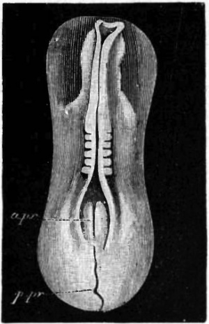
process the primitive streak undergoes important changes both in itself and in its relation to the embryo. Its anterior thicker part, which is embraced by the diverging medullary folds, soon becomes distinguished in structure from the posterior part, and is placed symmetrically in relation to the axis of the embryo, (Fig. 23 a.pr)-, at the same time the medullary folds, which at first simply diverge on each side of the primitive streak, bend in again and meet behind so as completely to enclose this front part of the primitive streak. The region, where the medullary folds diverge, is known as the sinus rhomboidalis of the embryo bird, though it has no connection with the similarly named structure in the adult.
This is a convenient place to notice remarkable appearances which present themselves close to the junction of the neural plate and the primitive streak. These are temporary passages leading from the hinder end of the neural groove or tube into the alimentary canal. They vary somewhat in different species of birds, and it is possible that in some species there may be several openings of the kind, which appear one after the other and then close again. They were first discovered by Gasser, and are spoken of as the neur "enteric passages or canals 1 . In all cases, with some doubtful exceptions, they lead round the posterior end of the notochord, or through the point where the notochord falls into the primitive streak.
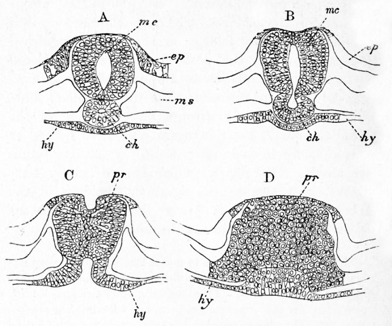
The largest of these passages is present in the embryo duck with twenty-six mesoblastic somites, and is represented in the series of sections (Fig. 25). The passage leads obliquely backwards and ventralwards from the hind end of the neural tube into the notochord, where the latter joins the primitive streak (B). A narrow diverticulum from this passage is continued forwards for a short distance along the axis of the notochord (A, cfi). After traversing the notochord, the passage is continued into a hypoblastic diverticulum, which opens ventrally into the future lumen of the alimentary tract (C). Shortly behind the point where the neurenteric passage communicates with the neural tube the latter structure opens dorsally, and a groove on the surface of the primitive streak is continued backwards from it for a short distance (C). The first part of this passage to appear is the hypoblastic diverticulum above mentioned.
(1 "Die Primitivstreifen bei Vogelembryonen." Schrift. d. Gesell. z. Beford d. Gesammten Naturwiss. zu Marburg. Vol. u. Supplement i. 1879.)
Fig. 25. Four transverse sections through the neurenteric passage and adjoining parts in a duck embryo with twenty-six mesoblastc somites.
- A. Section in front of the neurenteric canal, shewing a lumen in the notochord.
- B. Section through the passage from the medullary canal into the notochord.
- C. Section shewing the hypoblastic opening of the neurenteric canal, and the groove on the surface of the primitive streak, which opens in front into the medullary canal.
- D. Primitive streak immediately behind the opening of the neurenteric passage.
- me. medullary canal ; ep. epi blast ; hy. hypoblast ; ch. notochord ; pr. primitive streak.
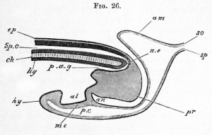
In the chick we have found in some cases an incomplete passage prior to the formation of the first somite. At a later stage there is a perforation on the floor of the neural canal, which is not so marked as those in the goose or duck, and never results in a complete continuity between the neural and alimentary tracts ; but simply leads from the floor of the neural canal into the tissues of the tail-swelling, and thence into a cavity in the posterior part of the notochord. The hinder diverticulum of the neural canal along the line of the primitive groove is, moreover, very considerable in the chick, and is not so soon obliterated as in the goose. The incomplete passage in the chick arises at a period when about twelve somites are present. The third passage is formed in the chick during the third day of incubation.
The anterior part of the primitive streak becomes converted into the tail-swelling ; the groove of the posterior part gradually shallows and finally disappears. The hinder part itself atrophies from behind forwards, and in the course of the folding off of the embryo from the yolk the part of the blastoderm where it was placed becomes folded in, so as to form part of the ventral wall of the embryo. The apparent hinder part of the primitive streak is therefore in reality ventral and anterior in relation to the embryo.
Since the commencement of incubation the area opaca has been spreading outwards over the surface of the yolk, and by the end of the first day has reached about the diameter of a sixpence. It appears more or less mottled over the greater part of its extent, but this is more particularly the case with the portion lying next to the pellucid area ; so much so, that around the pellucid area an inner ring of the opaque area may be distinguished from the rest by the difference of its aspect.
The mottled appearance of this inner ring is due to changes taking place in the mesoblast above the germinal wall changes which eventually result in the formation of what is called the vascular area, the outer border of which marks the extreme limit to which the mesoblast extends.
Summary Day 1
The changes then which occur during the first day may thus be briefly summarized :
- The hypoblast is formed as a continuous layer of plate-like cells from the lower layer of the segmentation spheres.
- The primitive streak is formed in the hinder part of the area pellucida as a linear proliferation of epiblast cells. These cells spread out as a layer on each side of the primitive streak, and form part of the mesoblast.
- The primitive groove is formed along the axis of the primitive streak.
- The pellucid area becomes pear-shaped, the broad end corresponding with the future head of the embryo. Its long axis lies at right angles to the long axis of the egg.
- The medullary plate with the medullary groove makes its appearance in front of the primitive groove.
- The primitive hypoblast in the region of the medullary plate gives rise to an axial rod of cells forming the notochord, and to two lateral plates of mesoblast. The innermost stratum of the primitive layer forms the permanent hypoblast.
- The development of the head-fold gives rise to the first definite appearance of the head.
- The medullary folds rise up and meet first in the region of the mid-brain to form the neural tube.
- By the cleavage of the mesoblast, the somatopleure separates from the splanchnopleure.
- One or more pairs of mesoblastic somites make their appearance in the vertebral portion of the mesoblastic plates.
- The first trace of the amnion appears in front of the head-fold.
- The vascular area begins to be distinguished from the rest of the opaque area.
The Elements of Embryology - Volume 1 (1883)
The History of the Chick: Egg structure and incubation beginning | Summary whole incubation | First day | Second day - first half | Second day - second half | Third day | Fourth day | Fifth day | Sixth day to incubation end | Appendix
| Historic Disclaimer - information about historic embryology pages |
|---|
| Pages where the terms "Historic" (textbooks, papers, people, recommendations) appear on this site, and sections within pages where this disclaimer appears, indicate that the content and scientific understanding are specific to the time of publication. This means that while some scientific descriptions are still accurate, the terminology and interpretation of the developmental mechanisms reflect the understanding at the time of original publication and those of the preceding periods, these terms, interpretations and recommendations may not reflect our current scientific understanding. (More? Embryology History | Historic Embryology Papers) |
Glossary Links
- Glossary: A | B | C | D | E | F | G | H | I | J | K | L | M | N | O | P | Q | R | S | T | U | V | W | X | Y | Z | Numbers | Symbols | Term Link
Cite this page: Hill, M.A. (2024, April 27) Embryology Book - The Elements of Embryology - Chicken 3. Retrieved from https://embryology.med.unsw.edu.au/embryology/index.php/Book_-_The_Elements_of_Embryology_-_Chicken_3
- © Dr Mark Hill 2024, UNSW Embryology ISBN: 978 0 7334 2609 4 - UNSW CRICOS Provider Code No. 00098G


